Chest Radiography. CRC 330 Cardiorespiratory Care University of South Alabama
|
|
|
- Gyles Gilmore
- 7 years ago
- Views:
Transcription
1 Chest Radiography CRC 330 Cardiorespiratory Care University of South Alabama
2 Objectives Cite the indications for a chest radiograph Describe the radiographic views Describe the systematic review of a chest radiograph Define, list clinical signs, and radiographic evidence of common respiratory problems Identify objects implanted in the chest by radiography
3 Indications for a Chest Radiograph Detect lung pathology Atelectasis, pneumothorax, confirm physical examination Determine appropriate therapy Hyperinflation, chest tube, PEEP Evaluate effectiveness of therapy Determine position of invasive tubes Vascular catheters, chest and N-G tube, ETT Trends disease progression

4 Radiographic Views Posterior-anterior Chest against the film Standard Correct heart size
5 Radiographic Views Anterior-posterior Back against film Portable Enlarges heart

6 Radiographic Views Lateral Decubitus Patient positioned on the side Identifies fluid levels

7 Radiographic Views Lateral Upright Left side against film Sharp view of LLL Identifies presence of fluid in the posterior sulcus Identifies hyperinflation/ increased A-P diameter Retrosternal air

8 Radiographic Views Lateral neck Evaluates periglottic edema Croup Epiglottitis
9 Chest Radiograph Evaluation Systematic Four densities Rotation Exposure Diaphragms Heart shadow Costophrenic angles Ribs Clavicles Lung and vascular markings Trachea and carina Stomach bubble Scapulas
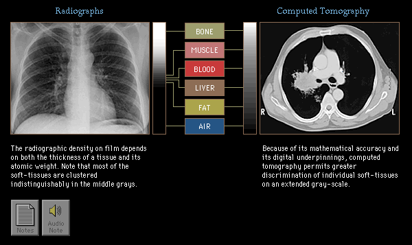
10 Densities
11 Rotation Film should be symmetrical Note position of clavicles Trachea should be midline
12 Exposure Determine the visibility of the intervertebral discs through the heart shadow Lucency of pulmonary blood vessels Overexposed= overdarkened Underexposed= lack of visualization of vertebral bodies through the heart Lungs whiter than normal
13 Normal Chest Radiograph Right diaphragm higher than left Heart shadow < ½ thoracic width Air-filled costophrenic angles
14 Normal Chest Radiograph Posterior ribs are most lucent and horizontal Clavicles are symmetric Lung and vascular markings
15 Normal Chest Radiograph Trachea and carina visible Stomach bubble under left diaphragm Scapulas produce a vertical line through lung fields
16 Atelectasis Definition Loss of air due to compression or absorption Clinical signs Tachypnea, decreased BS Radiology Fissure and mediastinal shift, vascular crowding, increased density of lung fields

17 Hyperinflation Definition Increased air in the chest Clinical signs Barrel chest, increased vocal fremitus, hyperresonance Radiology Increased lucency, depressed diaphragms, narrow heart. Increased retrosternal air

18 Interstitial Lung Disease Definition Diffuse, scarring, alveolar injury Clinical signs Dyspnea, dry cough, increased fremitus, fine crackles Radiography Alveolar filling, granular
19 Pulmonary Edema Definition Fluid in interstitial or alveolar space, diffuse, increased interstitial pressure or permeability Clinical signs Frothy sputum, dyspnea, crackles Radiology Fluffy, enlarged heart in CHF, increased hilar markings

20 Pleural Effusion Definition Fluid in the pleural space Clinical signs Tachypnea, decreased fremitus, decreased BS Radiology Blunted costophrenic angles, air-fluid line
21 Consolidation Definition Fluid or pus filled airspaces without alveolar collapse Clinical signs Tachypnea, fever, decreased fremitus, crackles Radiology Lobar, homogenous, air bronchograms

22 Silhouette Sign In consolidation, there is a loss of border between two objects of similar density Helps determine where the abnormality is Dotted lines indicate loss of border

23 Pneumothorax Definition Air in the pleural space Clinical signs Acute respiratory distress, asymmetrical chest movement, decreased fremitus, hyperresonance, decreased BS Radiology Vertical line, no lung markings outside the line

24 Endotracheal Tube Placement Tip should be 3-5 cm above the carina Here, tip is in Right mainstem Assure equal BS Care in advancing the tube

25 Pulmonary Artery Catheter Catheter proceeds through the VC, RA, RV, and into the PA Catheter should reside in zone 3

26 Chest (Pleural) Tube Inserted by the physician Evacuation of air and/or fluid 32 F Radiopaque stripe Note the ETT

27 Laryngotracheobronchitis (Croup) Definition Viral infection of the larynx and trachea Clinical signs 6mo-3yoa, brassy hoarse cough, stridor, low fever Radiology Narrowing and edema of subglottic trachea, ballooning of hypopharynx

28 Computerized Tomography (CT) Radiographs that focus on slices of the organ being scanned Computer enhanced to improve resolution Able to define small abnormalities and lesions (cancer)
Administrative. Patient name Date compare with previous Position markers R-L, upright, supine Technical quality
 CHEST X-RAY Administrative Patient name Date compare with previous Position markers R-L, upright, supine Technical quality AP or PA ( with x-ray beam entering from back of patient, taken at 6 feet) Good
The silhouette sign. Dr Etienne Leroy-Terquem Centre hospitalier de Meulan les Mureaux. France French-cambodian association for pneumology (OFCP)
") The silhouette sign Dr Etienne Leroy-Terquem Centre hospitalier de Meulan les Mureaux. France French-cambodian association for pneumology (OFCP) The silhouette sign When 2 opacities of the same density
The silhouette sign Dr Etienne Leroy-Terquem Centre hospitalier de Meulan les Mureaux. France French-cambodian association for pneumology (OFCP) The silhouette sign When 2 opacities of the same density
Chest X-ray STUDY GUIDE 2014-2015
 Internal Medicine Clerkship Chest X-ray STUDY GUIDE 2014-2015 10 Steps/ABC s for Reading Chest X-Rays & Table of Contents to be used with CXR CD-ROM #1 10 Steps / ABC s for Reading Chest X-Rays 0 Normal
Congestive Heart Failure
 William Herring, M.D. 2002 Congestive Heart Failure In Slide Show mode, to advance slides, press spacebar or click left mouse button Congestive Heart Failure Causes of Coronary artery disease Hypertension
Pulmonary Patterns VMA 976
 Pulmonary Patterns VMA 976 PULMONARY PATTERNS Which pulmonary patterns are commonly described in veterinary medicine? PULMONARY PATTERNS Normal Alveolar Interstitial Structured/Nodular Unstructured Bronchial
NORMAL CHEST RADIOGRAPHY. Front and lateral view
 NORMAL CHEST RADIOGRAPHY Front and lateral view Dr Etienne Leroy-Terquem Centre hospitalier de Meulan les Mureaux. France French-cambodian association for pneumology (OFCP) OFCP How to obtain a good quality
CERVICAL MEDIASTINOSCOPY WITH BIOPSY
 INFORMED CONSENT INFORMATION ADDRESSOGRAPH DATA CERVICAL MEDIASTINOSCOPY WITH BIOPSY You have decided to have an important procedure and we appreciate your selection of UCLA Healthcare to meet your needs.
Radiation-Induced Lung Injury
 May 2001 Radiation-Induced Lung Injury Warren Phipps, Harvard Medical School Year III Our Patient D.C. is a 50 year-old woman with a 30-pack year history of smoking who presented to the ED because she
Lung & Thorax Exams. Charlie Goldberg, M.D. Professor of Medicine, UCSD SOM cggoldberg@ucsd.edu
 Lung & Thorax Exams Charlie Goldberg, M.D. Professor of Medicine, UCSD SOM cggoldberg@ucsd.edu Lung Exam Includes Vital Signs & Cardiac Exam 4 Elements (cardiac & abdominal too) Observation Palpation Percussion
CHEST TUBES AND CHEST DRAINAGE SYSTEMS
 CHEST TUBES AND CHEST DRAINAGE SYSTEMS Central Nursing Orientation April 2008 Revised September 2011 OBJECTIVES Describe common tubes and indications for use at LHSC Review indications and contraindications,
Chapter 2 - Anatomy & Physiology of the Respiratory System
 Chapter 2 - Anatomy & Physiology of the Respiratory System Written by - AH Kendrick & C Newall 2.1 Introduction 2.2 Gross Anatomy of the Lungs, 2.3 Anatomy of the Thorax, 2.4 Anatomy and Histology of the
General Thoracic Surgery ICD9 to ICD10 Crosswalks. C34.11 Malignant neoplasm of upper lobe, right bronchus or lung
 ICD-9 Code ICD-9 Description ICD-10 Code ICD-10 Description 150.3 Malignant neoplasm of upper third of esophagus C15.3 Malignant neoplasm of upper third of esophagus 150.4 Malignant neoplasm of middle
Standards for Chest Radiography
 Standards for Chest Radiography First adopted in 1994 and revised in June 2000 These standards were prepared by the Chest Expert Advisory Panel of the CAR: Craig L. Coblentz, M.D., Chair, Frederick Matzinger,
PNEUMOMEDIASTINUM: A PATIENT PRESENTATION
 November 2002 PNEUMOMEDIASTINUM: A PATIENT PRESENTATION Alden Chip McDonald, III Harvard Medical School, Year III AGENDA I. Patient Presentation II. Diagnosis of Pneumomediastinum III. Causes of Pneumomediastinum
R/F. Efforts to Reduce Exposure Dose in Chest Tomosynthesis Targeting Lung Cancer Screening. 3. Utility of Chest Tomosynthesis. 1.
 R/F Efforts to Reduce Exposure Dose in Chest Tomosynthesis Targeting Lung Cancer Screening Department of Radiology, National Cancer Center Hospital East Kaoru Shimizu Ms. Kaoru Shimizu 1. Introduction
Disease/Illness GUIDE TO ASBESTOS LUNG CANCER. What Is Asbestos Lung Cancer? www.simpsonmillar.co.uk Telephone 0844 858 3200
 GUIDE TO ASBESTOS LUNG CANCER What Is Asbestos Lung Cancer? Like tobacco smoking, exposure to asbestos can result in the development of lung cancer. Similarly, the risk of developing asbestos induced lung
MECHINICAL VENTILATION S. Kache, MD
 MECHINICAL VENTILATION S. Kache, MD Spontaneous respiration vs. Mechanical ventilation Natural spontaneous ventilation occurs when the respiratory muscles, diaphragm and intercostal muscles pull on the
Chapter 2 Cardiac Interpretation of Pediatric Chest X-Ray
 Chapter 2 Cardiac Interpretation of Pediatric Chest X-Ray Ra-id Abdulla and Douglas M. Luxenberg Key Facts The cardiac silhouette occupies 50 55% of the chest width on an anterior posterior chest X-ray
Chest radiography is the most
 ... Chest Radiography for Radiologic Technologists DAN L. HOBBS, M.S.R.S., R.T.(R)(CT)(MR) The chest exam is performed more frequently than any other exam in the imaging department. It is important for
Lung Cancer. This reference summary will help you better understand lung cancer and the treatment options that are available.
 Lung Cancer Introduction Lung cancer is the number one cancer killer of men and women. Over 165,000 people die of lung cancer every year in the United States. Most cases of lung cancer are related to cigarette
General Information About Non-Small Cell Lung Cancer
 General Information About Non-Small Cell Lung Cancer Non-small cell lung cancer is a disease in which malignant (cancer) cells form in the tissues of the lung. The lungs are a pair of cone-shaped breathing
Management of Chest Tubes and Air Leaks after Lung Resection
 Management of Chest Tubes and Air Leaks after Lung Resection Emily Kluck PA-C The Johns Hopkins Hospital Baltimore, MD AATS 2014, Toronto, CAN April 2014 Management of Chest Tubes 1 Overview Review the
X-ray (Radiography) - Chest
 - Chest") Scan for mobile link. X-ray (Radiography) - Chest What is a Chest X-ray (Chest Radiography)? The chest x-ray is the most commonly performed diagnostic x-ray examination. A chest x-ray produces images of
Scan for mobile link. X-ray (Radiography) - Chest What is a Chest X-ray (Chest Radiography)? The chest x-ray is the most commonly performed diagnostic x-ray examination. A chest x-ray produces images of
N26 Chest Tubes 5/9/2012
 Thoracic cavity, pleural space 1 Conditions requiring chest drainage_1 Air between the pleurae is a pneumothorax Occurs when there is an opening on the surface of the lung or in the airways, y, in the
Lung Cancer: Diagnosis, Staging and Treatment
 PATIENT EDUCATION patienteducation.osumc.edu Lung Cancer: Diagnosis, Staging and Treatment Cancer begins in our cells. Cells are the building blocks of our tissues. Tissues make up the organs of the body.
April 2015 CALGARY ZONE CLINICAL REFERENCE PULMONARY CENTRAL ACCESS & TRIAGE
 April 2015 CALGARY ZONE CLINICAL REFERENCE CENTRAL ACCESS & TRIAGE Introduction Pulmonary consulting services are organized through the Calgary Zone Pulmonary Central Access and Triage (PCAT). Working
X-ray (Radiography), Chest
, Chest") X-ray (Radiography), Chest What is a Chest X-ray (Chest Radiography)? The chest x-ray is the most commonly performed diagnostic x-ray examination. A chest x-ray makes images of the heart, lungs, airways,
X-ray (Radiography), Chest What is a Chest X-ray (Chest Radiography)? The chest x-ray is the most commonly performed diagnostic x-ray examination. A chest x-ray makes images of the heart, lungs, airways,
RESPIRATORY VENTILATION Page 1
 Page 1 VENTILATION PARAMETERS A. Lung Volumes 1. Basic volumes: elements a. Tidal Volume (V T, TV): volume of gas exchanged each breath; can change as ventilation pattern changes b. Inspiratory Reserve
MEDICAL REPORT MEDICAL HISTORY QUESTIONS
 MEDICAL HISTORY QUESTIONS PAGE 1 OF 7 IF YOUR ANSWER IS YES TO ANY OF THE FOLLOWING QUESTIONS, PLEASE PROVIDE ADDITIONAL INFORMATION INCLUDING: DIAGNOSIS, DATE, AND TREATMENT (INCLUDING MEDICATIONS AND/OR
Linfoma maligno pulmonar tratado com Nerium oleander. http://www.drozel.org/eng/diagnosis_malignant_mg.htm CASE REPORT
 Linfoma maligno pulmonar tratado com Nerium oleander http://www.drozel.org/eng/diagnosis_malignant_mg.htm CASE REPORT Diagnosis: Malignant lymphoma, lung cancer A 60-year-old woman experienced pain in
Mesothelioma. 1995-2013, The Patient Education Institute, Inc. www.x-plain.com ocft0101 Last reviewed: 03/21/2013 1
 Mesothelioma Introduction Mesothelioma is a type of cancer. It starts in the tissue that lines your lungs, stomach, heart, and other organs. This tissue is called mesothelium. Most people who get this
Basic Data. 鍾 XX, female Age:59 y/o
 Basic Data 鍾 XX, female Age:59 y/o Chief complain for evaluation of the left pleural mass Clinical course This 59 years old lady Hypertension for 3 years, under herbal control. An episode of high BP up
UW MEDICINE PATIENT EDUCATION. Aortic Stenosis. What is heart valve disease? What is aortic stenosis?
 UW MEDICINE PATIENT EDUCATION Aortic Stenosis Causes, symptoms, diagnosis, and treatment This handout describes aortic stenosis, a narrowing of the aortic valve in your heart. It also explains how this
Neonatal Intubation. Purpose. Scope. Indications. Equipment Cardiorespiratory monitor SaO 2 monitor. Anatomic Considerations.
 Page 1 of 5 Purpose Scope Indications Neonatal Intubation To assure proper placement of endotracheal tubes for maximum ventilation using proper intubation procedures. The policy applies to all Respiratory
Evaluation and treatment of emphysema in a preterm infant
 ISPUB.COM The Internet Journal of Pediatrics and Neonatology Volume 11 Number 1 Evaluation and treatment of emphysema in a preterm infant T Saad, P Chess, W Pegoli, P Katzman Citation T Saad, P Chess,
Case 2. 30 year old involved in a MVA complaining of chest pain. Bruising over the right upper chest. Your Diagnosis
 Case 2 30 year old involved in a MVA complaining of chest pain. Bruising over the right upper chest. Your Diagnosis Diagnosis: Posterior Sterno-clavicular dislocation [PSCD] A posterior sterno-clavicular
ENT Emergencies. Injuries of the Neck. Registrar Dept Trauma and emergency Medicine Tygerberg Hospital
 ENT Emergencies Injuries of the Neck Registrar Dept Trauma and emergency Medicine Tygerberg Hospital Neck Injuries Blunt and Penetrating Trauma Blunt Injuries Blunt trauma direct/indirect Trauma to larynx
A. function: supplies body with oxygen and removes carbon dioxide. a. O2 diffuses from air into pulmonary capillary blood
 A. function: supplies body with oxygen and removes carbon dioxide 1. ventilation = movement of air into and out of lungs 2. diffusion: B. organization a. O2 diffuses from air into pulmonary capillary blood
HEALTH CARE FOR EXPOSURE TO ASBESTOS. 2010 The SafetyNet Centre for Occupational Health and Safety Research Memorial University www.safetynet.mun.
 HEALTH CARE FOR PATIENTS WITH EXPOSURE TO ASBESTOS 2010 The SafetyNet Centre for Occupational Health and Safety Research Memorial University www.safetynet.mun.ca HEALTH CARE FOR PATIENTS WITH EXPOSURE
SUMMARY OF S.B. 15 ASBESTOS/SILICA LITIGATION REFORM BILL
 SUMMARY OF S.B. 15 ASBESTOS/SILICA LITIGATION REFORM BILL S.B. 15, the asbestos/silica litigation reform bill, distinguishes between the claims of people who are physically impaired or sick due to exposure
 P R E S E N T S Dr. Mufa T. Ghadiali is skilled in all aspects of General Surgery. His General Surgery Services include: General Surgery Advanced Laparoscopic Surgery Surgical Oncology Gastrointestinal
Radiography provides a rapid, noninvasive
 P ro c e d u re s P ro I M G I N G Peer Reviewed Clifford R. erry, DVM, Diplomate CVR University of Florida Interpreting Small nimal Thoracic Radiographs Radiography provides a rapid, noninvasive mechanism
Acute heart failure may be de novo or it may be a decompensation of chronic heart failure.
 Management of Acute Left Ventricular Failure Acute left ventricular failure presents as pulmonary oedema due to increased pressure in the pulmonary capillaries. It is important to realise though that left
Online supplements are not copyedited prior to posting.
 Functional Impact of a Spectrum of Interstitial Lung Abnormalities in Rheumatoid Arthritis Tracy J. Doyle, MD, MPH; Paul F. Dellaripa, MD; Kerri Batra, MD; Michelle L. Frits, BA; Christine K. Iannaccone,
Respiratory Assessment for Nurses (part two)
") Respiratory Assessment for Nurses (part two) Introduction Part one of Respiratory Assessment for Nurses outlined the importance of appropriate respiratory assessment to improve care outcomes for the acutely
Respiratory Assessment for Nurses (part two) Introduction Part one of Respiratory Assessment for Nurses outlined the importance of appropriate respiratory assessment to improve care outcomes for the acutely
 Pulmonary Complications from Lung Cancer Treatment, Part 1: Chemotherapy-Induced Pneumonitis by Dr. Gerard Silvestri, Medical University of South Carolina Dr. West: Hello, and welcome to our webinar with
Basic techniques of pulmonary physical therapy (I) 100/04/24
 100/04/24") Basic techniques of pulmonary physical therapy (I) 100/04/24 Evaluation of breathing function Chart review History Chest X ray Blood test Observation/palpation Chest mobility Shape of chest wall Accessory
Basic techniques of pulmonary physical therapy (I) 100/04/24 Evaluation of breathing function Chart review History Chest X ray Blood test Observation/palpation Chest mobility Shape of chest wall Accessory
6. Histopathology of Alveoli 7. Surfactant 8. Blood supply of lungs 9. Lymphatics of Lungs 10. Nerve supply of Lungs 11. Pleura 12.
 ANATOMY OF LUNGS - 1. Gross Anatomy of Lungs 2. Surfaces and Borders of Lungs 3. Hilum and Root of Lungs 4. Fissures and Lobes of Lungs 5. Bronchopulmonary segments 6. Histopathology of Alveoli 7. Surfactant
Sample Learning Objectives for a Medical School Radiology Curriculum: Listed by Subjects
 Sample Learning Objectives for a Medical School Radiology Curriculum: Listed by Subjects This document lists sample learning objectives by subject matter The numerical ranking in parenthesis following
Non-Small Cell Lung Cancer
 Non-Small Cell Lung Cancer About Your Lungs and Lung Cancer How do your lungs work? To understand lung cancer it is helpful to understand your lungs. Your lungs put oxygen into the blood, which the heart
NEEDLE THORACENTESIS Pneumothorax / Hemothorax
 NEEDLE THORACENTESIS Pneumothorax / Hemothorax By: Steven Jones, NREMT-P Pneumothorax Pneumothorax is a collection of air or gas in the pleural space of the lung, causing the lung to collapse. Pneumothorax
The abnormal chest X-ray when to refer to a specialis t
 The abnormal chest X-ray when to refer to a specialis t An abnormal chest X-ray must be followed up. OLGA MZILENI, MB ChB, MMed (Int Med) Professor and Head of Internal Medicine and Pulmonology, University
New Cardiothoracic Surgery CPT Codes for 2013
 New Cardiothoracic Surgery CPT Codes for 2013 There were several changes to the cardiothoracic surgery CPT codes for 2013. There are five new codes in the general thoracic surgery section, with one revised
Peripherally Inserted Central Catheter (PICC) for Outpatient
 for Outpatient") Peripherally Inserted Central Catheter (PICC) for Outpatient Introduction A Peripherally Inserted Central Catheter, or PICC line, is a thin, long, soft plastic tube inserted into a vein of the arm. It
Peripherally Inserted Central Catheter (PICC) for Outpatient Introduction A Peripherally Inserted Central Catheter, or PICC line, is a thin, long, soft plastic tube inserted into a vein of the arm. It
Abdomen X-Ray (AXR) Collimation is ideally from diaphragms to lower border of the symphysis pubis and the lateral skin margins.
 Collimation is ideally from diaphragms to lower border of the symphysis pubis and the lateral skin margins.") Abdomen X-Ray (AXR) Collimation is ideally from diaphragms to lower border of the symphysis pubis and the lateral skin margins. LMP of child-bearing age female patients should be checked. 1. Acute abdomen
Abdomen X-Ray (AXR) Collimation is ideally from diaphragms to lower border of the symphysis pubis and the lateral skin margins. LMP of child-bearing age female patients should be checked. 1. Acute abdomen
Asbestos Disease: An Overview for Clinicians Asbestos Exposure
 Asbestos Asbestos Disease: An Overview for Clinicians Asbestos Exposure Asbestos: A health hazard Exposure to asbestos was a major occupational health hazard in the United States. The first large-scale
INTERNATIONAL TRAUMA LIFE SUPPORT
 INTERNATIONAL TRAUMA LIFE SUPPORT NEEDLE DECOMPRESSION OF TENSION PNEUMOTHORAX Roy Alson, MD, PhD, FACEP, FAAEM and Sabina Braithwaite, MD, MPH, FACEP INTRODUCTION The purpose of this document is to update
A Patient s Guide to Diffuse Idiopathic Skeletal Hyperostosis (DISH)
") A Patient s Guide to Diffuse Idiopathic Skeletal Hyperostosis (DISH) Introduction Diffuse Idiopathic Skeletal Hyperostosis (DISH) is a phenomenon that more commonly affects older males. It is associated
A Patient s Guide to Diffuse Idiopathic Skeletal Hyperostosis (DISH) Introduction Diffuse Idiopathic Skeletal Hyperostosis (DISH) is a phenomenon that more commonly affects older males. It is associated
Neoplasms of the LUNG and PLEURA
 Neoplasms of the LUNG and PLEURA 2015-2016 FCDS Educational Webcast Series Steven Peace, BS, CTR September 19, 2015 2015 Focus o Anatomy o SSS 2000 o MPH Rules o AJCC TNM 1 Case 1 Case Vignette HISTORY:
Minimally Invasive Spine Surgery
 Chapter 1 Minimally Invasive Spine Surgery 1 H.M. Mayer Primum non nocere First do no harm In the long history of surgery it always has been a basic principle to restrict the iatrogenic trauma done to
CHAPTER 1: THE LUNGS AND RESPIRATORY SYSTEM
 CHAPTER 1: THE LUNGS AND RESPIRATORY SYSTEM INTRODUCTION Lung cancer affects a life-sustaining system of the body, the respiratory system. The respiratory system is responsible for one of the essential
written by Harvard Medical School COPD It Can Take Your Breath Away www.patientedu.org/copd
 written by Harvard Medical School COPD It Can Take Your Breath Away www.patientedu.org/copd What Is COPD? COPD stands for chronic obstructive pulmonary disease. There are two major diseases included in
Small cell lung cancer
 Small cell lung cancer Small cell lung cancer is a disease in which malignant (cancer) cells form in the tissues of the lung. The lungs are a pair of cone-shaped breathing organs that are found within
Upper Cervical Spine - Occult Injury and Trigger for CT Exam
 Upper Cervical Spine - Occult Injury and Trigger for CT Exam Bakman M, Chan K, Bang C, Basu A, Seo G, Monu JUV Department of Imaging Sciences University of Rochester Medical Center, Rochester, NY Introduction
Computed tomographic atlas for the new international lymph node map for lung cancer: A radiation oncologist perspective
 Practical Radiation Oncology (2013) 3, 54 66 www.practicalradonc.org Special Article Computed tomographic atlas for the new international lymph node map for lung cancer: A radiation oncologist perspective
TRACHEOSTOMY TUBE PARTS
 Page1 NR 33 TRACHEOSTOMY CARE AND SUCTIONING Review ATI Basic skills videos: Tracheostomy care and Endotracheal suction using a closed suction set. TRACHEOSTOMY TUBE PARTS Match the numbers on the diagram
The WHO manual of diagnostic imaging. Radiographic Anatomy and Interpretation of the chest and the pulmonary System
 The WHO manual of diagnostic imaging Radiographic Anatomy and Interpretation of the chest and the pulmonary System The WHO manual of diagnostic imaging Radiographic Anatomy and Interpretation of the Chest
Radiologic Diagnosis of Spinal Metastases
 September 2002 Radiologic Diagnosis of Spinal Metastases Natalie J. M. Dailey, Harvard Medical Student Year III Our Patient s Presenting Story 70 year old male Presents to the hospital for laparascopic
PICC/Midclavicular/Midline Catheter
 47 PICC/Midclavicular/Midline Catheter Introduction- PICC/ MCV/ Midline You have a PICC/Midclavicular/Midline (peripherally inserted) catheter. This catheter should make receiving I.V. medicines or solutions
Diseases. Inflammations Non-inflammatory pleural effusions Pneumothorax Tumours
 Pleura Visceral pleura covers lungs and extends into fissures Parietal pleura limits mediastinum and covers dome of diaphragm and inner aspect of chest wall. Two layers between them (pleural cavity) contains
Understanding Pleural Mesothelioma
 Understanding Pleural Mesothelioma UHN Information for patients and families Read this booklet to learn about: What is pleural mesothelioma? What causes it? What are the symptoms? What tests are done to
Influenza (Flu) Influenza is a viral infection that may affect both the upper and lower respiratory tracts. There are three types of flu virus:
 Influenza is a viral infection that may affect both the upper and lower respiratory tracts. There are three types of flu virus:") Respiratory Disorders Bio 375 Pathophysiology General Manifestations of Respiratory Disease Sneezing is a reflex response to irritation in the upper respiratory tract and is associated with inflammation
Respiratory Disorders Bio 375 Pathophysiology General Manifestations of Respiratory Disease Sneezing is a reflex response to irritation in the upper respiratory tract and is associated with inflammation
The EMT Instructional Guidelines in this section include all the topics and material at the EMR level PLUS the following material:
 Airway Management, Respiration and Artificial Ventilation EMR Applies knowledge (fundamental depth, foundational breadth) of general anatomy and physiology to assure a patent airway, adequate mechanical
CT findings in Differential Diagnosis between Tuberculous Pleurisy and Malignant Effusion
 CT findings in Differential Diagnosis between Tuberculous Pleurisy and Malignant Effusion Poster No.: E-0084 Congress: ESTI 2012 Type: Scientific Exhibit Authors: S. S. Shim, Y. Kim; Seoul/KR Keywords:
COPD It Can Take Your Breath Away www.patientedu.org
 written by Harvard Medical School COPD It Can Take Your Breath Away www.patientedu.org What Is COPD? COPD stands for chronic obstructive pulmonary disease. There are 2 major diseases included in COPD:
Defending Lung Cancer Claims A Primer for Young Lawyers
 Defending Lung Cancer Claims A Primer for Young Lawyers Michael W. Drumke Swanson, Martin & Bell, LLP 330 North Wabash Avenue, Suite 3300 Chicago, IL 60611-3604 (312) 321-9100 (312) 321-0990 mdrumke@smbtrials.com
WORKPLACE SAFETY AND INSURANCE APPEALS TRIBUNAL DECISION NO. 1894/06
 WORKPLACE SAFETY AND INSURANCE APPEALS TRIBUNAL DECISION NO. 1894/06 BEFORE: R. Nairn : Vice-Chair HEARING: September 25, 2006 at Windsor Oral DATE OF DECISION: October 16, 2006 NEUTRAL CITATION: 2006
Defending the Rest Basics on Lung Cancer, Other Cancers and Asbestosis: Review of the B-Read and Pulmonary Function Testing
 Defending the Rest Basics on Lung Cancer, Other Cancers and Asbestosis: Review of the B-Read and Pulmonary Function Testing ASBESTOSIS November 2013 Bruce T. Bishop Lucy L. Brandon Willcox & Savage 440
Tina Mosaferi, Harvard Medical School Year III Gillian Lieberman, MD
 July 2014 Tina Mosaferi, Harvard Medical School Year III 1. Our Patient-Introduction 2. Asbestos Basics 3. Pulmonary Findings Manifestations demonstrated by companion patients 4. Our patient-conclusion
by Lee S. Newman, M.D., and Cecile S. Rose, M.D., M.P.H.
 OCCUPATIONAL ASBESTOSIS AND RELATED DISEASES by Lee S. Newman, M.D., and Cecile S. Rose, M.D., M.P.H. A 63-year-old man consulted an internist complaining of dyspnea on exertion. He reported the following:
Respiratory Emergencies. TEREM-APLS Course
 Respiratory Emergencies TEREM-APLS Course Case Study 1 Mother of 13-month-old boy found him choking and gagging next to container of spilled nuts. On arrival to register at TEREM, the following is noted:
2.06 Understand the functions and disorders of the respiratory system
 2.06 Understand the functions and disorders of the respiratory system 2.06 Understand the functions and disorders of the respiratory system Essential questions What are the functions of the respiratory
Your Lungs and COPD. Patient Education Pulmonary Rehabilitation. A guide to how your lungs work and how COPD affects your lungs
 Patient Education Your Lungs and COPD A guide to how your lungs work and how COPD affects your lungs Your lungs are organs that process every breath you take. They provide oxygen (O 2 ) to the blood and
Computed Tomography, Head Or Brain; Without Contrast Material, Followed By Contrast Material(S) And Further Sections
 And Further Sections") 1199SEIU BENEFIT AND PENSION FUNDS High Tech Diagnostic Radiology and s # 1 70336 Magnetic Resonance (Eg, Proton) Imaging, Temporomandibular Joint(S) 2 70450 Computed Tomography, Head Or Brain; Without
1199SEIU BENEFIT AND PENSION FUNDS High Tech Diagnostic Radiology and s # 1 70336 Magnetic Resonance (Eg, Proton) Imaging, Temporomandibular Joint(S) 2 70450 Computed Tomography, Head Or Brain; Without
Imaging of Thoracic Endovascular Stent-Grafts
 Imaging of Thoracic Endovascular Stent-Grafts Tariq Hameed, M.D. Department of Radiology and Imaging Sciences, Indiana University School of Medicine, Indianapolis, Indiana Disclosures: No relevant financial
Department of Surgery
 What is emphysema? 2004 Regents of the University of Michigan Emphysema is a chronic disease of the lungs characterized by thinning and overexpansion of the lung-like blisters (bullae) in the lung tissue.
Respiratory Concerns in Children with Down Syndrome
 Respiratory Concerns in Children with Down Syndrome Paul E. Moore, M.D. Associate Professor of Pediatrics and Pharmacology Director, Pediatric Allergy, Immunology, and Pulmonary Medicine Vanderbilt University
Lecture one lung pathology 4 th year MBBS. Dr Asgher Khan Demonstrator of pathology Rawalpindi Medical College Rwp.
 Lecture one lung pathology 4 th year MBBS Dr Asgher Khan Demonstrator of pathology Rawalpindi Medical College Rwp. Expectation at end of lecture Brief review of anatomy and physiology related to lungs
Asbestos: The Range of Its Ill-Effects. Ezra Cohen, MS III Dr. Gillian Lieberman, MD Core Radiology Clerkship, BIDMC April 16 th, 2010
 Asbestos: The Range of Its Ill-Effects Ezra Cohen, MS III Dr. Gillian Lieberman, MD Core Radiology Clerkship, BIDMC April 16 th, 2010 Outline of Presentation Patient Presentation Anatomy Defining the disease
Topic 2. Physical bases of ID (1) Bases of ultrasonography. Ultrasound (US). The Doppler effect. Interventionist ultrasonography.
 Bases of ultrasonography. Ultrasound (US). The Doppler effect. Interventionist ultrasonography.") SUBJECT GENERAL RADIOLOGY AND PHYSICAL MEDICINE CREDITS Total 6.5 Theory 3 Practical 3.5 GENERAL OBJECTIVES As part of the syllabus of the Faculty of Medicine, Radiology and Physical Medicine deals with
SUBJECT GENERAL RADIOLOGY AND PHYSICAL MEDICINE CREDITS Total 6.5 Theory 3 Practical 3.5 GENERAL OBJECTIVES As part of the syllabus of the Faculty of Medicine, Radiology and Physical Medicine deals with
Chest 1: Pulmonary Nodule Follow-up: Low-Dose Helical CT (Unenhanced) (Non-metastatic) Gantry Rotation Time. mas (Reg-Lg) 40-80
 (Non-metastatic) Gantry Rotation Time. mas (Reg-Lg) 40-80") Revisions Effective January 2012 Chest 1: Pulmonary Nodule Follow-up: Low-Dose Helical CT (Unenhanced) (Non-metastatic) Technologist Instructions Patient must cough several times prior to scan to clear
Revisions Effective January 2012 Chest 1: Pulmonary Nodule Follow-up: Low-Dose Helical CT (Unenhanced) (Non-metastatic) Technologist Instructions Patient must cough several times prior to scan to clear
CPT Code Changes for 2013
 CPT Code Changes for 2013 RADIOLOGY Cathy Woodall, CHC, CPC Nicholas Parish, CHC Compliance-Radiology McKesson Revenue Management Solutions This commentary is a summary prepared by McKesson s Revenue Management
SCAPULAR FRACTURES. Jai Relwani, Shoulder Fellow, Reading Shoulder Unit, Reading.
 SCAPULAR FRACTURES Jai Relwani, Shoulder Fellow, Reading Shoulder Unit, Reading. Aims Anatomy Incidence/Importance Mechanism Classification Principles of treatment Specific variations Conclusion Anatomy
Personal Injury Radiology: How Images Can Prove Your Case Despite Contrary Reports
 Personal Injury Radiology: How Images Can Prove Your Case Despite Contrary Reports LAJ Last Chance Meeting NOLA December 14, 2012 Mark D. Herbst, M.D., Ph.D. Mark D. Herbst, M.D., Ph.D. How to use a radiology
Imaging of the chest. Katarzyna Wypych Zbigniew Serafin
 Imaging of the chest Katarzyna Wypych Zbigniew Serafin Bronchial carcinoma Lung cancer (or frequently if somewhat incorrectly known as bronchogenic carcinoma) is the most common cause of cancer in men,
 September 2008 [KT 168] Sub. Code: 2063 M.D. DEGREE EXAMINATION Branch XVII Tuberculosis and Respiratory Diseases NON-TUBERCULOSIS CHEST DISEASES Common to Part II Paper II - (Old /New/Revised Regulations)
Lab 5 Overview of the Skeleton: Classification and Structure of Bones and Cartilages Exercise 9 The Axial Skeleton Exercise 10
 Lab 5 Overview of the Skeleton: Classification and Structure of Bones and Cartilages Exercise 9 The Axial Skeleton Exercise 10 Overview of the Skeleton Locate the important cartilages in the human skeleton
Preoperative Laboratory and Diagnostic Studies
 Preoperative Laboratory and Diagnostic Studies Preoperative Labratorey and Diagnostic Studies The concept of standardized testing in all presurgical patients regardless of age or medical condition is no
How To Treat Lung Cancer
 Case introduction 陳 先 生, 71y/0 96/10/9, Received TURP for BPH at 仁 愛 醫 院, pre-surgery CXR showed an abnormal mass over LUL. 96/10/19 TMUH, He went to Dr. 鍾 OPD for help Abnormal CXR finding Arrange chest
A. All cells need oxygen and release carbon dioxide why?
 I. Introduction: Describe how the cardiovascular and respiratory systems interact to supply O 2 and eliminate CO 2. A. All cells need oxygen and release carbon dioxide why? B. Two systems that help to