DIAGNOSTIC CYTOLOGY. ŽIVKA ERY MILANA PANJKOVIĆ Institute of Pulmonary. Diseases, Sr. Kamenica School of Medicine, University of Novi Sad
|
|
|
- Paul Giles Stokes
- 3 years ago
- Views:
Transcription

1 DIAGNOSTIC CYTOLOGY ŽIVKA ERY MILANA PANJKOVIĆ Institute of Pulmonary Diseases, Sr. Kamenica School of Medicine, University of Novi Sad

2 I AM CHARLY
3 Diagnostic Cytology Introduction Adventages and disadventages Samplings Stains Fluids FNAs Summary
4 Cytopathology refers to diagnostic techniques that are used to examine cells from various body sites to determine the cause or nature of disease

5 Cytopathology history
6 Cytopathology History The First Era 19th century The Second Era development and expansion Father of cytopathology Dr George Papanicolaou The Third Era consolidation Dr Leopold Koss Diagnostic Cytology The Fourth Era The Bethesda System for Reporting Cervical/Vaginal Cytology Diagnoses
7 Diagnostic Cytology Introduction Adventages and disadventages Samplings Stains Fluids FNAs Summary
8 Advantages of Cytopathology Samples can be: collected easily and qiuckly prepared, stained and interpreted quickly Inexpensive Little or no risk to the patient
9 Cytologic examinations identify disease process neoplasia vs inflammation specific vs nonspecific inflammation Direct therapy Form prognosis Determinate next diagnostic procedures
10 Disadvantages of Cytopathology IT IS NOT ALWAYS POSSIBLE TO: localize neoplastic lesion distinguishe preinvasive of invasive cancer distinguishe reactive of dysplastic and neoplastic changes determine tumor type

11
12 Advantages of Histopathology Microscopic examination usualy is much less demanding Ability to evaluate architecture Ability to cut additional section for special stains
13 Disadvantages of Histopathology Time required to create sections Identification of certain type of cells small cell carcinoma vs lymphoma
14 Always use histopathology!!!!!!!!!!!!!! To examine margines of resection To examine stromal invasion and deep of invasion Gross/cytopathology discrepancies
15 Cytopathology should not be compared to histopathology!!! Used together will provide rapid and most accurate diagnosis!!!!
16 Diagnostic Cytology Introduction Adventages and disadventages Samplings Stains Fluids FNAs Summary
17 Cytopathology Methods 1. Exfoliative cytology spontaneosly shed cells in body fluids 2. Abrasive cytology dislodges cells from body surfaces 3. Fine needle aspiration cytology FN, FNA, FNAB, FNAC
18 Cytopathology Methods 1. Exfoliative cytology spontaneosly shed cells in body fluids Urine CSF Sputum Effusions in body cavities (pleura, pericardium, peritoneum)
19 2. Abrasive cytology dislodges cells from body surfaces Imprint Scraping Endoscopic brushing of mucosal surfaces Washing (lavage) of mucosal or serosal surfaces Swab
20 3. Fine needle aspiration cytology FN, FNA, FNAB, FNAC Superficial nodules and organs - easily targeted Deep organs guidance of CT, US

21

22

23 Intraoperative Cytopathology
24 Intraoperative Cytopathology Accurate Fast More complete sampling Preserves tissue for permanent sections
25 Slide Preparation Conventional preparation Liquid based preparation Cell block
26 Diagnostic Cytology Introduction Adventages and disadventages Samplings Stains Fluids FNAs Summary
27 Stains Romanowsky type stains (for air dried slides) Papanicolaou stains (for immediate fixated slides)
28 Stains Romanowsky type stains (for air dried slides) Wright s stain Giemsa stain Wright s Giemsa stain May Grunwald Giemsa stain Diff- Quik stain

29 Diff-Quik stain Nuclear and nucleolar features are less preserved Cytoplasmatic features are better preserved
30 Papanicolaou stains for immediate fixated slides considerable time!!!

31 Papanicolaou stain Nuclear and nucleolar features are better preserved Cytoplasmic changes and microorganisms are not demonstrated
32 Fixation and Staining Effects Artifact Nuclear/ cytoplasmic ratio Chromatin pattern and color Nucleolar appearance Cytoplasmic features Extracellular matrix visiability and color

33 Additional Stains Cytochemistry Ziehl-Neelsen stain PAS stain

34 Immunocytochemistry

35
36 Diagnostic Cytology Introduction Adventages and disadventages Samplings Stains Fluids FNAs Summary

37 Diagnostic Cytology Fluids
38 Cavity Fluids Abdominal Pleural Pericardial Synovial CSF
39 Cavity Fluids Sampling techiques appearance during collection EDTA to prevent clotting direct smear - delayed processing Cell concentration Protein concentration
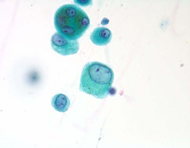
40 Cavity Fluids TRANSUDATE EXUDATE MODIFIED TRANSUDATE
41 Cavity Fluids Cell concentration Making slides Staining ( keep one or two slides in reserve, esspecialy if visiable clumps of cells)
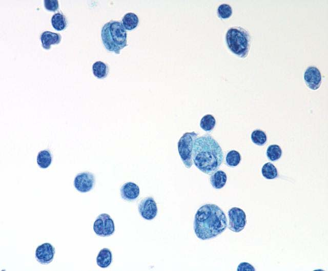
42 clusters
43 Fluid Examination Gross appearance of stained slide Scan using low magnification Examine details using oil lens Ad special stains as indicated
44 Description Adequacy on site Background necrotic, mucinous... Cell concentration high, low... Cell preservation lysis... Inflammatory cells which? dominant? Lining cells mesothelial, epithelial... Cells of interest tumor cells...
45 TRANSUDATE Protein concentration<2,5g/dl TNCC <1500 cells/µg MACROPHAGES
46 hemosiderophage macrophage
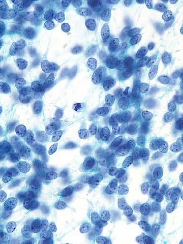
47 TRANSUDATE MACROPHAGES MESOTHELIAL CELLS

48 mesothelial cells
49 TRANSUDATE MACROPHAGES MESOTHELIAL CELLS LYMPHOCYTES
50 lymphocytes
51 MODIFIED TRANSUDATE Moderate protein concentration 2,5-7,5g/dl Moderate cellularity cells/µg Cardiovascular disease Neoplastic disease FIP Rupture of urinary bleadder Hepatic disease

52 EXUDATE
53 EXUDATE High protein concentration > 3,0 g/dl High TNCC > 7000 cells/µg - NONSPECIFIC - SPECIFIC

54 NONSPECIFIC EXUDATE
55 NONSPECIFIC EXUDATE Terminology: - acute, subacute, chronic - predominant cells

56 neutrophilic effusion

57 eosinophilic effusion

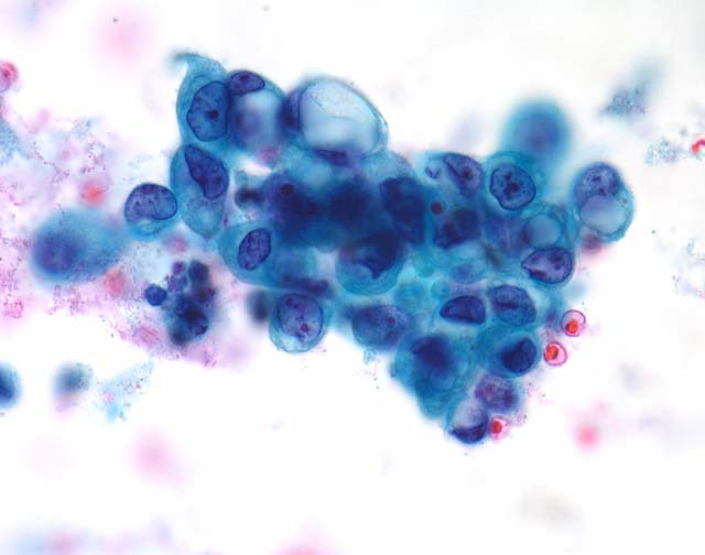
58 mixed cellularity effusion

59 lymphocytic effusion

60 Giant multinucleated cell Langhans type
61 Feline Infectious Peritonitis - FIP Abdominal and/or thoracic effusion in cats High protein concentration > 3,5 Low-moderate number of cell Cytopathology: eosinophilic background large number of neutrophils lesser number of macrophages, mesothelial cells, lymphocytes and plasma cells

62 SPECIFIC EXUDATE
63 SPECIFIC EXUDATE Lupus erythematosus SLE

64 LE cell
65 Malignant Effusion Primary tumors: MESOTHELIOMA

66 mesothelioma mesothelioma mesothelioma EMA stain - mesothelioma
67 LYMPHOPROLIFERATIVE DISORDERS

68 acute lymphoblastic leukemia non-hodgkin lymphoma multiple myeloma large cell lymphoma
69 Secondary tumors METASTATIC TUMORS

70 adenocarcinoma adenocarcinoma liposarcoma malignant melanoma
71 Positivity of fluids 60% - 70% - 90%
72 Hemorrhagic Effusions Presence of hemosiderophages Absence of platelets Hemostatic defect Trauma Neoplasia
73 Diagnostic Cytology Introduction Adventages and disadventages Samplings Stains Fluids FNAs Summary
74 Fine Needle Aspiration CytologyC Gross appearance of the stained slide Scan using low magnification - cellularity Examine areas of interest: background (erytrocytes, necrosis, preservation of cells) cell types, distribution, organisation request special stains if required
75 Cell Types Epithelial glandular squamous Stromal mesenchymal fibro chondro osteo neuroendocrine Inflammatory cells

76 squamous glandular mesenchymal neuroendocrine
77 The first important decision is: INFLAMMATION VS NEOPLASIA
78 Infective Agents

79 cryptoccocus parasytic eggs pneumocystis carinii worm
80 Cellular Changes

81 Cellular changes induced by Herpes simplex virus infection

82 TUMORS
83 Tumor Cell Types Round to caudate large cells epithelial tumors

84 round caudate
85 Tumor Cell Types Spindle to stelate small to medium cells mesenchymal tumors

86 spindle spindle
87 Tumor Cell Types Discrete small to medium round cells lymphoproliferative diseases, neuroendocrine tumors, poorly differentiated tumors
88 round small round small
89 Cell Organisation Papillary structures Clusters Sheets Glandular formations Honey combing Moulding

90 papillary cluster honey combing moulding
91 Second important decision is: BENIGN VS MALIGNANT
92 BENIGN TUMORS

93 adenoma papilloma
94 Criteria of Malignancy Nuclear features Hyperchromasia Anisokaryosis High N/C ratio Multinucleation Mitotic figures increases/abnormal Nucleoli large/variable shaped

95 Hyperchromasia Anisokaryosis High N/C ratio

96 multinucleation
97 atypical mitosis

98 nucleolar features
99 Cytoplasmic features Vacuolisation Keratinization Cannibalism
100 vacuolisation

101 keratinization

102 cannibalism
103 FNA Positivity 90%-100% Our results (798 cases of lung FNA) 54% positive for malignancy 36% negative for malignancy 10% inadequate
104 Lesions that can mimic many criteria of malignancy: Hyperplasia Reactive changes Regenerative and reparative changes

105 MIMIC MALIGNANCY OR REAL MALIGNANCY?????

106 hyperplasia reactive changes reactive changes repair
107 SPRIGGS I BODDINGTON (1989) THERE IS NO KNOWN CRITERION NOR CONSTELLATION OF CRITERIA WHICH ARE UNIVERSALLY DIAGNOSTIC OF MALIGNANCY
108 Future Challenges for Veterinary Cytopathology Image guided FNA cytology Telecytology Use of additional l stains Cytochemistry Immunocytochemistry
109 Diagnostic Cytology Introduction Adventages and disadventages Samplings Stains Fluids FNAs Summary
110 Summary Cytology is diagnostic method Cytology is quick, inexpensive and accurate method, with a little risk to patient Requires good communication with clinicians and correlation with other diagnostic methods Requires continual learning and education Enjoy!!!!!!!

111 33th EUROPIAN CONGRESS OF CYTOLOGY MADRID October
Cytology : first alert of mesothelioma? Professor B. Weynand, UCL Yvoir, Belgium
 Cytology : first alert of mesothelioma? Professor B. Weynand, UCL Yvoir, Belgium Introduction 3 cavities with the same embryologic origin the mesoderme Pleura Exudates Pleura Peritoneum Pericardium 22%
Diagnostic Challenge. Department of Pathology,
 Cytology of Pleural Fluid as a Diagnostic Challenge Paavo Pääkkö,, MD, PhD Chief Physician and Head of the Department Department of Pathology, Oulu University Hospital,, Finland Oulu University Hospital
How To Diagnose And Treat A Tumour In An Effusion
 Effusions of the Serous Cavities Annika Dejmek Professor/Consultant in Cytopathology Clinical Pathology; Department of Laboratory Medicine, Malmö, Lund University 5th EFCS Tutorial Trondheim 2012 Pleura
INFLAMMATION AND REACTIVE CHANGES IN CERVICAL EPITHELIUM
 INFLAMMATION AND REACTIVE CHANGES IN CERVICAL EPITHELIUM Inflammation is a response of a tissue to injury, often caused by invading microorganisms. The suffix which indicates inflammation is "-itis" (the
Cytology of Lymph Nodes
 Indications Cytology of Lymph Nodes Lymph node enlargement That was easy Mary Anna Thrall Don Meuten Indications Lymph node enlargement Suspect metastasis Normal sized lymph nodes are Normal Do NOT aspirate
Practical Effusion Cytology
 Practical Effusion Cytology A Community Pathologist s Approach to Immunocytochemistry in Body Fluid Cytology Emily E. Volk, MD William Beaumont Hospital Troy, MI College of American Pathologists 2004.
Cytopathology Case Presentation #8
 Cytopathology Case Presentation #8 Emily E. Volk, MD William Beaumont Hospital, Troy, MI Jonathan H. Hughes, MD Laboratory Medicine Consultants, Las Vegas, Nevada Clinical History 44 year old woman presents
Immunohistochemistry on cytology specimens from pleural and peritoneal fluid
 Immunohistochemistry on cytology specimens from pleural and peritoneal fluid Dr Naveena Singh Consultant Pathologist Bart health NHS Trust London United Kingdom Disclosures and Acknowledgements I have
Pleural and Pericardial fluids a one year analysis
 Pleural and Pericardial fluids a one year analysis Dr Olafsdottir Dr J Williams Department of Cytology, Llandough Hospital Contents Background Standards Aims/Questions Investigation group Results Conclusions
Diseases. Inflammations Non-inflammatory pleural effusions Pneumothorax Tumours
 Pleura Visceral pleura covers lungs and extends into fissures Parietal pleura limits mediastinum and covers dome of diaphragm and inner aspect of chest wall. Two layers between them (pleural cavity) contains
Explanation of your PAP smear
 Explanation of your PAP smear Approximately 5-10% of PAP smears in the United States are judged to be abnormal. Too often, the woman who receives this news worries that she already has, or will develop,
The develpemental origin of mesothelium
 Mesothelioma Tallinn 14.12.06 Henrik Wolff Finnish Institute of Occupational Health The develpemental origin of mesothelium Mesodermal cavities (pleura, peritoneum and pericardium ) are lined with mesenchymal
Disclosures. Learning Objectives. Effusion = Confusion. Diagnosis Of Serous Cavity Effusions - Beware The Mesothelial Cell!
 Disclosures Diagnosis Of Serous Cavity Effusions - Beware The Mesothelial Cell! No Relevant Financial Relationships with Commercial Interests Syed Z. Ali, M.D. Syed Z. Ali, M.D. Associate Professor of
Urinalysis and Body Fluids CRg. Synovial Fluid. Synovial Fluid. Unit 4. Composition and formation. Functions. Reasons for analysis.
 Urinalysis and Body Fluids CRg Unit 4 Synovial Fluid Synovial Fluid Composition and formation Secreted by cells of synovial membrane Very viscous, clear ultrafiltrate of plasma Contains Hyaluronic acid
Effusions: Mesothelioma and Metastatic Cancers
 Effusions: Mesothelioma and Metastatic Cancers Malignant Mesothelioma Incidence: 2,500 cases/year ~60-80% pts with pleural MM relationship with asbestos exposure Other risk factors: radiation, other carcinogens,
Update on Mesothelioma
 November 8, 2012 Update on Mesothelioma Intro incidence and nomenclature Update on Classification Diagnostic specimens Morphologic features Epithelioid Histology Biphasic Histology Immunohistochemical
Cytology of Effusion Fluids. Cytology of Effusion Fluids. Types of Effusion Fluids. Anatomy. Causes of Effusions. Sampling of Effusion Fluids
 Cytology of Effusion Fluids John W. Wong, MD, FRCPC Sunnybrook Health Sciences Centre Assistant Professor, Laboratory Medicine and Pathobiology Faculty of Medicine, University of Toronto November 10, 2012
Male. Female. Death rates from lung cancer in USA
 Male Female Death rates from lung cancer in USA Smoking represents an interesting combination of an entrenched industry and a clearly drug-induced cancer Tobacco Use in the US, 1900-2000 5000 100 Per Capita
MALIGNANT MESOTHELIOMA UPDATE ON PATHOLOGY AND IMMUNOHISTOCHEMISTRY
 MALIGNANT MESOTHELIOMA UPDATE ON PATHOLOGY AND IMMUNOHISTOCHEMISTRY Sisko Anttila, MD, PhD Jorvi Hospital Laboratory of Pathology Helsinki University Hospital Espoo, Finland 2nd Nordic Conference on Applied
ATLAS OF HEAD AND NECK PATHOLOGY THYROID PAPILLARY CARCINOMA
 Papillary carcinoma is the most common of thyroid malignancies and occurs in all age groups but particularly in women under 45 years of age. There is a high rate of cervical metastatic disease and yet
Thoracic Cavity. Photo: This normal canine lung collapsed when the thorax was opened and the negative pressure was lost in the thorax.
 Thoracic Cavity There are significant anatomical differences in the mediastinum of domestic animals. For instance, bovines, like humans, have well-developed mediastinal separation between the left and
Introduction: Tumor Swelling / new growth / mass. Two types of growth disorders: Non-Neoplastic. Secondary / adaptation due to other cause.
 Disorders of Growth Introduction: Tumor Swelling / new growth / mass Two types of growth disorders: Non-Neoplastic Secondary / adaptation due to other cause. Neoplastic. Primary growth abnormality. Non-Neoplastic
Case of the. Month October, 2012
 Case of the Month October, 2012 Case The patient is a 47-year-old male with a 3-week history of abdominal pain. A CT scan of the abdomen revealed a suggestion of wall thickening at the tip of the appendix
MALIGNANT MESOTHELIOMA UPDATE ON PATHOLOGY AND IMMUNOHISTOCHEMISTRY
 MALIGNANT MESOTHELIOMA CLASSIFICATION MALIGNANT MESOTHELIOMA UPDATE ON PATHOLOGY AND IMMUNOHISTOCHEMISTRY Sisko Anttila, MD, PhD Jorvi Hospital Laboratory of Pathology Helsinki University Hospital Espoo,
The Diagnosis of Cancer in the Pathology Laboratory
 The Diagnosis of Cancer in the Pathology Laboratory Dr Edward Sheffield Christmas Select 74 Meeting, Queen s Hotel Cheltenham, 3 rd December 2014 Agenda Overview of the pathology of cancer How specimens
How To Test For Cancer
 Diagnosis Of Serous Cavity Effusions - Beware The Mesothelial Cell! Effusion = Confusion Syed Z. Ali, M.D. Professor of Pathology and Radiology The Johns Hopkins Hospital Baltimore, Maryland Diagnostic
Case Report Predominantly Fibrous Malignant Mesothelioma in a Cat
 SAGE-Hindawi Access to Research Volume 2010, Article ID 396794, 4 pages doi:10.4061/2010/396794 Case Report Predominantly Fibrous Malignant Mesothelioma in a Cat Alexander Th. A. Weiss, Afonso B. da Costa,
Malignant Lymphomas and Plasma Cell Myeloma
 Malignant Lymphomas and Plasma Cell Myeloma Dr. Bruce F. Burns Dept. of Pathology and Lab Medicine Overview definitions - lymphoma lymphoproliferative disorder plasma cell myeloma pathogenesis - translocations
The Diagnostic Value of Pleural Fluid Cytology in Benign and Malignant Pleural Effusions
 Med. J. Cairo Univ., Vol. 80, No. 2, June: 95-103, 2012 www.medicaljournalofcairouniversity.com The Diagnostic Value of Pleural Fluid Cytology in Benign and Malignant Pleural Effusions SAMAR A. EL-SHEIKH,
Effusion cytology. Dr Alpha Tsui Royal Melbourne Hospital 2008
 Effusion cytology Dr Alpha Tsui Royal Melbourne Hospital 2008 General points: -large unilateral effusion (>1 litre) in the elderly is highly suspicious for malignancy -effusions associated with malignancies
YOUR LUNG CANCER PATHOLOGY REPORT
 UNDERSTANDING YOUR LUNG CANCER PATHOLOGY REPORT 1-800-298-2436 LungCancerAlliance.org A GUIDE FOR THE PATIENT 1 CONTENTS What is a Pathology Report?...3 The Basics...4 Sections of a Pathology Report...7
PRIMARY SEROUS CARCINOMA OF PERITONEUM: A CASE REPORT
 PRIMARY SEROUS CARCINOMA OF PERITONEUM: A CASE REPORT Dott. Francesco Pontieri (*) U.O. di Anatomia Patologica P.O. di Rossano (CS) Dott. Gian Franco Zannoni Anatomia Patologica Facoltà di Medicina e Chirurgia
Pathology of lung cancer
 Pathology of lung cancer EASO COURSE ON LUNG CANCER AND MESOTHELIOMA DAMASCUS (SYRIA), MAY 3-4, 2007 Gérard ABADJIAN MD Pathologist Associate Professor, Saint Joseph University Pathology Dept. Hôtel-Dieu
PATHOLOGY OF THE PLEURA: Mesothelioma and mimickers Necessity of Immunohistochemistry. M. Praet
 PATHOLOGY OF THE PLEURA: Mesothelioma and mimickers Necessity of Immunohistochemistry M. Praet Pathology of the Pleura Normal serosa: visceral and parietal layers Inflammation Neoplasia: Primary: mesothelioma
ALTHOUGH excellent accounts have been published in recent years of
 THE EXFOLIATIVE CYTOLOGY OF DIFFUSE MALIGNANT MESOTHELIOMA BERNARD NAYLOR Department of Pathology, University of Michigan, Ann Arbor, Michigan, U.S.A. PLATE~ LXXXVI-XCI ALTHOUGH excellent accounts have
DESMOPLASTIC SMALL ROUND CELL TUMOR: A RARE PATHOLOGY PUZZLE
 DESMOPLASTIC SMALL ROUND CELL TUMOR: A RARE PATHOLOGY PUZZLE Ryan Granger University of Rhode Island Cytotechnology program May 2, 2015 ASCT Annual Meeting Nashville, Tennessee DESMOPLASTIC SMALL ROUND
A. Pericardial smear. Examination of the pericardial aspirate can provide useful diagnostic information.
 5. PERICARDIUM Heart is encased by the pericardium which has a visceral layer (a) covering the heart and the parietal layer (b). In normal states it is thin, transparent and the myocardium can be seen
Outline. Workup for metastatic breast cancer. Metastatic breast cancer
 Metastatic breast cancer Immunostain Update: Diagnosis of metastatic breast carcinoma, emphasizing distinction from GYN primary 1/3 of breast cancer patients will show metastasis 1 st presentation or 20-30
INFLAMMATORY PLEURAL EFFUSION
 PLEURA- LESIONS LESIONS OF PLEURA Primary Intra pleural bacterial infections Neoplasm (mesothelioma) Secondary A complication of some underlying disease PLEURAL EFFUSION Common manifestation of both primary
Basic Professional Training Program for Associate Medical Technologist
 Basic Professional Training Program for Associate Medical Technologist Basic Cytology Part 2 (Preparartion and normal morphology) Normal Morphology in Liquid based Gynecologic Cytology Speaker: Mr. Fung
Carcinosarcoma of the Ovary
 Carcinosarcoma of the Ovary A Rare Finding Presented By: Kathryn Kiely Anisa I. Kanbour School of Cytotechnology of the University of Pittsburgh Medical Center Pittsburgh, PA Patient History 55 year old
Leukemias and Lymphomas: A primer
 Leukemias and Lymphomas: A primer Normal blood contains circulating white blood cells, red blood cells and platelets 700 red cells (oxygen) 1 white cell Neutrophils (60%) bacterial infection Lymphocytes
LYMPHOMA IN DOGS. Diagnosis/Initial evaluation. Treatment and Prognosis
 LYMPHOMA IN DOGS Lymphoma is a relatively common cancer in dogs. It is a cancer of lymphocytes (a type of white blood cell) and lymphoid tissues. Lymphoid tissue is normally present in many places in the
MALIGNANT MESOTHELIOMA: A TYPICAL PRESENTATION IN AN ATYPICAL PATIENT
 MALIGNANT MESOTHELIOMA: A TYPICAL PRESENTATION IN AN ATYPICAL PATIENT Written by: Karyn Varley MS, SCT(ASCP) The donating laboratory would like to remain anonymous. PATIENT HISTORY 28 year old female Lived
The Value of Thyroid Transcription Factor-1 in Cytologic Preparations as a Marker for Metastatic Adenocarcinoma of Lung Origin
 Anatomic Pathology / TTF-1 IN CYTOLOGY OF BODY FLUIDS The Value of Thyroid Transcription Factor-1 in Cytologic Preparations as a Marker for Metastatic Adenocarcinoma of Lung Origin Jonathan L. Hecht, MD,
Primary -Benign - Malignant Secondary
 TUMOURS OF THE LUNG Primary -Benign - Malignant Secondary The incidence of lung cancer has been increasing almost logarithmically and is now reaching epidemic levels. The overall cure rate is very low
Polyps. Hyperplasias. CAP 2011: Course AP104. The High Risk Benign Endometrium. Mutter and Nucci 1
 Course AP104 Endometrial Hyperplasia A morphologic Definition Hyperplasias Hormonal Effect or Precancer? George L. Mutter, MD Harvard Medical School and Brigham and Women s Hospital Boston, MA Endometrial
ORIGINAL ARTICLES. Materials and Methods
 ORIGINAL ARTICLES Cytomorphologic Features of Metastatic Urothelial Carcinoma in Serous Effusions Cheng Cheng Huang, M.D., PH.D., 1 Anoja Attele, M.D., 1 and Claire W. Michael, M.D. 2 * Metastatic urothelial
TUBERCULOSIS PLEURAL EFFUSION - MANAGEMENT
 TUBERCULOSIS PLEURAL EFFUSION - MANAGEMENT Introduction : ETB 15-20% Pleural effusion 20% in non HIV Under reporting because of AFB negative in fluid In HIV patients: EPTB 20% PTB + EPTB 50% Pleural Effusion
Neoplasms of the LUNG and PLEURA
 Neoplasms of the LUNG and PLEURA 2015-2016 FCDS Educational Webcast Series Steven Peace, BS, CTR September 19, 2015 2015 Focus o Anatomy o SSS 2000 o MPH Rules o AJCC TNM 1 Case 1 Case Vignette HISTORY:
CASE 34 (SLIDE C15-00344) PERICARDIAL EFFUSION IN A DOG
 PERICARDIAL EFFUSION IN A DOG") CASE 34 (SLIDE C15-00344) PERICARDIAL EFFUSION IN A DOG JONATHAN LEBOVITZ, DVM & MPH CANDIDATE, CLASS OF 2016 NICOLE WEINSTEIN, DVM, DACVP VIRGINIA-MARYLAND COLLEGE OF VETERINARY MEDICINE HISTORY AND PHYSICAL
CASE 34 (SLIDE C15-00344) PERICARDIAL EFFUSION IN A DOG JONATHAN LEBOVITZ, DVM & MPH CANDIDATE, CLASS OF 2016 NICOLE WEINSTEIN, DVM, DACVP VIRGINIA-MARYLAND COLLEGE OF VETERINARY MEDICINE HISTORY AND PHYSICAL
Changes in Breast Cancer Reports After Second Opinion. Dr. Vicente Marco Department of Pathology Hospital Quiron Barcelona. Spain
 Changes in Breast Cancer Reports After Second Opinion Dr. Vicente Marco Department of Pathology Hospital Quiron Barcelona. Spain Second Opinion in Breast Pathology Usually requested when a patient is referred
The Use of Immunohistochemistry to Distinguish Reactive Mesothelial Cells From Malignant Mesothelioma in Cytologic Effusions
 The Use of Immunohistochemistry to Distinguish Reactive Mesothelial Cells From Malignant Mesothelioma in Cytologic Effusions Farnaz Hasteh, MD 1 ; Grace Y. Lin, MD, PhD 1 ; Noel Weidner, MD 1 ; and Claire
Fine Needle Aspiration Cytologic Features of Well-Differentiated Papillary Mesothelioma in the Pleura
 The Korean Journal of Pathology 2009; 43: 583-8 DOI: 10.4132/KoreanJPathol.2009.43.6.583 Fine Needle Aspiration Cytologic Features of Well-Differentiated Papillary Mesothelioma in the Pleura - A Case Report
Rotation Specific Goals & Objectives: University Health Network-Princess Margaret Hospital/ Sunnybrook Breast/Melanoma
 Rotation Specific Goals & Objectives: University Health Network-Princess Margaret Hospital/ Sunnybrook Breast/Melanoma Medical Expert: Breast Rotation Specific Competencies/Objectives 1.0 Medical History
Classificazioni citologiche: verso uno schema internazionale unificato?
 Cytology and molecular biology for thyroid nodules diagnos6c categories to clinical ac6ons From Classificazioni citologiche: verso uno schema internazionale unificato? A. Crescenzi Diagnostic categories
Pancreas 23 rd Annual Seminar in Pathology Pittsburgh, PA. Disclosures. Outline
 Pancreas 23 rd Annual Seminar in Pathology Pittsburgh, PA Gladwyn Leiman Director of Cytopathology, Fletcher Allen Health Care Professor of Pathology, University of Vermont Disclosures None Outline Very
PROTOCOL OF THE RITA DATA QUALITY STUDY
 PROTOCOL OF THE RITA DATA QUALITY STUDY INTRODUCTION The RITA project is aimed at estimating the burden of rare malignant tumours in Italy using the population based cancer registries (CRs) data. One of
NEOPLASMS C00 D49. Presented by Jan Halloran CCS
 NEOPLASMS C00 D49 Presented by Jan Halloran CCS 1 INTRODUCTION A neoplasm is a new or abnormal growth. In the ICD-10-CM classification system, neoplastic disease is classified in categories C00 through
Invasive Epithelial Mesothelioma in a Dog
 77 Vet Pathol 42:77 81 (2005) Invasive Epithelial Mesothelioma in a Dog F. REGGETI, B.BRISSON, K.RUOTSALO, E.SOUTHORN, AND D. BIENZLE Abstract. This report describes the gross, microscopic, and immunohistochemical
General Rules SEER Summary Stage 2000. Objectives. What is Staging? 5/8/2014
 General Rules SEER Summary Stage 2000 Linda Mulvihill Public Health Advisor NCRA Annual Meeting May 2014 National Center for Chronic Disease Prevention and Health Promotion Division of Cancer Prevention
TUMORS OF THE TESTICULAR ADNEXA and SPERMATIC CORD
 TUMORS OF THE TESTICULAR ADNEXA and SPERMATIC CORD Victor E. Reuter, MD Memorial Sloan-Kettering Cancer Center reuterv@mskcc.org 66 th Annual Pathology Seminar California Society of Pathologists Short
Oncology. Topic-Bile Duct Carcinoma. Topic-Adenocarcinoma, Lung. Topic-Hemangiosarcoma, Spleen and Liver
 Topic-Adenocarcinoma, Lung Figure 1. Pulmonary adenocarcinoma. Figure 2. Pulmonary bronchiolar carcinoma. Figure 3. Pulmonary fibrosarcoma. Topic-Adenocarcinoma, Skin (Sweat Gland, Sebaceous) Figure 1.
Notice of Faculty Disclosure
 The Diagnosis of Malignant Mesothelioma Andrew Churg, MD Department of Pathology University of British Columbia Vancouver, BC, Canada achurg@mail.ubc.ca Notice of Faculty Disclosure In accordance with
Histopathology of Colorectal Cancer after Neoadjuvant Chemoradiation Therapy
 The Open Pathology Journal, 2009, 3, 91-98 91 Open Access Histopathology of Colorectal Cancer after Neoadjuvant Chemoradiation Therapy Maura O Neil * and Ivan Damjanov Department of Pathology and Laboratory
The Role of Genetic Testing in the Evaluation of Thyroid Nodules. Thyroid Cancer and FNA. Thyroid Cancer. Pure Follicular Cancers.
 Where does Molecular Analysis of FNA Specimens fit into the evaluation of thyroid nodules? The Role of Genetic Testing in the Evaluation of Thyroid Nodules Ultrasound TSH Risk factors Jill E. Langer, MD
3-F. Pathology of Mesothelioma
 3-F. Pathology of Mesothelioma Kouki Inai Professor of Department of Pathology, Graduate School of Biomedical Science, Hiroshima University Introduction Mesothelioma is a peculiar type of malignancy, which
Today s Topics. Tumors of the Peritoneum in Women
 Today s Topics Tumors of the Peritoneum in Women Charles Zaloudek, M.D. Department of Pathology 505 Parnassus Ave., M563 University of California, San Francisco San Francisco, CA USA charles.zaloudek@ucsf.edu
J of Evidence Based Med & Hlthcare, pissn- 2349-2562, eissn- 2349-2570/ Vol. 2/Issue 33/Aug. 17, 2015 Page 5063
 PERITONEAL MALIGNANT MESOTHELIOMA: A RARE S. R. Dhamotharan 1, S. Shanthi Nirmala 2, F. Celine Foustina Mary 3, M. Arul Raj Kumar 4, R. Vinothprabhu 5 HOW TO CITE THIS ARTICLE: S. R. Dhamotharan, S. Shanthi
Malignant Mesothelioma in Body Fluids - with Special Reference to Differential Diagnosis from Metastatic Adenocarcinoma -
 The Korean Journal of Pathology 2009; 43: 458-66 DOI: 10.4132/KoreanJPathol.2009.43.5.458 Malignant Mesothelioma in Body Fluids - with Special Reference to Differential Diagnosis from Metastatic Adenocarcinoma
Lung Carcinomas New 2015 WHO Classification. Spasenija Savic Pathology
 Lung Carcinomas New 2015 WHO Classification Spasenija Savic Pathology ***EXPECTED SPRING 2015*** This authoritative, concise reference book provides an international standard for oncologists and pathologists
The diagnostic usefulness of tumour markers CEA and CA-125 in pleural effusion
 Malaysian J Path01 2002; 24(1) : 53-58 The diagnostic usefulness of tumour markers CEA and CA-125 in pleural effusion Pavai STHANESHWAR MD, Sook-Fan YAP FRCPath, FRCPA and Gita JAYARAM MDPath, MRCPath
Nicole Kounalakis, MD
 Breast Disease: Diagnosis and Management Nicole Kounalakis, MD Assistant Professor of Surgery Goal of Breast Evaluation The goal of breast evaluation is to classify findings as: normal physiologic variations
Video Microscopy Tutorial 5
 Video Microscopy Tutorial 5 Lool Alikes in Effusion Cytology:Review of Diagnostic Challenges Claire Michael, MD There are no disclosures necessary. Look-Alikes in Effusion Cytology: Review of Diagnostic
Interesting Case Review. Renuka Agrawal, MD Dept. of Pathology City of Hope National Medical Center Duarte, CA
 Interesting Case Review Renuka Agrawal, MD Dept. of Pathology City of Hope National Medical Center Duarte, CA History 63 y/o male with h/o CLL for 10 years Presents with worsening renal function and hypercalcemia
ThinPrep Non-Gyn Lecture Series. Body Fluid Cytology
 ThinPrep Non-Gyn Lecture Series Body Fluid Cytology Benefits of ThinPrep Technology The use of ThinPrep Non-Gyn for body fluid specimens: Optimizes cell preservation Standardizes specimen preparation Simplifies
Disease/Illness GUIDE TO ASBESTOS LUNG CANCER. What Is Asbestos Lung Cancer? www.simpsonmillar.co.uk Telephone 0844 858 3200
 GUIDE TO ASBESTOS LUNG CANCER What Is Asbestos Lung Cancer? Like tobacco smoking, exposure to asbestos can result in the development of lung cancer. Similarly, the risk of developing asbestos induced lung
Diagnosis of Mesothelioma Pitfalls and Practical Information
 Diagnosis of Mesothelioma Pitfalls and Practical Information Mary Beth Beasley, M.D. Mt Sinai Medical Ctr Dept of Pathology One Gustave L Levy Place New York, NY 10029 (212) 241-5307 mbbeasleymd@yahoo.com
Case Report of Renal Cell Carcinoma Diagnosed in Voided Urine Confirmed by CD10 Immunocytochemistry
 Novel Insights from Clinical Practice DOI: 10.1159/000326940 Received: January 6, 2011 Accepted: February 21, 2011 Published online: July 22, 2011 Case Report of Renal Cell Carcinoma Diagnosed in Voided
Lung Cancer. Ossama Tawfik, MD, PhD Professor, Vice Chairman Director of Anatomic &Surgical Pathology University of Kansas School of Medicine
 Lung Cancer Ossama Tawfik, MD, PhD Professor, Vice Chairman Director of Anatomic &Surgical Pathology University of Kansas School of Medicine Alexandria, Egypt July 1-1 3, 2008 OBJECTIVES Describe and
Exercise 9: Blood. Readings: Silverthorn 5 th ed, 547 558, 804 805; 6 th ed, 545 557, 825 826.
 Exercise 9: Blood Readings: Silverthorn 5 th ed, 547 558, 804 805; 6 th ed, 545 557, 825 826. Blood Typing The membranes of human red blood cells (RBCs) contain a variety of cell surface proteins called
EDUCATIONAL COMMENTARY - GRANULOCYTE FORMATION AND CHRONIC MYELOCYTIC LEUKEMIA
 LEUKEMIA Educational commentary is provided through our affiliation with the American Society for Clinical Pathology (ASCP). To obtain FREE CME/CMLE credits click on Earn CE Credits under Continuing Education
Validation of BRAF Mutational Analysis in Thyroid Fine Needle Aspirations: A Morphologic- Molecular Approach
 Validation of BRAF Mutational Analysis in Thyroid Fine Needle Aspirations: A Morphologic- Molecular Approach Kerry C. Councilman, MD Assistant Professor University of Colorado Denver Goals: BRAF Mutation
What is Mesothelioma?
 What is Mesothelioma? Mesothelioma is a rare type of cancer that develops in the mesothelial cells found in one s body. These cells form membranous linings that surround and protect the body s organs and
LYMPHOMA. BACHIR ALOBEID, M.D. HEMATOPATHOLOGY DIVISION PATHOLOGY DEPARTMENT Columbia University/ College of Physicians & Surgeons
 LYMPHOMA BACHIR ALOBEID, M.D. HEMATOPATHOLOGY DIVISION PATHOLOGY DEPARTMENT Columbia University/ College of Physicians & Surgeons Normal development of lymphocytes Lymphocyte proliferation and differentiation:
WHICH SAMPLES SHOULD BE SUBMITTED WHEN LYMPHOID NEOPLASIA IS SUSPECTED?
 WHICH SAMPLES SHOULD BE SUBMITTED WHEN LYMPHOID NEOPLASIA IS SUSPECTED? Which test should be submitted? The answer to this depends on the clinical signs, and the diagnostic question you are asking. If
Frozen Section Diagnosis
 Frozen Section Diagnosis Dr Catherine M Corbishley Honorary Consultant Histopathologist St George s Healthcare NHS Trust and lead examiner final FRCPath Practical 2008-2011 Frozen Section Diagnosis The
Gladwyn Leiman, MCCCh, FIAC, FRCPath Scott Anderson, MD
 Cytology Works shop #5 Gladwyn Leiman, MCCCh, FIAC, FRCPath Scott Anderson, MD Disclosur re information The speakers have no relationship that represents a possible conflict of interest with respect to
20 Diagnostic Cytopathology, Vol 36, No 1 ' 2007 WILEY-LISS, INC.
 Utility of WT-1, p63, MOC31, Mesothelin, and Cytokeratin (K903 and CK5/6) Immunostains in Differentiating Adenocarcinoma, Squamous Cell Carcinoma, and Malignant Mesothelioma in Effusions Robert T. Pu,
Something Old, Something New.
 Something Old, Something New. Michelle A. Fajardo, D.O. Loma Linda University Medical Center Clinical Presentation 6 year old boy, presented with hematuria Renal mass demonstrated by ultrasound & CT scan
Mesothelioma: Questions and Answers
 CANCER FACTS N a t i o n a l C a n c e r I n s t i t u t e N a t i o n a l I n s t i t u t e s o f H e a l t h D e p a r t m e n t o f H e a l t h a n d H u m a n S e r v i c e s Mesothelioma: Questions
Diagnostic Cytology of Body Cavity Fluids in Dogs and Cats
 The 3 rd Annual Vet Education Online Veterinary Conference - July 2012 Diagnostic Cytology of Body Cavity Fluids in Dogs and Cats Dr Jenny Mills BVSc Dipl. Clin Path Dip Ed PhD MSc Murdoch University,
SMALL CELL LUNG CANCER
 Protocol for Planning and Treatment The process to be followed in the management of: SMALL CELL LUNG CANCER Patient information given at each stage following agreed information pathway 1. DIAGNOSIS New
This module consists of four units which will provide the user a basic knowledge of cancer as a disease.
 Module 5: What is Cancer? This module consists of four units which will provide the user a basic knowledge of cancer as a disease. After completing this module, cancer abstractors will be able to: Define
Cystic Neoplasms of the Pancreas: A multidisciplinary approach to the prevention and early detection of invasive pancreatic cancer.
 This lecture is drawn from the continuing medical education program Finding Hope: Prevention, Early Detection and Treatment of Pancreatic Cancer, Nov, 2011. Robert P. Jury, MD Cystic Neoplasms of the Pancreas:
More than 2,500 people are diagnosed with mesothelioma in the UK each year.
 This information is an extract from the booklet Understanding mesothelioma. You may find the full booklet helpful. We can send you a free copy see page 5. Contents Introduction Pleural mesothelioma Peritoneal
Hematology Morphology Critique
 Survey Slide: History: 60-year-old female presenting with pneumonia Further Laboratory Data: Hgb : 90 g/l RBC : 2.92 10 12 /L Hct : 0.25 L/L MCV : 87 fl MCH : 30.8 pg MCHC : 355 g/l RDW : 17.7 % WBC :
http://www.springer.com/3-540-22006-2
 http://www.springer.com/3-540-22006-2 1 Molecular Pathology Laboratory of the Future Christopher A. Moskaluk 1.1 The Past The integration of laboratory analysis with human medicine has traditionally been
Protocol applies to all primary borderline and malignant epithelial tumors, and malignant mesothelial neoplasms of the peritoneum.
 Peritoneum Protocol applies to all primary borderline and malignant epithelial tumors, and malignant mesothelial neoplasms of the peritoneum. Protocol revision date: January 2004 No AJCC/UICC staging system