Differentiating Wide Complex Tachycardia MANOLATOS DIMITRIOS M.D
|
|
|
- Denis Cross
- 7 years ago
- Views:
Transcription
1 Differentiating Wide Complex Tachycardia MANOLATOS DIMITRIOS M.D
2 Definitions Causes Electrocardiographic features Diagnostic Criteria
3 Wide Complex Tachycardias (WCT) -QRS duration > 120 ms and heart rate > 100 beats/miin Ventricular Tachycardia (VT) - 3 or more consecutive ventricular beats with a rate of 100 beats/min or more Nonsustained VT (NSVT) tachycardia < 30 seconds duration
4 Sustained VT: Last > 30 seconds Causes significant hemodynamic symptoms Requires therapeutic intervention for termination Monomorphic VT uniform and stable QRS appearance in any given lead Polymorphic VT continuouslly varying QRS morphology and/or axis in a single lead during an episode
5 Supraventricular tachycardia (SVT) any tachycardia using the normal AV conduction system for ventricular excitation, w/ the tachycardia originating in the atria or the AV node and requiring the AV node for its maintenance Aberrant conduction (aberrancy) conduction delay or block in the His-Purkinje system during antegrade conduction of impulses over the normal AV-conduction system resulting in a wide, abnormal QRS
6 Pre-excitation excitation syndrome AV conduction can occur via the normal conduction system and via an accessory AV pathway The two pathways create tthe substrate for a reentrant circuit, facillittating AV reentrant tachycardia (AVRT) Orthodromic AVRT antegrade conduction over the AV node and retrograde conduction via the accessory pathway; QRS is narrow unless there is aberrant conduction Antidromic AVRT antegrade conduction over the accessory pathway and retrograde conduction over the AV node or a second accessory pathway; QRS is wide Pre-excited excited tachycardias ventricular activation occurs predominately or exclusively via an accessory pathway
7 VT - most common cause of WCT (80% of cases in unselected populations and more than 95% in pts w/ structural heart disease Can originate anywhere below the AV node: His bundle,, bundlle branches,, fascicles,, Purkinje fibers,, and ventricular myocardial tissue Fixed conduction block when there is a baseline block during sinus rhythm due to pathological lesions in the conduction system Seen as baseline left bundle branch block (LBBB),, right bundle branch block (RBBB),, or a nonspecific intraventricular conduction delay (IVCD)
8 Orthodromic AVRT w/ aberrancy Pre-excitation excitation tachycardias Antidromic AVRT Relatively uncommon, occurring in about 6% of cases Difficult to differentiate from VT because ventricular activation begins outside the normal intraventricular conduction system in both Atrial fibrillation with an accessory pathway Tachycardia's w/ two accessory pathways Pacemakers and cardiac resynchronization therapy
9 Electrocardiograms Rate: limited use in distinguishing VT from SVT as there is too much overlap Consider Atrial flutter when HR is ~150 beats/min Regularity: VT is generally regular, though there can be slight variation in the RR intervals Slight irregularity at the onset ( warm( warm--up phenomenon )-favors VT Grossly irregular WCT likely represents: 1) AF w/ aberrant conduction, 2) AF w/ conduction over an accessory pathway,, 3) polymorphic VT Uniformity of the RR intervals' favors SVT
10 Axis: mean QRS axis in the normal range favors SVTw/ aberration Right superior axis of -90 to ± 180 strongly suggests VT(sometimes called northwest or extreme right axis) Should compare the axis during SR,, an axis shift during WCT of > 40 favors VT In RBBB-like WCT, axis to the left of -30 suggest VT In LBBB-like WCT,axis to the right of +90 suggests VT
11 QRS duration: generally, a wider QRS favors VT In RBBB-- --like WCT,, duration > 140 msec suggests VT In LBBB-- --like WCT,, duration > 160 msec suggest VT QRS duration > 160 msec is a strong predictor of VT regardless of bundle-- --branch block morphology Except in cases of SVT w/ an AV accessory pathway and the presence of drugs capable of slowing intraventricular conduction Such as class 1A or class 1C agents or amiodarone QRS duration < 140 msec does not exclude VT VT originating from the septum or w/in the His-Purkinje system
12 Concordance: present when QRS complexes in all 6 precordial leads (V1-V6) V6) are monophasic w/ the same polarity (90% specificity for VT) Positive concordance All entirely positive w/ totally,monophonic R waves Most often due to VT but can also occur in rare cases of antidromic AVRT w/ a left posterior accessory pathway Negative concordance -- All entirely negative w/ deep monophonic QS complexes Concordance is not present if any of the 6 leads has a biphasic QRS (qr( or RS complexes) Absence of concordance is not diagnostically helpful
13 AV dissociation: atrial activity that is independent of ventricular activity occurs in 20-50% off VT and almost never in SVT When the atrial rate is slower than the ventricular rate, this strongly suggest VT Atrial rate faster than the ventricular rate suggests a SVT, such as atrial flutter or AT w/ 2:1 AV conduction There is a consistent relationship between the P waves and the QRS complexes
14 Presence of this largely establishes the diagnosis of VT but its absence is not helpful May sometimes not be evident on ECG Some cases of VT, the ventricular impulses conduct retrograde through the AV node and capture the atrium, preventing dissociation
15 Echo beat VT impulse conducted retrogradely through the AV node to produce atrial capture that, on to the ventricles, produces a narrow QRS complex Fusion beat when one impulse originating from the ventricle and a second supraventricular impulse simultaneously activate the ventricular myocardium Morphology is intermediate between that of a sinus beat and a purely ventricular complex Capture beat normal conduction momentarily captured control of ventricular activation from the VT focus
16 QRS Morphology: most approaches involve classifying the WCT as having RBBB like pattern or LBBB-- --like pattern RBBB-like pattern: QRS polarity is positive in leads V1 and V2 LBBB-like pattern: QRS polarity is negative in leads V1 and V2
17 RBBB pattern associations: Lead V1: monophasic R or biphasic qr complex favors VT Triphasic RSR or RsR complex ( rabbit( rabbit--ear sign) ) favors SVT,, except if the left peak of the RsR complex is taller than the right peak Lead V6: rs complex (R wave smaller than S wave) favors VT Rs (R wave larger than S wave) complex favors SVT
18 LBBB pattern associations: Lead V1 or V2:: broad initial R wave of >30 msec duration favors s VT Slurred or notched downstroke of the S wave favors VT Duration from the onset of the QRS complex to the nadir of the QS or S wave of M 60 msec favors VT Absence of an initial R wave or a small initial R wave of < 30 msec favors SVT A swift, smooth downstroke of the S wave in w/ a duration of < 60 msec favors SVT Lead V6: any Q or QS wave favors VT Absence of a Q wave favors SVT
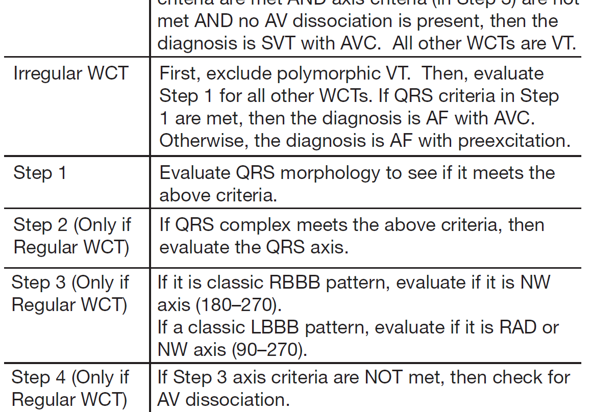
19 Brugada Criteria Stepwise approach in which four criteria for VT are sequentially evaluated and if any are satisfied,the diagnosis off VT is made; if none are fulfilled then diagnosis off SVT is made by exclusion 1)Lead V1-V6 V6 are inspected for an RS complex, if none than concordance is present 2)If an RS complex is present, the longest interval in any lead between the onset of the R wave and the nadir of the S wave (RS interval) is measured VT if the RS interval is > 100 msec
20 3)If the RS interval is < 100 msec, then evaluate for the presence of AV dissociation to diagnose VT 4)If the RS interval is < 100 msec and AV dissociation is not evident, then evaluate the QRS morphology for V1-positive and V1-negative WCT If either the V1-V2 V2 or the V6 criteria are not consistent w/ VT, then SVT is assumed
21
22
23 New avr Algorithm Hypothesized reasons for using avr: During SVT w/ BBB,, the initial septal activation and the latter main ventricular activation waterfront move away from lead avr,, creating a negative QRS complex in lead avr Exception to this generalization is occurs in inferior myocardial infarction where there is the loss of the initial inferiorly directed forces creating an initial r wave (rs( complex) during NSR or SVT Because an initial dominant R wave in avr is incompatible w/ SVT,, its presence suggest VT,, typiically originating from the inferior or apical region
24 Useful for detecting VT originating from sites other than the inferior or apical wall, but would not show an initial R wave in avr Would rather show a slow,, initial upward vector of variable size pointing toward avr even if the main vector of the VT points downward and creates a predominately negative QRS in lead avr Exceptions would be VT originating from the most basal sites of the interventricular septum or free wall
25 Stepwise approach similar to the Brugada criteria used to analyze monophonic WCT: 1) Evaluate for the presence of an initial R wave 2) Evaluate for the presence of an initial r or q wave with width M 40 msec 3)Evaluate for notching on the descending limb of a negative onset, predominately negative QRS complex
26

27
28 lead II algorithm We analyzed the RWPT at lead II, defined as the duration of the QRS from the initiation of depolarization until the first change of the polarity, independent of whether the QRS deflection was positive or negative. A R-wave peak time 50 ms at DII is a simple and highly sensitive criterion that discriminates VT from SVT in patients with wide QRS complex tachycardias. Pava LF, et al. Heart Rhythm 2010;7:922 6.


29
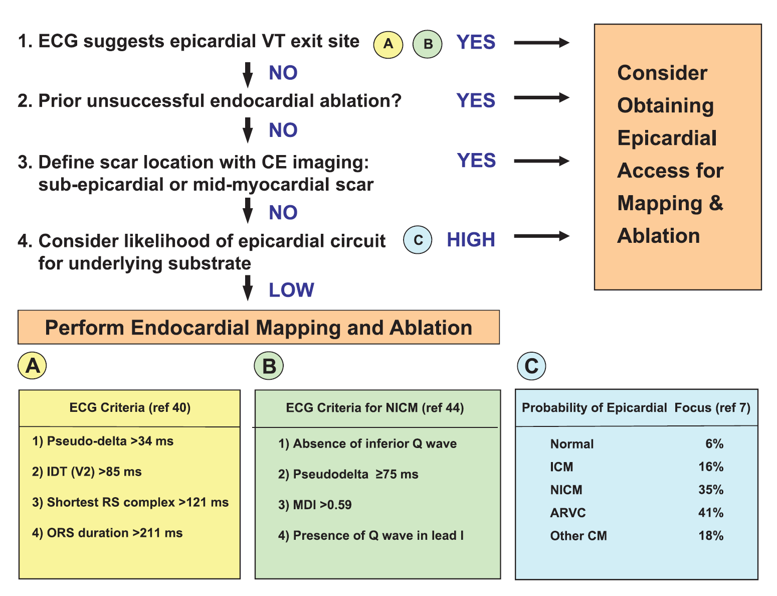
30 ECG criteria for epicardial VT
31
32
33
34 Case 1 65 yo CAD (anterior MI), LVEF: 35%, NYHA I bb, ACE, spironolactone, amiodarone incessant wide QRS tachycardia
35 Case 1
36 Case 1 1. VT left ventricular origin (apex) 2. SVT with aberration 3. VT right ventricular origin (apex) 4. Antidromic SVT via an AP
37 mid-diastolic diastolic potentials
38 Case 2 60 yo man prior inferior MI LVEF: 45%
39 Case 2
40 Case 2 1. VT left ventricular origin 2. SVT with aberration 3. VT right ventricular origin 4. Antidromic SVT via an AP
41 Case 2, activation mapping
42 Case 3 17 yo female ECG during SR: unremarkable Transthoracic echocardiography: normal LV and RV
43 Case 3
44 Case 3 1. RVOT VT 2. SVT with aberration 3. LVOT VT 4. Idiopathic left fascicular VT
45 Case 4 35 yo man Syncope during exercise ECG during SR: negative T waves V1-3 Transthoracic echocardiography: RV dilation
46 Case 4
47 1. SVT with aberration Case 4 2. RVOT VT due to ARVC 3. Epicardial VT due to ARVC 4. Mitral annulus VT
48 Case 5 27 yo man ECG during SR: unremarkable Holter monitoring: sustained tachycardia Verapamil terminates the tachycardia
49 Case 5
50 Case 5 1. Mitral annulus VT 2. SVT with aberration 3. LVOT VT 4. Idiopathic left fascicular VT
51 Case 5 presystolic potential
52 Case 5
53
54 Case 6 67 yo no structural heart disease Transthoracic echocardiography and coronary angiography ruled out structural heart disease MRI was normal referred to our hospital for EPS/ablation
55 Case 6
56 Case 6 1. VT left ventricular origin (apex) 2. SVT with aberration 3. VT right ventricular origin (apex) 4. Antidromic SVT via an AP
57 Case 6 pace mapping of 12 leads
58 Case 6 propagation map voltage map
59 Case 7 41 yo no structural heart disease Transthoracic echocardiography and exercise test ruled out structural heart disease. Referred to our hospital for EPS/ablation
60 Case 7
61 Case 7 1. VT left ventricular origin 2. Idiopathic left fascicular VT 3. VT right ventricular origin (apex) 4. Antidromic SVT via an AP (Mahaim fiber)
62 ΣΑΣ ΕΥΧΑΡΙΣΤΩ
QRS Complexes. Fast & Easy ECGs A Self-Paced Learning Program
 6 QRS Complexes Fast & Easy ECGs A Self-Paced Learning Program Q I A ECG Waveforms Normally the heart beats in a regular, rhythmic fashion producing a P wave, QRS complex and T wave I Step 4 of ECG Analysis
BIPOLAR LIMB LEADS UNIPOLAR LIMB LEADS PRECORDIAL (UNIPOLAR) LEADS VIEW OF EACH LEAD INDICATIVE ECG CHANGES
 LEADS VIEW OF EACH LEAD INDICATIVE ECG CHANGES") BIPOLAR LIMB LEADS Have both a distinctive positive and negative pole. Lead I LA (positive) RA (negative) Lead II LL (positive) RA (negative) Lead III LL (positive) LA (negative) UNIPOLAR LIMB LEADS Have
BIPOLAR LIMB LEADS Have both a distinctive positive and negative pole. Lead I LA (positive) RA (negative) Lead II LL (positive) RA (negative) Lead III LL (positive) LA (negative) UNIPOLAR LIMB LEADS Have
Introduction to Electrocardiography. The Genesis and Conduction of Cardiac Rhythm
 Introduction to Electrocardiography Munther K. Homoud, M.D. Tufts-New England Medical Center Spring 2008 The Genesis and Conduction of Cardiac Rhythm Automaticity is the cardiac cell s ability to spontaneously
NEONATAL & PEDIATRIC ECG BASICS RHYTHM INTERPRETATION
 NEONATAL & PEDIATRIC ECG BASICS & RHYTHM INTERPRETATION VIKAS KOHLI MD FAAP FACC SENIOR CONSULATANT PEDIATRIC CARDIOLOGY APOLLO HOSPITAL MOB: 9891362233 ECG FAX LINE: 011-26941746 THE BASICS: GRAPH PAPER
the basics Perfect Heart Institue, Piyavate Hospital
 ECG INTERPRETATION: the basics Damrong Sukitpunyaroj MD Damrong Sukitpunyaroj, MD Perfect Heart Institue, Piyavate Hospital Overview Conduction Pathways Systematic Interpretation Common abnormalities in
Systematic Approach to 12 Lead EKG Interpretation
 Systematic Approach to 12 Lead EKG Interpretation Maureen Knechtel MPAS, PA-C Wellmont CVA Heart Institute Disclosure Statement of Financial Interest I, Maureen Knechtel, do not have a financial interest/arrangement
12-Lead EKG Interpretation. Judith M. Haluka BS, RCIS, EMT-P
 12-Lead EKG Interpretation Judith M. Haluka BS, RCIS, EMT-P ECG Grid Left to Right = Time/duration Vertical measure of voltage (amplitude) Expressed in mm P-Wave Depolarization of atrial muscle Low voltage
The P Wave: Indicator of Atrial Enlargement
 Marquette University e-publications@marquette Physician Assistant Studies Faculty Research and Publications Health Sciences, College of 8-12-2010 The P Wave: Indicator of Atrial Enlargement Patrick Loftis
Electrophysiology Introduction, Basics. The Myocardial Cell. Chapter 1- Thaler
 Electrophysiology Introduction, Basics Chapter 1- Thaler The Myocardial Cell Syncytium Resting state Polarized negative Membrane pump Depolarization fundamental electrical event of the heart Repolarization
Wide-Complex Tachycardias in the ED: Myths and Pitfalls
 Wide-Complex Tachycardias in the ED: Myths and Pitfalls, FACEP, FAAEM Professor and Vice Chair Director, Emergency Cardiology Fellowship Department of Emergency Medicine University of Maryland School of
MULTIPLE CHOICE. Choose the one alternative that best completes the statement or answers the question.
 Exam Name MULTIPLE CHOICE. Choose the one alternative that best completes the statement or answers the question. 1) What term is used to refer to the process of electrical discharge and the flow of electrical
ECG made extra easy. medics.cc
 ElectroCardioGraphyraphy ECG made extra easy Overview Objectives for this tutorial What is an ECG? Overview of performing electrocardiography on a patient Simple physiology Interpreting the ECG Objectives
An Introduction to Tachyarrhythmias R. A. Seyon MN, NP, CCN(C) & Dr. R. G. Williams
 & Dr. R. G. Williams") Arrhythmias 1 An Introduction to Tachyarrhythmias R. A. Seyon MN, NP, CCN(C) & Dr. R. G. Williams Things to keep in mind when analyzing arrhythmias: Electrical activity recorded in 12 and 15 leads Examine
Arrhythmias 1 An Introduction to Tachyarrhythmias R. A. Seyon MN, NP, CCN(C) & Dr. R. G. Williams Things to keep in mind when analyzing arrhythmias: Electrical activity recorded in 12 and 15 leads Examine
Signal-averaged electrocardiography late potentials
 SIGNAL AVERAGED ECG INTRODUCTION Signal-averaged electrocardiography (SAECG) is a special electrocardiographic technique, in which multiple electric signals from the heart are averaged to remove interference
Understanding the Electrocardiogram. David C. Kasarda M.D. FAAEM St. Luke s Hospital, Bethlehem
 Understanding the Electrocardiogram David C. Kasarda M.D. FAAEM St. Luke s Hospital, Bethlehem Overview 1. History 2. Review of the conduction system 3. EKG: Electrodes and Leads 4. EKG: Waves and Intervals
Introduction to Electrophysiology. Wm. W. Barrington, MD, FACC University of Pittsburgh Medical Center
 Introduction to Electrophysiology Wm. W. Barrington, MD, FACC University of Pittsburgh Medical Center Objectives Indications for EP Study How do we do the study Normal recordings Abnormal Recordings Limitations
HTEC 91. Topic for Today: Atrial Rhythms. NSR with PAC. Nonconducted PAC. Nonconducted PAC. Premature Atrial Contractions (PACs)
") HTEC 91 Medical Office Diagnostic Tests Week 4 Topic for Today: Atrial Rhythms PACs: Premature Atrial Contractions PAT: Paroxysmal Atrial Tachycardia AF: Atrial Fibrillation Atrial Flutter Premature Atrial
HTEC 91 Medical Office Diagnostic Tests Week 4 Topic for Today: Atrial Rhythms PACs: Premature Atrial Contractions PAT: Paroxysmal Atrial Tachycardia AF: Atrial Fibrillation Atrial Flutter Premature Atrial
ST Segment Elevation Nothing is ever as hard (or easy) as it looks
 as it looks") ST Segment Elevation Nothing is ever as hard (or easy) as it looks Cameron Guild, MD Division of Cardiology University of Mississippi Medical Center February 17, 2012 Objectives 1. Describe the electrical
ST Segment Elevation Nothing is ever as hard (or easy) as it looks Cameron Guild, MD Division of Cardiology University of Mississippi Medical Center February 17, 2012 Objectives 1. Describe the electrical
Tachyarrhythmias (fast heart rhythms)
") Patient information factsheet Tachyarrhythmias (fast heart rhythms) The normal electrical system of the heart The heart has its own electrical conduction system. The conduction system sends signals throughout
Patient information factsheet Tachyarrhythmias (fast heart rhythms) The normal electrical system of the heart The heart has its own electrical conduction system. The conduction system sends signals throughout
Diagnosis Code Crosswalk : ICD-9-CM to ICD-10-CM Cardiac Rhythm and Heart Failure Diagnoses
 Diagnosis Code Crosswalk : to 402.01 Hypertensive heart disease, malignant, with heart failure 402.11 Hypertensive heart disease, benign, with heart failure 402.91 Hypertensive heart disease, unspecified,
By the end of this continuing education module the clinician will be able to:
 EKG Interpretation WWW.RN.ORG Reviewed March, 2015, Expires April, 2017 Provider Information and Specifics available on our Website Unauthorized Distribution Prohibited 2015 RN.ORG, S.A., RN.ORG, LLC Developed
Electrocardiographic recognition and ablation of outflow tract ventricular tachycardia
 VIEWPOINT Electrocardiographic recognition and ablation of outflow tract ventricular tachycardia Rupa Bala, MD, Francis E. Marchlinski, MD From the Hospital of the University of Pennsylvania, Philadelphia,
INTRODUCTORY GUIDE TO IDENTIFYING ECG IRREGULARITIES
 INTRODUCTORY GUIDE TO IDENTIFYING ECG IRREGULARITIES NOTICE: This is an introductory guide for a user to understand basic ECG tracings and parameters. The guide will allow user to identify some of the
Objectives. The ECG in Pulmonary and Congenital Heart Disease. Lead II P-Wave Amplitude during COPD Exacerbation and after Treatment (50 pts.
 The ECG in Pulmonary and Congenital Heart Disease Gabriel Gregoratos, MD Objectives Review the pathophysiology and ECG signs of pulmonary dysfunction Review the ECG findings in patients with: COPD (chronic
Interpreting a rhythm strip
 3 Interpreting a rhythm strip Just the facts In this chapter, you ll learn: the components of an ECG complex and their significance and variations techniques for calculating the rate and rhythm of an ECG
Copyright 2006 Blaufuss Multimedia. All rights reserved. Page 1
 Copyright 2006 Blaufuss Multimedia. All rights reserved. Page 1 002 Sinus Rhythm, atrial rate 90 Mobitz II AVB, Ventricular rate 50 Left Atrial Enlargement Left Ventricular Hypertrophy RBBB a) Long R-R
ECG Measurments and Interpretation Programs
 ECG Measurments and Interpretation Programs Physician s Guide Distributed by Welch Allyn 4341 State Street Road, PO Box 220 Skaneateles Falls, NY 13153-0220 www.welchallyn.com Sales and Service information:
Evaluation and Initial Treatment of Supraventricular Tachycardia
 T h e n e w e ngl a nd j o u r na l o f m e dic i n e clinical practice Evaluation and Initial Treatment of Supraventricular Tachycardia Mark S. Link, M.D. This Journal feature begins with a case vignette
BASIC CARDIAC ARRHYTHMIAS Revised 10/2001
 BASIC CARDIAC ARRHYTHMIAS Revised 10/2001 A Basic Arrhythmia course is a recommended prerequisite for ACLS. A test will be given that will require you to recognize cardiac arrest rhythms and the most common
Lead avr: The Neglected Lead
 Chapter 22 Lead avr: The Neglected Lead M Chenniappan INTRODUCTION Lead avr, one of the 12 electrocardiographic leads, is frequently ignored in clinical medicine. In fact, many clinicians refer to the
ACLS Chapter 3 Rhythm Review Instructor Lesson Plan to Accompany ACLS Study Guide 3e
 ACLS Chapter 3 Rhythm Review Lesson Plan Required reading before this lesson: ACLS Study Guide 3e Textbook Chapter 3 Materials needed: Multimedia projector, computer, ACLS Chapter 3 Recommended minimum
How to read the ECG in athletes: distinguishing normal form abnormal
 How to read the ECG in athletes: distinguishing normal form abnormal Antonio Pelliccia, MD Institute of Sport Medicine and Science www.antoniopelliccia.it Cardiac adaptations to Rowing Vagotonia Sinus
JAPI VOL. 52 NOVEMBER 2004 www.japi.org 883
 Review Article Wide Complex Tachycardia : Recognition and Management in the Emergency Room IB Ray Abstract Cardiac arrhythmias often present as urgent medical conditions requiring immediate care. Patient
Electrocardiography Review and the Normal EKG Response to Exercise
 Electrocardiography Review and the Normal EKG Response to Exercise Cardiac Anatomy Electrical Pathways in the Heart Which valves are the a-v valves? Closure of the a-v valves is associated with which heart
Scott Hubbell, MHSc, RRT-NPS, C-NPT, CCT Clinical Education Coordinator/Flight RRT EagleMed
 Scott Hubbell, MHSc, RRT-NPS, C-NPT, CCT Clinical Education Coordinator/Flight RRT EagleMed Identify the 12-Lead Views Explain the vessels of occlusion Describe the three I s Basic Interpretation of 12-Lead
The abbreviation EKG, for electrocardiogram,
 CLIN PEDIATR OnlineFirst, published on January 28, 2010 as doi:10.1177/0009922809336206 Simplified Pediatric Electrocardiogram Interpretation Clinical Pediatrics Volume XX Number X Month XXXX xx-xx 2009
Premature Ventricular Contractions. Ralph Augostini, MD FACC FHRS
 Premature Ventricular Contractions Ralph Augostini, MD FACC FHRS Orlando, Florida October 7-9, 2011 Premature Ventricular Contractions: ACC/AHA/ESC 2006 Guidelines for Management of Patients With Ventricular
Diagnostic and Therapeutic Procedures
 Diagnostic and Therapeutic Procedures Diagnostic and therapeutic cardiovascular s are central to the evaluation and management of patients with cardiovascular disease. Consistent with the other sections,
Atrial & Junctional Dysrhythmias
 Atrial & Junctional Dysrhythmias Atrial & Junctional Dysrhythmias Atrial Premature Atrial Complex Wandering Atrial Pacemaker Atrial Tachycardia (ectopic) Multifocal Atrial Tachycardia Atrial Flutter Atrial
«Δυσλειτουργία βηματοδότη. Πως μπορούμε να την εκτιμήσουμε στο ιατρείο.» Koσσυβάκης Χάρης Καρδιολογικό Τμήμα Γ.Ν.Α. «Γ. ΓΕΝΝΗΜΑΤΑΣ
 «Δυσλειτουργία βηματοδότη. Πως μπορούμε να την εκτιμήσουμε στο ιατρείο.» Koσσυβάκης Χάρης Καρδιολογικό Τμήμα Γ.Ν.Α. «Γ. ΓΕΝΝΗΜΑΤΑΣ Diagnostic tools History: symptoms, physical examination 12 leads ECG,
GERIATRYCZNE PROBLEMY KLINICZNE/GERIATRICS MEDICAL PROBLEMS
 65 G E R I A T R I A 2011; 5: 65-69 GERIATRYCZNE PROBLEMY KLINICZNE/GERIATRICS MEDICAL PROBLEMS Otrzymano/Submitted: 24.02.2011 Poprawiono/Corrected: 01.03.2011 Zaakceptowano/Accepted: 06.03.2011 Akademia
22 Arrhythmias. C. Scharf and F. Duru. Siegenthaler, Differential Diagnosis in Internal Medicine (ISBN9783131421418), 2007 Georg Thieme Verlag
, 2007 Georg Thieme Verlag") 22 22 Arrhythmias C. Scharf and F. Duru 22 712 Arrhythmias 22.1 Differential Diagnosis of Arrhythmias 714 Medical History 714 Clinical Examination 714 Electrocardiogram (ECG) 715 Additional Tools for the
22 22 Arrhythmias C. Scharf and F. Duru 22 712 Arrhythmias 22.1 Differential Diagnosis of Arrhythmias 714 Medical History 714 Clinical Examination 714 Electrocardiogram (ECG) 715 Additional Tools for the
Electrocardiography I Laboratory
 Introduction The body relies on the heart to circulate blood throughout the body. The heart is responsible for pumping oxygenated blood from the lungs out to the body through the arteries and also circulating
Heart and Vascular System Practice Questions
 Heart and Vascular System Practice Questions Student: 1. The pulmonary veins are unusual as veins because they are transporting. A. oxygenated blood B. de-oxygenated blood C. high fat blood D. nutrient-rich
Activity 4.2.3: EKG. Introduction. Equipment. Procedure
 Activity 4.2.3: EKG The following is used with permission of Vernier Software and Technology. This activity is based on the experiment Analyzing the Heart with EKG from the book Human Physiology with Vernier,
ACLS RHYTHM TEST. 2. A 74-year-old woman with chest pain. Blood pressure 192/90 and rates her pain 9/10.
 ACLS RHYTHM TEST Name Date Choose the best answer for each of the following questions. Each of the following strips is 6 seconds in length. 1. Identify the following rhythm a. Sinus bradycardia with 2
Wolff-Parkinson-White Syndrome
 Wolff-Parkinson-White Syndrome Introduction and epidemiology Supraventricular tachycardias (SVTs) denote all tachyarrhythmias that originate from supraventricular tissue or require it to be a part of the
CARDIAC ELECTROPHYSIOLOGY, ARRHYTHMIAS AND PACING. Medical Knowledge. Goals and Objectives PF EF MF LF Aspirational
 Know the histology and gross anatomy of the normal sinoatrial node, atrial conduction pathways, atrioventricular (AV) junction and nod, His bundle, conduction fascicles and terminal intra-ventricular conduction
Medtronic Cardiac Rhythm and Heart Failure ICD-10 Coding for Physicians
 Medtronic Cardiac Rhythm and Heart Failure ICD-10 Coding for Physicians May 19, 2015 Disclaimer This presentation is intended for educational use. Any duplication is prohibited without written consent
HEART HEALTH WEEK 3 SUPPLEMENT. A Beginner s Guide to Cardiovascular Disease HEART FAILURE. Relatively mild, symptoms with intense exercise
 WEEK 3 SUPPLEMENT HEART HEALTH A Beginner s Guide to Cardiovascular Disease HEART FAILURE Heart failure can be defined as the failing (insufficiency) of the heart as a mechanical pump due to either acute
HOW TO READ AN ECG. Rate = 300 / big squares 1 line = 300 2 line = 150 3 line = 75 4 line = 60 5 line = 50 6 line = 42 7 line = 38
 HOW TO READ AN ECG Pathophysiology Pacemaker Rates: SAN 60-100 AVN 40-60 Ventricle 20-40 Areas of ECG Horizontal scale: 1mm = 0.04s 5mm = 0.2s Calculating Rate Rate = 300 / big squares 1 line = 300 2 line
Table of Contents Error! Bookmark not defined.
 Table of Contents EKG TRACING...1 Figure 1 - EKG Tracing... Error! Bookmark not defined. STEP 1...1 Rate... 1 Figure 2 - Determining the Rate... 1 Step 2...2 Rhythm... 2 Figure 3 - Determining the Rhythm
PVC s / PAC s What Do They Mean? What Should You Do? Jeffrey H. Neuhauser, D.O.,F.A.C.C. BHHI Primary Care Symposium February 27, 2015
 PVC s / PAC s What Do They Mean? What Should You Do? Jeffrey H. Neuhauser, D.O.,F.A.C.C. BHHI Primary Care Symposium February 27, 2015 Financial disclosures Paid speaker for Pfizer Learning Objectives
Tips and Tricks to Demystify 12 Lead ECG Interpretation
 Tips and Tricks to Demystify 12 Lead ECG Interpretation Mission: Lifeline North Dakota Regional EMS and Hospital Conference Samantha Kapphahn, DO Essentia Health- Interventional Cardiology June 5th, 2014
Interpreting AV (Heart) Blocks: Breaking Down the Mystery
 Blocks: Breaking Down the Mystery") Interpreting AV (Heart) Blocks: Breaking Down the Mystery 2 Contact Hours Copyright 2012 by RN.com. All Rights Reserved. Reproduction and distribution of these materials is prohibited without the express
Interpreting AV (Heart) Blocks: Breaking Down the Mystery 2 Contact Hours Copyright 2012 by RN.com. All Rights Reserved. Reproduction and distribution of these materials is prohibited without the express
Normal Sinus Rhythm. Sinus Bradycardia. Sinus Tachycardia. Rhythm ECG Characteristics Example (NSR) & consistent. & consistent.
 & consistent. & consistent.") Normal Sinus Rhythm (NSR) Rate: 60-100 per minute Rhythm: R- R = P waves: Upright, similar P-R: 0.12-0.20 second & consistent P:qRs: 1P:1qRs Sinus Tachycardia Exercise Hypovolemia Medications Fever Hypoxia
Normal Sinus Rhythm (NSR) Rate: 60-100 per minute Rhythm: R- R = P waves: Upright, similar P-R: 0.12-0.20 second & consistent P:qRs: 1P:1qRs Sinus Tachycardia Exercise Hypovolemia Medications Fever Hypoxia
Electrophysiology Daymar College. Lisa H. Young, RN, BSN, MAE 2011
 Electrophysiology Daymar College Lisa H. Young, RN, BSN, MAE 2011 Electrical Conduction Pathway Chemical Basis for Impulse Formation Cardiac Action Potential Phases http://www.youtube.com/watch?v=oqpffilde0e
Treating AF: The Newest Recommendations. CardioCase presentation. Ethel s Case. Wayne Warnica, MD, FACC, FACP, FRCPC
 Treating AF: The Newest Recommendations Wayne Warnica, MD, FACC, FACP, FRCPC CardioCase presentation Ethel s Case Ethel, 73, presents with rapid heart beating and mild chest discomfort. In the ED, ECG
ECG INTERPRETATION MANUAL
 Lancashire & South Cumbria Cardiac Network ECG INTERPRETATION MANUAL THE ABNORMAL ECG Lancashire And South Cumbria Cardiac Physiologist Training Manual AV NODAL BLOCKS (HEART BLOCKS) Disturbances of intra
Electrodes placed on the body s surface can detect electrical activity, APPLIED ANATOMY AND PHYSIOLOGY. Circulatory system
 4 READING AND INTERPRETING THE ELECTROCARDIOGRAM Electrodes placed on the body s surface can detect electrical activity, which occurs in the heart. The recording of these electrical events comprises an
EKG Abnormalities. I. Early repolarization abnormality:
 I. Early repolarization abnormality: EKG Abnormalities A. A normal variant. Early repolarization is most often seen in healthy young adults. Look for ST elevation, tall QRS voltage, "fishhook" deformity
Equine Cardiovascular Disease
 Equine Cardiovascular Disease 3 rd most common cause of poor performance in athletic horses (after musculoskeletal and respiratory) Cardiac abnormalities are rare Clinical Signs: Poor performance/exercise
Comparison of bipolar and unipolar programmed electrical stimulation for the initiation of ventricular arrhythmias: significance of anodal excitation
 DIAGNOSTIC METHODS ELECTROPHYSIOLOGY Comparison of bipolar and unipolar programmed electrical stimulation for the initiation of ventricular arrhythmias: significance of anodal excitation during bipolar
PRO-CPR. 2015 Guidelines: PALS Algorithm Overview. (Non-AHA supplementary precourse material)
") PRO-CPR 2015 Guidelines: PALS Algorithm Overview (Non-AHA supplementary precourse material) Please reference Circulation (from our website), the ECC Handbook, or the 2015 ACLS Course Manual for correct
PRO-CPR 2015 Guidelines: PALS Algorithm Overview (Non-AHA supplementary precourse material) Please reference Circulation (from our website), the ECC Handbook, or the 2015 ACLS Course Manual for correct
2 Clinical Cardiac Electrophysiology
 2 Clinical Cardiac Electrophysiology Andrew C. Rankin F. Russell Quinn Alan P. Rae 2.1 Introduction... 54 2.2 History of Clinical Electrophysiology... 54 2.3 Methodology... 55 2.3.1 Electrophysiological
Unusual Electrocardiogram During Cardiac Resynchronization. What is the Mechanism?
 Unusual Electrocardiogram During Cardiac Resynchronization. What is the Mechanism? S. Serge BAROLD, M.D. Tampa General Hospital, Tampa, FL, USA ABSTRACT Figure 1 shows the ECG of a patient with a cardiac
Feature Vector Selection for Automatic Classification of ECG Arrhythmias
 Feature Vector Selection for Automatic Classification of ECG Arrhythmias Ch.Venkanna 1, B. Raja Ganapathi 2 Assistant Professor, Dept. of ECE, G.V.P. College of Engineering (A), Madhurawada, A.P., India
RAPID INTERPRETATION OF. EKG s
 Personal Quick Reference Sheets 333 (pages 333 to 346) There is no need to remove these reference pages from your book. To download and print them in full color, go to: www.themdsite.com Reference Sheets
Catheter Ablation. A Guided Approach for Treating Atrial Arrhythmias
 Catheter Ablation A Guided Approach for Treating Atrial Arrhythmias A P A T I E N T H A N D B O O K This brochure will provide an overview of atrial arrhythmias (heart rhythm problems affecting the upper
An ECG Primer. Quick Look. I saw it, but I did not realize it. Elizabeth Peabody
 4 An ECG Primer Quick Look Cardiac Monitoring System - p. 64 ECG Paper - p. 73 Lead Polarity and Vectors - p. 77 Basic ECG Components - p. 79 Heart Rate and Pulse Rate - p. 91 Summary - p. 94 Chapter Quiz
Exchange solutes and water with cells of the body
 Chapter 8 Heart and Blood Vessels Three Types of Blood Vessels Transport Blood Arteries Carry blood away from the heart Transport blood under high pressure Capillaries Exchange solutes and water with cells
Current Management of Atrial Fibrillation DISCLOSURES. Heart Beat Anatomy. I have no financial conflicts to disclose
 Current Management of Atrial Fibrillation Mary Macklin, MSN, APRN Concord Hospital Cardiac Associates DISCLOSURES I have no financial conflicts to disclose Book Women: Fit at Fifty. A Guide to Living Long.
The 7 th international Scientific Congress of The Egyptian Cardiac Rhythm Association ECRA 2010. Highlights of the Conference VT
 The 7 th international Scientific Congress of The Egyptian Cardiac Rhythm Association ECRA 2010 0 Highlights of the Conference VT Highlights of VT Two sessions for VT 9 lectures 1 state of art 3 debates
Atrial Fibrillation An update on diagnosis and management
 Dr Arvind Vasudeva Consultant Cardiologist Atrial Fibrillation An update on diagnosis and management Atrial fibrillation (AF) remains the commonest disturbance of cardiac rhythm seen in clinical practice.
Atrial Fibrillation in the Wolff-Parkinson-White Syndrome. John Whitaker, Conn Sugihara and Michael Cooklin (Guy s and St Thomas NHS Foundation Trust)
") Atrial Fibrillation in the Wolff-Parkinson-White Syndrome John Whitaker, Conn Sugihara and Michael Cooklin (Guy s and St Thomas NHS Foundation Trust) A 38 year old paramedic was admitted with symptoms
Atrial Fibrillation in the Wolff-Parkinson-White Syndrome John Whitaker, Conn Sugihara and Michael Cooklin (Guy s and St Thomas NHS Foundation Trust) A 38 year old paramedic was admitted with symptoms
Left Posterior Fascicular Block in Canine and Primate Hearts
 Left Posterior Fascicular Block in Canine and Primate Hearts n Electrocardiographic Study By THOMS B. WTT, JR., M.D., PH.D., ND RYMOND D. PRuirr, M.D., M.S. SUMMRY To determine the electrocardiographic
NAME OF THE HOSPITAL: 1. Coronary Balloon Angioplasty: M7F1.1/ Angioplasty with Stent(PTCA with Stent): M7F1.3
: M7F1.3") 1. Coronary Balloon Angioplasty: M7F1.1/ Angioplasty with Stent(PTCA with Stent): M7F1.3 1. Name of the Procedure: Coronary Balloon Angioplasty 2. Select the Indication from the drop down of various indications
1. Coronary Balloon Angioplasty: M7F1.1/ Angioplasty with Stent(PTCA with Stent): M7F1.3 1. Name of the Procedure: Coronary Balloon Angioplasty 2. Select the Indication from the drop down of various indications
How should we treat atrial fibrillation in heart failure
 Advances in Cardiac Arrhhythmias and Great Innovations in Cardiology Torino, 23/24 Ottobre 2015 How should we treat atrial fibrillation in heart failure Matteo Anselmino Dipartimento Scienze Mediche Città
The new generation in ECG interpretation
 The new generation in ECG interpretation Philips DXL ECG Algorithm, Release PH100B The Philips DXL ECG Algorithm, developed by the Advanced Algorithm Research Center, uses sophisticated analytical methods
2 ECG basics. Leads and planes. Leads. Planes. from different perspectives, which are called leads and planes.
 558302.qxp 3/14/12 10:47 PM Page 12 2 ECG basics One of the most valuable diagnostic tools available, an electrocardiogram (ECG) records the heart s electrical activity as waveforms. By interpreting these
GUIDELINE 11.9 MANAGING ACUTE DYSRHYTHMIAS. (To be read in conjunction with Guideline 11.7 Post-Resuscitation Therapy in Adult Advanced Life Support)
") AUSTRALIAN RESUSCITATION COUNCIL GUIDELINE 11.9 MANAGING ACUTE DYSRHYTHMIAS (To be read in conjunction with Guideline 11.7 Post-Resuscitation Therapy in Adult Advanced Life Support) The term cardiac arrhythmia
AUSTRALIAN RESUSCITATION COUNCIL GUIDELINE 11.9 MANAGING ACUTE DYSRHYTHMIAS (To be read in conjunction with Guideline 11.7 Post-Resuscitation Therapy in Adult Advanced Life Support) The term cardiac arrhythmia
Supraventricular tachycardia (SVT), by definition, includes. Supraventricular Tachycardia: Diagnosis and Management
, by definition, includes. Supraventricular Tachycardia: Diagnosis and Management") SYMPOSIUM SUPRAVENTRICULAR ON TACHYCARDIA: CARDIOVASCULAR DIAGNOSIS AND MANAGEMENT DISEASES Supraventricular Tachycardia: Diagnosis and Management DAVID J. FOX, BMSC, MBCHB, MRCP; ALEXANDER TISCHENKO,
SYMPOSIUM SUPRAVENTRICULAR ON TACHYCARDIA: CARDIOVASCULAR DIAGNOSIS AND MANAGEMENT DISEASES Supraventricular Tachycardia: Diagnosis and Management DAVID J. FOX, BMSC, MBCHB, MRCP; ALEXANDER TISCHENKO,
ECG Measurement and Interpretation
 ECG Measurement and Interpretation Statement of accuracy for analysing ECG units *2.530036* Physicians Guide Sales and Service Information The SCHILLER sales and service centre network is world-wide. For
Automatic External Defibrillators
 Last Review Date: May 27, 2016 Number: MG.MM.DM.10dC2 Medical Guideline Disclaimer Property of EmblemHealth. All rights reserved. The treating physician or primary care provider must submit to EmblemHealth
Evaluation copy. Analyzing the Heart with EKG. Computer
 Analyzing the Heart with EKG Computer An electrocardiogram (ECG or EKG) is a graphical recording of the electrical events occurring within the heart. In a healthy heart there is a natural pacemaker in
PSIO 603/BME 511 1 Dr. Janis Burt February 19, 2007 MRB 422; 626-6833 jburt@u.arizona.edu. MUSCLE EXCITABILITY - Ventricle
 SIO 63/BME 511 1 Dr. Janis Burt February 19, 27 MRB 422; 626-6833 MUSCLE EXCITABILITY - Ventricle READING: Boron & Boulpaep pages: 483-57 OBJECTIVES: 1. Draw a picture of the heart in vertical (frontal
The heart then repolarises (or refills) in time for the next stimulus and contraction.
 in time for the next stimulus and contraction.") Atrial Fibrillation BRIEFLY, HOW DOES THE HEART PUMP? The heart has four chambers. The upper chambers are called atria. One chamber is called an atrium, and the lower chambers are called ventricles. In
Atrial Fibrillation BRIEFLY, HOW DOES THE HEART PUMP? The heart has four chambers. The upper chambers are called atria. One chamber is called an atrium, and the lower chambers are called ventricles. In
Guideline for the management of arrhythmias
 Guideline for the management of arrhythmias The following guideline is approved only for use at University College London Hospitals NHS Foundation Trust. It is provided as supporting information for the
Relax and Learn at the FARM 2012. History of Cardiac Monitoring. History of Cardiac Monitoring 10/17/2012
 Relax and Learn at the FARM 2012 Session 3: Bedside Cardiac Monitoring: Arrhythmias, ST Segments, and QT Interval Karen MarzlinDNP, RN, CCNS, CCRN-CMC, CHFN Cardiovascular Nursing Education Associates
ECG Signal Analysis Using Wavelet Transforms
 Bulg. J. Phys. 35 (2008) 68 77 ECG Signal Analysis Using Wavelet Transforms C. Saritha, V. Sukanya, Y. Narasimha Murthy Department of Physics and Electronics, S.S.B.N. COLLEGE (Autonomous) Anantapur 515
8 Peri-arrest arrhythmias
 8 Peri-arrest arrhythmias Introduction Cardiac arrhythmias are relatively common in the peri-arrest period. They are common in the setting of acute myocardial infarction and may precipitate ventricular
Cardiac Arrhythmias. Introduction. Sinus Rhythms. Premature Beats. Secondary article. John A Kastor, University of Maryland, Baltimore, Maryland, USA
 John A Kastor, University of Maryland, Baltimore, Maryland, USA Cardiac arrhythmias are disturbances in the rhythm of the heart manifested by irregularity or by abnormally fast rates ( tachycardias ) or
COVERAGE GUIDANCE: ABLATION FOR ATRIAL FIBRILLATION
 COVERAGE GUIDANCE: ABLATION FOR ATRIAL FIBRILLATION Question: How should the EGBS Coverage Guidance regarding ablation for atrial fibrillation be applied to the Prioritized List? Question source: Evidence
Biology 347 General Physiology Lab Advanced Cardiac Functions ECG Leads and Einthoven s Triangle
 Biology 347 General Physiology Lab Advanced Cardiac Functions ECG Leads and Einthoven s Triangle Objectives Students will record a six-lead ECG from a resting subject and determine the QRS axis of the
THE HEART Dr. Ali Ebneshahidi
 THE HEART Dr. Ali Ebneshahidi Functions is of the heart & blood vessels 1. The heart is an essential pumping organ in the cardiovascular system where the right heart pumps deoxygenated blood (returned
NCDR ICD Registry V2.1 Data Collection Form Generator & Leads
 A. DEMOGRAPHICS Last Name 2000 : First Name 2010 : Middle Name 2020 : SSN 2030 : - - SSN N/A 2031 Patient ID 2040 : (auto) Other ID 2045 : Birth Date 2050 : Race: B. EPISODE OF CARE (ADMISSION) Sex 2060
Electrophysiology Heart Study - EPS -
 Electrophysiology Heart Study - EPS - What is an EPS? EPS is short for ElectroPhysiology heart Study. This procedure looks at the electrical system of your heart. An EPS will show if you have a heart rhythm
Adult Cardiac Surgery ICD9 to ICD10 Crosswalks
 164.1 Malignant neoplasm of heart C38.0 Malignant neoplasm of heart 164.1 Malignant neoplasm of heart C45.2 Mesothelioma of pericardium 198.89 Secondary malignant neoplasm of other specified sites C79.89
The heart walls and coronary circulation
 CHAPTER 1 The heart walls and coronary circulation The heart is located in the central-left part of the thorax (lying on the diaphragm) and is oriented anteriorly, with the apex directed forward, downward,
The Electrocardiogram (ECG)
") The Electrocardiogram (ECG) Preparation for RWM Lab Experiment The first ECG was measured by Augustus Désiré Waller in 1887 using Lippmann's capillary electrometer. Recorded ECG: http://www.youtube.com/watch_popup?v=q0jmfivadue&vq=large
The Electrocardiogram (ECG) Preparation for RWM Lab Experiment The first ECG was measured by Augustus Désiré Waller in 1887 using Lippmann's capillary electrometer. Recorded ECG: http://www.youtube.com/watch_popup?v=q0jmfivadue&vq=large