Special sensory receptors Distinct, localized receptor cells in head Vision Taste Smell Hearing Equilibrium
|
|
|
- Milton Lloyd
- 7 years ago
- Views:
Transcription
1 Special sensory receptors Distinct, localized receptor cells in head Vision Taste Smell Hearing Equilibrium 1
2 Special sensory receptors Distinct, localized receptor cells in head Vision Taste Smell Hearing Equilibrium 2
3 70% of body's sensory receptors in eye Visual processing by ~ half cerebral cortex Most of eye protected by cushion of fat and bony orbit 3
4 Protect the eye and aid eye function Eyebrows Eyelids (palpebrae) Conjunctiva Lacrimal apparatus Extrinsic eye muscles 4
5 Overlie supraorbital margins Function Shade eye from sunlight Prevent perspiration from reaching eye 5
6 Protect eye anteriorly Separated at palpebral fissure Meet at medial and lateral commissures Lacrimal caruncle At medial commissure Contains oil and sweat glands Tarsal plates supporting connective tissue 6
7 Eyelashes Nerve endings of follicles initiate reflex blinking Lubricating glands associated with eyelids Tarsal (Meibomian) glands Modified sebaceous glands Oily secretion lubricates lid and eye Ciliary glands between hair follicles Modified sweat glands 7
8 Levator palpebrae superioris Gives upper eyelid mobility Blink reflexively every 3-7 seconds Protection Spread secretions to moisten eye 8
9 Transparent mucous membrane Produces a lubricating mucous secretion Palpebral conjunctiva lines eyelids Bulbar conjunctiva covers white of eyes Conjunctival sac between palpebral and bulbar conjunctiva Where contact lens rests 9
10 Lacrimal gland and ducts that drain into nasal cavity Lacrimal gland in orbit above lateral end of eye Lacrimal secretion (tears) Dilute saline solution containing mucus, antibodies, and lysozyme Blinking spreads tears toward 10
11 medial commissure Tears enter paired lacrimal canaliculi via lacrimal puncta Then drain into lacrimal sac and nasolacrimal duct 10
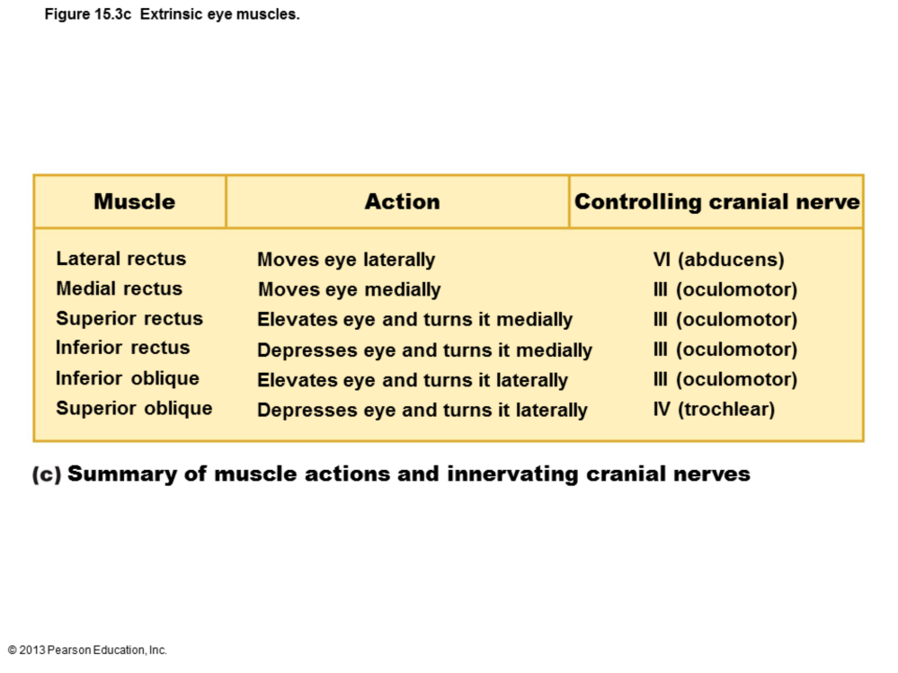
12 Six straplike extrinsic eye muscles Originate from bony orbit; insert on eyeball Enable eye to follow moving objects; maintain shape of eyeball; hold in orbit Four rectus muscles originate from common tendinous ring; names indicate movements 11
13 Superior, inferior, lateral, medial rectus muscles Two oblique muscles move eye in vertical plane and rotate eyeball Superior and inferior oblique muscles 11
14 Six straplike extrinsic eye muscles Originate from bony orbit; insert on eyeball Enable eye to follow moving objects; maintain shape of eyeball; hold in orbit Four rectus muscles originate from common tendinous ring; names indicate movements 12
15 Superior, inferior, lateral, medial rectus muscles Two oblique muscles move eye in vertical plane and rotate eyeball Superior and inferior oblique muscles 12
16 13
17 Wall of eyeball contains three layers Fibrous Vascular Inner Internal cavity filled with fluids called humors Lens separates internal cavity into anterior and posterior segments (cavities) 14
18 Outermost layer; dense avascular connective tissue Two regions: sclera and cornea 1. Sclera Opaque posterior region Protects, shapes eyeball; anchors extrinsic eye muscles Continuous with dura mater of brain posteriorly 15
19 2. Cornea Transparent anterior 1/6 of fibrous layer Bends light as it enters eye Sodium pumps of corneal endothelium on inner face help maintain clarity of cornea Numerous pain receptors contribute to blinking and tearing reflexes 16
20 Middle pigmented layer Three regions: choroid, ciliary body, and iris 1. Choroid region Posterior portion of uvea Supplies blood to all layers of eyeball Brown pigment absorbs light to prevent light scattering and visual confusion 17
21 3. Iris Colored part of eye 2. Ciliary body Ring of tissue surrounding lens Smooth muscle bundles (ciliary muscles) control lens shape Capillaries of ciliary processes secrete fluid Ciliary zonule (suspensory ligament) holds lens in position 18
22 3. Iris Colored part of eye Pupil central opening that regulates amount of light entering eye Close vision and bright light sphincter pupillae (circular muscles) contract; pupils constrict Distant vision and dim light dilator pupillae (radial muscles) contract; pupils dilate sympathetic fibers 19
23 Changes in emotional state pupils dilate when subject matter is appealing or requires problem-solving skills 19
24 Originates as outpocketing of brain Delicate two-layered membrane Outer Pigmented layer Single-cell-thick lining Absorbs light and prevents its scattering Phagocytize photoreceptor cell fragments Stores vitamin A Optic disc (blind spot) Site where optic nerve leaves eye Lacks photoreceptors Quarter-billion photoreceptors of two types Rods Cones 20
25 Inner Neural layer Transparent Composed of three main types of neurons Photoreceptors, bipolar cells, ganglion cells Signals spread from photoreceptors to bipolar cells to ganglion cells Ganglion cell axons exit eye as optic nerve 21
26 Rods Cones Dim light, peripheral vision receptors More numerous, more sensitive to light than cones No color vision or sharp images Numbers greatest at periphery Vision receptors for bright light High-resolution color vision Macula lutea exactly at posterior pole Mostly cones Fovea centralis Tiny pit in center of macula with all cones; best vision 22
27 Two sources of blood supply Choroid supplies outer third (photoreceptors) Central artery and vein of retina supply inner two-thirds Enter/exit eye in center of optic nerve Vessels visible in living person 23
28 The lens and ciliary zonule separate eye into two segments Anterior and posterior segments Posterior segment contains vitreous humor that Transmits light Supports posterior surface of lens Holds neural layer of retina firmly against pigmented layer Contributes to intraocular pressure 24
29 Forms in embryo; lasts lifetime Anterior segment composed of two chambers Anterior chamber between cornea and iris Posterior chamber between iris and lens 24
30 Anterior segment contains aqueous humor Plasma like fluid continuously formed by capillaries of ciliary processes Drains via scleral venous sinus (canal of Schlemm) at scleracornea junction Supplies nutrients and oxygen 25
31 mainly to lens and cornea but also to retina, and removes wastes Glaucoma: blocked drainage of aqueous humor increases pressure and causes compression of retina and optic nerve blindness 25
32 Biconvex, transparent, flexible, and avascular Changes shape to precisely focus light on retina Two regions Lens epithelium anteriorly; Lens fibers form bulk of lens Lens fibers filled with transparent 26
33 protein crystallin Lens becomes more dense, convex, less elastic with age cataracts (clouding of lens) consequence of aging, diabetes mellitus, heavy smoking, frequent exposure to intense sunlight 26
34 Eyes respond to visible light Light Small portion of electromagnetic spectrum Wavelengths of nm Packets of energy (photons or quanta) that travel in wavelike fashion at high speeds Color of light objects reflect determines color eye perceives 27
35 Refraction Bending of light rays Due to change in speed when light passes from one transparent medium to another Occurs when light meets surface of different medium at an oblique angle Curved lens can refract light 28
36 Light passing through convex lens (as in eye) is bent so that rays converge at focal point Image formed at focal point is upside-down and reversed right to left Concave lenses diverge light Prevent light from focusing 29
37 Pathway of light entering eye: cornea, aqueous humor, lens, vitreous humor, entire neural layer of retina, photoreceptors Light refracted three times along pathway Entering cornea Entering lens 30
38 Leaving lens Majority of refractory power in cornea Change in lens curvature allows for fine focusing 30
39 Eyes best adapted for distant vision Far point of vision Distance beyond which no change in lens shape needed for focusing 20 feet for emmetropic (normal) eye Cornea and lens focus light precisely on retina Ciliary muscles relaxed Lens stretched flat by tension in ciliary zonule 31
40 Light from close objects (<6 m) diverges as approaches eye Requires eye to make active adjustments using three simultaneous processes Accommodation of lenses Constriction of pupils Convergence of eyeballs 32
41 Accommodation Changing lens shape to increase refraction Near point of vision Closest point on which the eye can focus Presbyopia loss of accommodation over age 50 Constriction 33
42 Accommodation pupillary reflex constricts pupils to prevent most divergent light rays from entering eye Convergence Medial rotation of eyeballs toward object being viewed 33
43 34
44 Myopia (nearsightedness) Focal point in front of retina, e.g., eyeball too long Corrected with a concave lens Hyperopia (farsightedness) Focal point behind retina, e.g., eyeball too short Corrected with a convex lens 35
45 Astigmatism Unequal curvatures in different parts of cornea or lens Corrected with cylindrically ground lenses or laser procedures 35
46 36
47 Rods and cones Modified neurons Receptive regions called outer segments Contain visual pigments (photopigments) Molecules change shape as absorb light Inner segment of each joins cell body 37
48 38
49 Vulnerable to damage Degenerate if retina detached Destroyed by intense light Outer segment renewed every 24 hours Tips fragment off and are phagocytized 39
50 Vulnerable to damage Degenerate if retina detached Destroyed by intense light Outer segment renewed every 24 hours Tips fragment off and are phagocytized 40
51 rods Functional characteristics Very sensitive to light Best suited for night vision and peripheral vision Contain single pigment Perceived input in gray tones only Pathways converge, causing fuzzy, indistinct images 41
52 Cones Functional characteristics Need bright light for activation (have low sensitivity) React more quickly Have one of three pigments for colored view Nonconverging pathways result in detailed, high-resolution vision Color blindness lack of one or more cone pigments 42
53 rods Functional characteristics Very sensitive to light Best suited for night vision and peripheral vision Contain single pigment Perceived input in gray tones only Pathways converge, causing fuzzy, indistinct images 43
54 Cones Functional characteristics Need bright light for activation (have low sensitivity) React more quickly Have one of three pigments for colored view Nonconverging pathways result in detailed, high-resolution vision Color blindness lack of one or more cone pigments 44
55 Retinal Light-absorbing molecule that combines with one of four proteins (opsins) to form visual pigments Synthesized from vitamin A Retinal isomers: 11-cis-retinal (bent form) and all-trans-retinal (straight form) Bent form straight form when pigment absorbs light Conversion of bent to straight initiates reactions electrical impulses along optic nerve 45
56 Deep purple pigment of rods rhodopsin 11-cis-retinal + opsin rhodopsin Three steps of rhodopsin formation and breakdown Pigment synthesis Pigment bleaching Pigment regeneration 46
 Pigment regeneration All-trans retinal converted to 11-cis isomer Rhodopsin regenerated in outer segments 47")
57 Pigment synthesis Rhodopsin forms and accumulates in dark Pigment bleaching When rhodopsin absorbs light, retinal changes to all-trans isomer Retinal and opsin separate (rhodopsin breakdown) Pigment regeneration All-trans retinal converted to 11-cis isomer Rhodopsin regenerated in outer segments 47
58 Light-activated rhodopsin activates G protein transducin Transducin activates PDE, which breaks down cyclic GMP (cgmp) In dark, cgmp holds channels of outer segment open Na + and Ca 2+ depolarize cell In light cgmp breaks down, channels close, cell hyperpolarizes Hyperpolarization is signal! 48
59 Similar as process in rods Cones far less sensitive to light Takes higher-intensity light to activate cones 49

60 Photoreceptors and bipolar cells only generate graded potentials (EPSPs and IPSPs) 50
 depolarize, release neurotransmitter onto ganglion cells Ganglion cells generate APs transmitted in optic nerve to")
61 When light hyperpolarizes photoreceptor cells Stop releasing inhibitory neurotransmitter glutamate Bipolar cells (no longer inhibited) depolarize, release neurotransmitter onto ganglion cells Ganglion cells generate APs transmitted in optic nerve to brain 51
62 Move from darkness into bright light Both rods and cones strongly stimulated Pupils constrict Large amounts of pigments broken down instantaneously, producing glare Visual acuity improves over 5 10 minutes as: Rod system turns off Retinal sensitivity decreases Cones and neurons rapidly adapt 52
63 Move from bright light into darkness Cones stop functioning in low-intensity light Rod pigments bleached; system turned off Rhodopsin accumulates in dark Transducin returns to outer segments Retinal sensitivity increases within minutes Pupils dilate 53
64 Axons of retinal ganglion cells form optic nerve Medial fibers of optic nerve decussate at optic chiasma Most fibers of optic tracts continue to lateral geniculate body of thalamus Fibers from thalamic neurons form optic radiation and project to primary visual cortex in occipital lobes 54
65 Fibers from thalamic neurons form optic radiation Optic radiation fibers connect to primary visual cortex in occipital lobes 55
66 Fibers from thalamic neurons form optic radiation Optic radiation fibers connect to primary visual cortex in occipital lobes Other optic tract fibers send branches to midbrain, ending in superior colliculi (initiating visual reflexes) 56
67 A small subset of ganglion cells in retina contain melanopsin (circadian pigment), which projects to: Pretectal nuclei (involved with pupillary reflexes) Suprachiasmatic nucleus of hypothalamus, timer for daily biorhythms 57
68 Both eyes view same image from slightly different angles Depth perception (three-dimensional vision) results from cortical fusion of slightly different images Requires input from both eyes 58
69 59
70 60
71 Retinal cells split input into channels Color, brightness, angle, direction, speed of movement of edges (sudden changes of brightness or color) Lateral inhibition decodes "edge" information Job of amacrine and horizontal cells 61
72 Lateral geniculate nuclei of thalamus Process for depth perception, cone input emphasized, contrast sharpened Primary visual cortex (striate cortex) Neurons respond to dark and bright edges, and object orientation Provide form, color, motion inputs to visual association areas (prestriate cortices) 62
73 Occipital lobe centers (anterior prestriate cortices) continue processing of form, color, and movement Complex visual processing extends to other regions "What" processing identifies objects in visual field Ventral temporal lobe 63
74 "Where" processing assesses spatial location of objects Parietal cortex to postcentral gyrus Output from both passes to frontal cortex Directs movements 63
The Eye ACCESSORY STRUCTURES
 The Eye The eye forms a visual image and projects it onto the sensory receptors (photoreceptors) of the retina. ACCESSORY STRUCTURES Anatomy and Physiology Text and Laboratory Workbook, Stephen G. Davenport,
Help maintain homeostasis by capturing stimuli from the external environment and relaying them to the brain for processing.
 The Sense Organs... (page 409) Help maintain homeostasis by capturing stimuli from the external environment and relaying them to the brain for processing. Ex. Eye structure - protected by bony ridges and
Applications in Dermatology, Dentistry and LASIK Eye Surgery using LASERs
 Applications in Dermatology, Dentistry and LASIK Eye Surgery using LASERs http://www.medispainstitute.com/menu_laser_tattoo.html http://www.life123.com/bm.pix/bigstockphoto_close_up_of_eye_surgery_catar_2264267.s600x600.jpg
BIOL 1108 Vertebrate Anatomy Lab
 BIOL 1108 Vertebrate Anatomy Lab This lab explores major organs associated with the circulatory, excretory, and nervous systems of mammals. Circulatory System Vertebrates are among the organisms that have
SHEEP EYE DISSECTION PROCEDURES
 SHEEP EYE DISSECTION PROCEDURES The anatomy of the human eye can be better shown and understood by the actual dissection of an eye. One eye of choice for dissection, that closely resembles the human eye,
What role does the nucleolus have in cell functioning? Glial cells
 Nervous System Lab The nervous system of vertebrates can be divided into the central nervous system, which consists of the brain and spinal cord, and the peripheral nervous system, which contains nerves,
Senses 3. The optics of the eye Accommodation of the eye Ammetropias The eyeground Visual field
 Senses 3 The optics of the eye Accommodation of the eye Ammetropias The eyeground Visual field Practical tasks Purkinje s images Keratoscopy Ophthalmoscopy Purkinje s flash figure Determination of the
Sensory Organs (Receptors) Sensory Physiology. Sensory Adaptation. Four Steps to Sensation. Types of Sensors Structural Design
 Sensory Physiology. Sensory Adaptation. Four Steps to Sensation. Types of Sensors Structural Design") Sensory Organs (Receptors) Sensory Physiology Chapter 10 Monitor the internal and external environment Transmit peripheral signals to CNS for processing Critical for homeostasis Types of Sensors Structural
Sensory Organs (Receptors) Sensory Physiology Chapter 10 Monitor the internal and external environment Transmit peripheral signals to CNS for processing Critical for homeostasis Types of Sensors Structural
Orbit & Cranial Nerves II, III, IV, & VI
 Orbit & Cranial Nerves II, III, IV, & VI PCC Year 1, Spring Quarter Lawrence M. Witmer, PhD Life Sciences Building 123 OBJECTIVES: to understand the anatomy of the bony orbit and its contents, in particular,
Chapter 17 The Special Senses Lecture Outline
 Chapter 17 The Special Senses Lecture Outline Five special senses Olfaction = smell Gustation = taste Vision = sight Hearing Equilibrium Special sensory receptors: 1. Distinct cells 2. Complex organ /
1 Cornea 6 Macula 2 Lens 7 Vitreous humor 3 Iris 8 Optic disc 4 Conjunctiva 9 Ciliary muscles 5 Sclera 10 Choroid
 Anatomy and Physiology Quiz 1 Sample Question Answers Use the following table to answer Questions 1 2. 1 Cornea 6 Macula 2 Lens 7 Vitreous humor 3 Iris 8 Optic disc 4 Conjunctiva 9 Ciliary muscles 5 Sclera
SPECIAL SENSES. Introduction: Activity 1: Observation of the Human Eye Model
 SPECIAL SENSES Introduction: The special senses include vision, hearing, equilibrium (balance), taste and smell. In these activities you will be performing a series of physiological tests for each of these
18. What is limbic system? A. The inner parts of cerebral hemispheres associated with deep structures and from a complex structure. 19.
 CHAPTER 21 NEURAL CONTROL AND COORDINATION One mark Questions: 1. Name the structural and functional unit of nervous system? A. Neuron. 2. What does central Nervous System consists of? A. Brain and spinal
PUPILS AND NEAR VISION. Akilesh Gokul PhD Research Fellow Department of Ophthalmology
 PUPILS AND NEAR VISION Akilesh Gokul PhD Research Fellow Department of Ophthalmology Iris Anatomy Two muscles: Radially oriented dilator (actually a myo-epithelium) - like the spokes of a wagon wheel Sphincter/constrictor
The Physiology of the Senses Lecture 1 - The Eye www.tutis.ca/senses/
 The Physiology of the Senses Lecture 1 - The Eye www.tutis.ca/senses/ Contents Objectives... 2 Introduction... 2 Accommodation... 3 The Iris... 4 The Cells in the Retina... 5 Receptive Fields... 8 The
An Overview of Anatomy and Physiology of the Eye
 An Overview of Anatomy and Physiology of the Eye Changes in the eyelid fissure A, Graves disease stare. B, Myathenia gravis. C, Congenital ptosis of right eye. D, Levator disinsertion. E, Horner syndrome
Lab Exercise 9. Nervous Tissue. Brain. Cranial Nerves. Spinal Cord. Spinal Nerves
 Lab Exercise 9 Nervous Tissue Brain Cranial Nerves Spinal Cord Spinal Nerves Textbook Reference: See Chapter 11 for histology of nerve tissue and spinal cord See Chapter 12 for brain and spinal cord anatomy
Vision: Receptors. Modes of Perception. Vision: Summary 9/28/2012. How do we perceive our environment? Sensation and Perception Terminology
 How do we perceive our environment? Complex stimuli are broken into individual features, relayed to the CNS, then reassembled as our perception Sensation and Perception Terminology Stimulus: physical agent
Keeping Your Eyes Healthy after Treatment for Childhood Cancer
 Keeping Your Eyes Healthy after Treatment for Childhood Cancer High doses of radiation to the brain, eye, or eye socket (orbit) during treatment for childhood cancer can have a long-lasting affect on the
The light. Light (normally spreads out straight... ... and into all directions. Refraction of light
 The light Light (normally spreads out straight...... and into all directions. Refraction of light But when a light ray passes from air into glas or water (or another transparent medium), it gets refracted
IMAGE ASSISTANT: OPHTHALMOLOGY
 IMAGE ASSISTANT: OPHTHALMOLOGY Summary: The Image Assistant has been developed to provide medical doctors with a software tool to search, display, edit and use medical illustrations of their own specialty,
Integration and Coordination of the Human Body. Nervous System
 I. General Info Integration and Coordination of the Human Body A. Both the and system are responsible for maintaining 1. Homeostasis is the process by which organisms keep internal conditions despite changes
Processing the Image or Can you Believe what you see? Light and Color for Nonscientists PHYS 1230
 Processing the Image or Can you Believe what you see? Light and Color for Nonscientists PHYS 1230 Optical Illusions http://www.michaelbach.de/ot/mot_mib/index.html Vision We construct images unconsciously
Nervous System Organization. PNS and CNS. Nerves. Peripheral Nervous System. Peripheral Nervous System. Motor Component.
 Nervous System Organization PNS and CNS Chapters 8 and 9 Peripheral Nervous System (PNS) connects CNS to sensory receptors, muscles and glands Central Nervous System (CNS) control/integrating center brain
Eye Diseases. 1995-2014, The Patient Education Institute, Inc. www.x-plain.com otf30101 Last reviewed: 05/21/2014 1
 Eye Diseases Introduction Some eye problems are minor and fleeting. But some lead to a permanent loss of vision. There are many diseases that can affect the eyes. The symptoms of eye diseases vary widely,
The Visual Cortex 0 http://www.tutis.ca/neuromd/index.htm 20 February 2013
 T he Visual Cortex 0 Chapter contents Contents Chapter 2... 0 T he Visual Cortex... 0 Chapter Contents... 1 Introduction... 2 Optic Chiasm... 2 Where do the eye's ganglion cells project to?... 3 To where
Chapter 7: The Nervous System
 Chapter 7: The Nervous System Objectives Discuss the general organization of the nervous system Describe the structure & function of a nerve Draw and label the pathways involved in a withdraw reflex Define
BSM Connection elearning Course
 BSM Connection elearning Course Anatomy of the Eye 2004, BSM Consulting All Rights Reserved. Table of Contents OVERVIEW...1 THE MECHANISM OF SIGHT... 1 EXTERNAL STRUCTURE OF THE EYE... 1 INTERNAL STRUCTURE
Nervous System: Special Senses (Chapter 17) Lecture Materials for Amy Warenda Czura, Ph.D. Suffolk County Community College
 Lecture Materials for Amy Warenda Czura, Ph.D. Suffolk County Community College") Nervous System: Special Senses (Chapter 17) Lecture Materials for Amy Warenda Czura, Ph.D. Suffolk County Community College Primary Sources for figures and content: Eastern Campus Marieb, E. N. Human Anatomy
Nervous System: Special Senses (Chapter 17) Lecture Materials for Amy Warenda Czura, Ph.D. Suffolk County Community College Primary Sources for figures and content: Eastern Campus Marieb, E. N. Human Anatomy
Eye Injuries. The Eyes The eyes are sophisticated organs. They collect light and focus it on the back of the eye, allowing us to see.
 Eye Injuries Introduction The design of your face helps protect your eyes from injury. But injuries can still damage your eyes. Sometimes injuries are severe enough that you could lose your vision. Most
Chapter 15. Autonomic Nervous System (ANS) and Visceral Reflexes. general properties Anatomy. Autonomic effects on target organs
 and Visceral Reflexes. general properties Anatomy. Autonomic effects on target organs") Chapter 15 Autonomic Nervous System (ANS) and Visceral Reflexes general properties Anatomy Autonomic effects on target organs Central control of autonomic function 15-1 Copyright (c) The McGraw-Hill Companies,
Chapter 15 Autonomic Nervous System (ANS) and Visceral Reflexes general properties Anatomy Autonomic effects on target organs Central control of autonomic function 15-1 Copyright (c) The McGraw-Hill Companies,
THE EYES IN MARFAN SYNDROME
 THE EYES IN MARFAN SYNDROME Marfan syndrome and some related disorders can affect the eyes in many ways, causing dislocated lenses and other eye problems that can affect your sight. Except for dislocated
2401 : Anatomy/Physiology
 Dr. Chris Doumen Week 7 2401 : Anatomy/Physiology The Brain Central Nervous System TextBook Readings Pages 431 through 435 and 463-467 Make use of the figures in your textbook ; a picture is worth a thousand
Thyroid Eye Disease. Anatomy: There are 6 muscles that move your eye.
 Thyroid Eye Disease Your doctor thinks you have thyroid orbitopathy. This is an autoimmune condition where your body's immune system is producing factors that stimulate enlargement of the muscles that
Nervous System: PNS and CNS
 Nervous System: PNS and CNS Biology 105 Lecture 10 Chapter 8 Outline I. Central Nervous System vs Peripheral Nervous System II. Peripheral Nervous System A. Somatic Nervous System B. Autonomic Nervous
Review Vocabulary spectrum: a range of values or properties
 Standards 7.3.19: Explain that human eyes respond to a narrow range of wavelengths of the electromagnetic spectrum. 7.3.20: Describe that something can be seen when light waves emitted or reflected by
Chapter 15 Anatomy and Physiology Lecture
 1 THE AUTONOMIC NERVOUS SYSTEM Chapter 15 Anatomy and Physiology Lecture 2 THE AUTONOMIC NERVOUS SYSTEM Autonomic Nervous System (ANS) regulates the activity of smooth muscles, cardiac muscles, and certain
A concise guide to Safety Glasses, the different standards and the effects of light on the eye. Contents. Links. Year of publication: 2010
 A concise guide to Safety Glasses, the different standards and the effects of light on the eye Year of publication: 2010 Produced by the leading supplier of Safety Glasses in the UK. All Rights Reserved.
Vitreo-Retinal and Macular Degeneration Frequently Asked Questions
 Vitreo-Retinal and Macular Degeneration Frequently Asked Questions What is a Vitreo-Retinal specialist? Retinal specialists are eye physicians and surgeons who focus on diseases in the back of the eye
LASIK. What is LASIK? Eye Words to Know. Who is a good candidate for LASIK?
 2014 2015 LASIK What is LASIK? LASIK (laser in situ keratomileusis) is a type of refractive surgery. This kind of surgery uses a laser to treat vision problems caused by refractive errors. You have a refractive
iocutouchtm for ipad Contents of Videos and Still Images Anatomy 906. Normal Eye and Orbit - no labels 907. Normal Eye and Orbit - with labels
 iocutouchtm for ipad Contents of Videos and Still Images Anatomy 906. Normal Eye and Orbit - no labels 907. Normal Eye and Orbit - with labels Normal Eye 474. Normal Eye overview 476. Cornea - overview
...You Need to know about
 What......You Need to know about LASIK Our Eyes Eyes are the windows to our world. They are so important to us that for many years we have looked for better ways to fix visual problems and improve our
The Eye: Anatomy, Histology & Histopathology. Gillian Shaw, DVM, MS September 9, 2011 gilliancshaw@gmail.com
 The Eye: Anatomy, Histology & Histopathology Gillian Shaw, DVM, MS September 9, 2011 gilliancshaw@gmail.com Overview Macroscopic Anatomy Some clinical presentations of eye disease Microscopic Anatomy Normal
MODERN CLINICAL OPTOMETRY BILLING & CODING THE MEDICAL EYE EXAMINATION. Definitions of Eye Examinations. Federal Government Definition
 MODERN CLINICAL OPTOMETRY BILLING & CODING THE MEDICAL EYE EXAMINATION Craig Thomas, O.D. 3900 West Wheatland Road Dallas, Texas 75237 972-780-7199 thpckc@yahoo.com Definitions of Eye Examinations Optometry
HEALTHY EYES. Type 2 diabetes. Information for patients. Disease Management Programme
 HEALTHY EYES Information for patients Type 2 diabetes Disease Management Programme WHAT CAN I DO FOR HEALTHY EYES DESPITE MY TYPE 2 DIABETES? More and more people suffer from type 2 diabetes; among them,
MAZAHERI LASIK METHOD FOR VISUAL ENHANCEMENT TECHNICAL FIELD OF THE INVENTION. [0001] The present invention is directed, in general, to
![MAZAHERI LASIK METHOD FOR VISUAL ENHANCEMENT TECHNICAL FIELD OF THE INVENTION. [0001] The present invention is directed, in general, to](/thumbs/26/8162223.jpg "MAZAHERI LASIK METHOD FOR VISUAL ENHANCEMENT TECHNICAL FIELD OF THE INVENTION. [0001] The present invention is directed, in general, to") MAZAHERI LASIK METHOD FOR VISUAL ENHANCEMENT TECHNICAL FIELD OF THE INVENTION [0001] The present invention is directed, in general, to a surgical procedure and, more particularly, to surgical procedure
MAZAHERI LASIK METHOD FOR VISUAL ENHANCEMENT TECHNICAL FIELD OF THE INVENTION [0001] The present invention is directed, in general, to a surgical procedure and, more particularly, to surgical procedure
Information and consent for patients preparing for refractive surgery LASIK Laser Eye Center Kubati
 1. General information Not long ago, the WHO - World Health Organization has described ametropy (medical term for diopter) as a category of disability creating a solution to the needs of many with ametropy
Seeing Beyond the Symptoms
 Seeing Beyond the Symptoms Cataracts are one of the leading causes of vision impairment in the United States. 1 However, because cataracts form slowly and over a long period of time, many people suffer
The Human Eye. Structure and Function. Clyde W. Oyster. Sinauer Associates, Inc. Publishers Sunderland, Massachusetts
 The Human Eye Structure and Function Clyde W. Oyster The University of Alabama at Birmingham Sinauer Associates, Inc. Publishers Sunderland, Massachusetts Brief Contents Preface xxiii Acknowledgments xxviii
Sheep Brain Dissection
 Sheep Brain Dissection http://www.carolina.com/product/preserved+organisms/preserved+animals+%28mammal s%29/sheep+organs/preserved+sheep+dissection.do Michigan State University Neuroscience Program Brain
The Cornea, The Lens & Refractive Surgery. John S. Berestka, M.D. Timothy J. Ehlen, M.D. Matthew R. Ramsey, M.D. Stanley D. Walker, M.D.
 The Cornea, The Lens & Refractive Surgery John S. Berestka, M.D. Timothy J. Ehlen, M.D. Matthew R. Ramsey, M.D. Stanley D. Walker, M.D. 1 TABLE OF CONTENTS Cornea 3 Five layers of the Cornea 4 Refractive
CHAPTER 6 PRINCIPLES OF NEURAL CIRCUITS.
 CHAPTER 6 PRINCIPLES OF NEURAL CIRCUITS. 6.1. CONNECTIONS AMONG NEURONS Neurons are interconnected with one another to form circuits, much as electronic components are wired together to form a functional
Welcome to Lesson 11 of the Basic Human Anatomy Course. Today, we ll be studying the Human Nervous System.
 Basic Human Anatomy Lesson 11: Nervous System Welcome to Lesson 11 of the Basic Human Anatomy Course. Today, we ll be studying the Human Nervous System. I have 19 goals for you in this lesson: 1. Name
North Bergen School District Benchmarks
 Grade: 10,11, and 12 Subject: Anatomy and Physiology First Marking Period Define anatomy and physiology, and describe various subspecialties of each discipline. Describe the five basic functions of living
Animal Tissues. I. Epithelial Tissue
 Animal Tissues There are four types of tissues found in animals: epithelial tissue, connective tissue, muscle tissue, and nervous tissue. In this lab you will learn the major characteristics of each tissue
Physical and Mental Conditions Guidelines VISION CONDITIONS AND ACTIONS Page 5.4
 Physical and Mental Conditions Guidelines VISION CONDITIONS AND ACTIONS Page 5.4 AMBLYOPIA (Lazy Eye) A reduction in the acuteness of vision without apparent eye disease. This condition cannot be entirely
Table of Contents. Page. Introducing Crystalens 2. What is a cataract 3. How the eye works 4. Vision restoration options 5
 P/N 50-0065, Revision B, p.1 PATIENT LABELING Table of Contents Introducing Crystalens 2 Page What is a cataract 3 How the eye works 4 Vision restoration options 5 Are you a candidate for the Crystalens?
COW S EYE dissection. Dissecting a Cow s Eye Step-by-Step Instructions. Safety first!
 COW S EYE dissection Dissecting a Cow s Eye Step-by-Step Instructions One way to figure out how something works is to look inside it. To learn about how your eyes work, you can dissect, or take apart,
Autonomic Nervous System Dr. Ali Ebneshahidi
 Autonomic Nervous System Dr. Ali Ebneshahidi Nervous System Divisions of the nervous system The human nervous system consists of the central nervous System (CNS) and the Peripheral Nervous System (PNS).
Optical Illusion Eye Tricks. This one will make you dizzy. Try focusing on just the center circle while moving your head.
 Optical Illusion Eye Tricks This one will make you dizzy. Try focusing on just the center circle while moving your head. Photocopy from original for best quality. H A N D O U T The Visual System The properly,
Parts of the Brain. Chapter 1
 Chapter 1 Parts of the Brain Living creatures are made up of cells. Groups of cells, similar in appearance and with the same function, form tissue. The brain is a soft mass of supportive tissues and nerve
3) Cerebral Cortex & Functions of the 4 LOBES. 5) Cranial Nerves (Nerves In the Cranium, i.e., Head)
 Cerebral Cortex & Functions of the 4 LOBES. 5) Cranial Nerves (Nerves In the Cranium, i.e., Head)") Lecture 5 (Oct 8 th ): ANATOMY and FUNCTION OF THE NERVOUS SYSTEM Lecture Outline 1) Basic Divisions (CNS vs. PNS, Somatic vs. Autonomic) and Directional Terms 2) The Brain (Hindbrain/ Midbrain/ Forebrain)
Lecture 5 (Oct 8 th ): ANATOMY and FUNCTION OF THE NERVOUS SYSTEM Lecture Outline 1) Basic Divisions (CNS vs. PNS, Somatic vs. Autonomic) and Directional Terms 2) The Brain (Hindbrain/ Midbrain/ Forebrain)
ReLEx smile Minimally invasive vision correction Information for patients
 ReLEx smile Minimally invasive vision correction Information for patients Seeing is living Our eyes are our most important sensory organ. The human brain obtains over 80 % of its information via the sense
Chapter 17: Light and Image Formation
 Chapter 17: Light and Image Formation 1. When light enters a medium with a higher index of refraction it is A. absorbed. B. bent away from the normal. C. bent towards from the normal. D. continues in the
A Center for VisionCare 4418 Vineland Avenue, Suite 106 North Hollywood, CA 91607 818/762-0647
 A Center for VisionCare 4418 Vineland Avenue, Suite 106 North Hollywood, CA 91607 818/762-0647 Alan I. Mandelberg, M.D. Donald I. Goldstein, M.D. Glossary of Refractive Surgery Terms Aberration: An irregularity
Chapter 15. Neurotransmitters of the ANS
 Chapter 15 Neurotransmitters of the ANS Neurotransmitters and Receptors How can the same ANS neurons create different effects on different target tissue? Variety of neurotransmitters Secondly, different
Science In Action 8 Unit C - Light and Optical Systems. 1.1 The Challenge of light
 1.1 The Challenge of light 1. Pythagoras' thoughts about light were proven wrong because it was impossible to see A. the light beams B. dark objects C. in the dark D. shiny objects 2. Sir Isaac Newton
CENTRAL NERVOUS SYSTEM. Sensory Pathway (PNS) OVERVIEW OF SPINAL CORD ANATOMY OF THE SPINAL CORD FUNCTIONS OF THE SPINAL CORD
 OVERVIEW OF SPINAL CORD ANATOMY OF THE SPINAL CORD FUNCTIONS OF THE SPINAL CORD") CENTRAL NERVOUS SYSTEM Central nervous system (CNS) brain and spinal cord enclosed in bony coverings Functions of the spinal cord spinal cord reflexes integration ti (summation of inhibitory and excitatory)
CENTRAL NERVOUS SYSTEM Central nervous system (CNS) brain and spinal cord enclosed in bony coverings Functions of the spinal cord spinal cord reflexes integration ti (summation of inhibitory and excitatory)
Fundus Photograph Reading Center
 Modified 7-Standard Field Digital Color Fundus Photography (7M-D) 8010 Excelsior Drive, Suite 100, Madison WI 53717 Telephone: (608) 410-0560 Fax: (608) 410-0566 Table of Contents 1. 7M-D Overview... 2
Light and Sound. Pupil Booklet
 Duncanrig Secondary School East Kilbride S2 Physics Elective Light and Sound Name: Pupil Booklet Class: SCN 3-11a - By exploring the refraction of light when passed through different materials, lenses
LASIK. Cornea. Iris. Vitreous
 LASIK Introduction LASIK surgery is a procedure that improves vision and can decrease or eliminate the need for eyeglasses or contact lenses. If you and your doctor decide that LASIK surgery is right for
Vascular System The heart can be thought of 2 separate pumps from the right ventricle, blood is pumped at a low pressure to the lungs and then back
 Vascular System The heart can be thought of 2 separate pumps from the right ventricle, blood is pumped at a low pressure to the lungs and then back to the left atria from the left ventricle, blood is pumped
Nervous System Divisions of the Nervous system
 Nervous System Divisions of the Nervous system Central Nervous System relays messages, processes information, and analyzes information; Consists of brain and spinal cord Meninges connective tissue around
Trulign Toric Accommodating Intraocular Lenses PATIENT INFORMATION BROCHURE
 Trulign Toric Accommodating Intraocular Lenses PATIENT INFORMATION BROCHURE Table of Contents Page Glossary 2 Introducing Trulign Toric Accommodating IOL 5 What is a cataract 5 What is corneal astigmatism
D.U.C. Assist. Lec. Faculty of Dentistry General Physiology Ihsan Dhari. The Autonomic Nervous System
 The Autonomic Nervous System The portion of the nervous system that controls most visceral functions of the body is called the autonomic nervous system. This system helps to control arterial pressure,
refractive surgery a closer look
 2011-2012 refractive surgery a closer look How the eye works Light rays enter the eye through the clear cornea, pupil and lens. These light rays are focused directly onto the retina, the light-sensitive
Tucson Eye Care, PC. Informed Consent for Cataract Surgery And/Or Implantation of an Intraocular Lens
 Tucson Eye Care, PC Informed Consent for Cataract Surgery And/Or Implantation of an Intraocular Lens INTRODUCTION This information is provided so that you may make an informed decision about having eye
Skeletal, Muscular, and Integumentary Systems
 Chapter 36 Skeletal, Muscular, and Integumentary Systems Section 36 1 The Skeletal System (pages 921 925) This section describes the skeletal system and its functions. Introduction (page 921) 1. What forms
DISTANCE LEARNING COURSE. Anatomy of the Patient Exam. 2008 2012, BSM Consulting All rights reserved.
 DISTANCE LEARNING COURSE Anatomy of the Patient Exam 2008 2012, BSM Consulting All rights reserved. Table of Contents OVERVIEW... 1 PRELIMINARY WORK-UP... 1 OTHER DIAGNOSTIC TESTING... 3 DIAGNOSIS... 4
Patient Information Content
 KORNMEHL LASER EYE ASSOCIATES Patient Information Content Near Vision Loss Understanding Near Vision Loss Why is My Near Vision Changing? In our 40 s and 50 s, we begin to experience the naturally frustrating
Verisyse Phakic IOL. Facts You Need to Know About Implantation of the Verisyse Phakic IOL (-5 to -20 D) for the Correction of Myopia (Nearsightedness)
 for the Correction of Myopia (Nearsightedness)") Verisyse Phakic IOL Facts You Need to Know About Implantation of the Verisyse Phakic IOL (-5 to -20 D) for the Correction of Myopia (Nearsightedness) Patient Information Brochure This brochure is designed
Verisyse Phakic IOL Facts You Need to Know About Implantation of the Verisyse Phakic IOL (-5 to -20 D) for the Correction of Myopia (Nearsightedness) Patient Information Brochure This brochure is designed
Cassie Schroeder Refractive Surgery Coordinator Boozman-Hof Regional Eye Clinic (479) 246-1820 (479) 531-3937
 246-1820 (479) 531-3937") Thank you for your interest in refractive surgery here at Boozman-Hof Regional Eye Clinic. Enclosed is a bio on Dr. Cole and articles about LASEK. There is also information on our financing company that
Thank you for your interest in refractive surgery here at Boozman-Hof Regional Eye Clinic. Enclosed is a bio on Dr. Cole and articles about LASEK. There is also information on our financing company that
DISSECTION OF THE SHEEP'S BRAIN
 DISSECTION OF THE SHEEP'S BRAIN Introduction The purpose of the sheep brain dissection is to familiarize you with the threedimensional structure of the brain and teach you one of the great methods of studying
Making Vision a Health Priority. Speaker s Guide
 Making Vision a Health Priority Speaker s Guide SLIDE 1 Introduction of the speaker. The purpose of this presentation is to provide information about vision changes that can occur with age and to talk
Visual Acuity, Impairments and Vision Insurance Plan Provisions. Stuart West Specialty Sales Manager Virginia CE Forum 2009 Course # 201718
 Visual Acuity, Impairments and Vision Insurance Plan Provisions Stuart West Specialty Sales Manager Virginia CE Forum 2009 Course # 201718 How Vision Works Light passes through the cornea & lens Light
Physics 1230: Light and Color
 Physics 1230: Light and Color The Eye: Vision variants and Correction http://www.colorado.edu/physics/phys1230 What does 20/20 vision mean? Visual acuity is usually measured with a Snellen chart Snellen
Cataract Surgery Patient Information
 Cataract Patient Information 1. Within the human eye, there is a normal structure called the lens. In youth, this lens is clear, and light rays pass through and are focused by this lens as well as the
Information For Consent For Cataract Surgery
 Information For Consent For Cataract Surgery Your Ophthalmologist has diagnosed you with a visually significant cataract. The following handout will explain your condition and give you the information
Chapter 9 Nervous System
 Chapter 9 Nervous System Nervous System function: The nervous system is composed of neurons and neuroglia. at the ends of peripheral nerves gather information and convert it into nerve impulses. When sensory
waves rays Consider rays of light from an object being reflected by a plane mirror (the rays are diverging): mirror object
: mirror object") PHYS1000 Optics 1 Optics Light and its interaction with lenses and mirrors. We assume that we can ignore the wave properties of light. waves rays We represent the light as rays, and ignore diffraction.
PHYS1000 Optics 1 Optics Light and its interaction with lenses and mirrors. We assume that we can ignore the wave properties of light. waves rays We represent the light as rays, and ignore diffraction.
Hearing and Vision Program. Public Health Muskegon County
 Hearing and Vision Program Public Health Muskegon County H&V Screening: Two of the Required Public Health Services in Michigan The Hearing and Vision programs are required by the Michigan Public Health
Heart and Vascular System Practice Questions
 Heart and Vascular System Practice Questions Student: 1. The pulmonary veins are unusual as veins because they are transporting. A. oxygenated blood B. de-oxygenated blood C. high fat blood D. nutrient-rich
BIO130 Chapter 14 The Brain and Cranial Nerves Lecture Outline
 BIO130 Chapter 14 The Brain and Cranial Nerves Lecture Outline Brain structure 1. Cerebrum Hemispheres: left & right Cerebral cortex Gyri Sulci Fissures Longitudinal fissure Corpus callosum Lobes Central
3. The neuron has many branch-like extensions called that receive input from other neurons. a. glia b. dendrites c. axons d.
 Chapter Test 1. A cell that receives information and transmits it to other cells via an electrochemical process is called a(n) a. neuron b. hormone c. glia d. endorphin Answer: A difficulty: 1 factual
Vision Correction Surgery Patient Information
 Vision Correction Surgery Patient Information Anatomy of the eye: The eye is a complex organ composed of many parts, and normal vision requires these parts to work together. When a person looks at an object,
The Tissue Level of Organization
 The Tissue Level of Organization Tissues A groups of similar cells, usually having similar embryonic origin and specialized function Histology: the study of tissues Four general types Epithelial Muscle
How To Know If You Can See Without Glasses Or Contact Lense After Lasik
 The LASIK experience I WHO CAN HAVE LASIK? To be eligible for LASIK you should be at least 21 years of age, have healthy eyes and be in good general health. Your vision should not have deteriorated significantly
Informed Consent For Cataract Surgery And/Or Implantation of an Intraocular Lens
 Informed Consent For Cataract Surgery And/Or Implantation of an Intraocular Lens INTRODUCTION This information is given to you so that you can make an informed decision about having eye surgery. Take as
SECONDARY GLAUCOMA: Pseudoexfoliation (PXF), Pigmentary Dispersion Syndrome (PDS), Neovascular (NV), Uveitic
, Pigmentary Dispersion Syndrome (PDS), Neovascular (NV), Uveitic") SECONDARY GLAUCOMA: Pseudoexfoliation (PXF), Pigmentary Dispersion Syndrome (PDS), Neovascular (NV), Uveitic Many roads can lead to glaucoma. With the exception of primary open-angle glaucoma (POAG) and
SECONDARY GLAUCOMA: Pseudoexfoliation (PXF), Pigmentary Dispersion Syndrome (PDS), Neovascular (NV), Uveitic Many roads can lead to glaucoma. With the exception of primary open-angle glaucoma (POAG) and
Student Academic Learning Services Page 1 of 8 Nervous System Quiz
 Student Academic Learning Services Page 1 of 8 Nervous System Quiz 1. The term central nervous system refers to the: A) autonomic and peripheral nervous systems B) brain, spinal cord, and cranial nerves