Preface ULTRASOUND CLINICS. Gary J. Whitman, MD
|
|
|
- Clarence Rodgers
- 7 years ago
- Views:
Transcription
1
2 xi ULTRASOUND CLINICS Preface Ultrasound Clin 1 (2007) xi xii Gary J. Whitman, MD Department of Diagnostic Radiology The University of Texas M. D. Anderson Cancer Center P.O. Box , Unit 1350 Houston, TX 77230, USA address: gwhitman@di.mdacc.tmc.edu Gary J. Whitman, MD Guest Editor Breast sonography is indispensable. Most breast lesions, other than small clusters of calcifications, can and may be evaluated with ultrasound. In the last decade and a half, we have witnessed tremendous progress in breast ultrasound. The days of using breast ultrasound solely to differentiate cysts from solid masses are gone forever. Breast sonography is commonly used to evaluate mammographic and palpable abnormalities, and ultrasound is often used to evaluate findings initially noted on magnetic resonance imaging (MRI). Sonography also plays a role in screening for breast cancer and in evaluating the extent of disease in the breast and the regional lymph nodes. Nearly all breast masses are sampled with ultrasound-guided biopsy. Core biopsies, vacuumassisted biopsies, and fine-needle aspirations are commonly performed with sonographic guidance, often followed by ultrasound-guided marker placement. In addition, sonography can be used to help guide catheters to drain abscesses or deliver radiation therapy. In some centers, ultrasound is guiding cryoablation and radiofrequency ablation of breast lesions. Breast ultrasound can be performed just about anywhere. Although most ultrasound machines are housed in sonography rooms, breast ultrasound can be and has been performed in mammography rooms, operating rooms, surgical holding areas, emergency rooms, examination rooms, patient hospital rooms, and intensive care units. Breast ultrasound plays a major role in the daily management of women with breast diseases. In this issue of Ultrasound Clinics, we explore some of the common uses of breast sonography. Drs. Berg and Woel cover mammographic-sonographic correlation in a comprehensive manner, and Dr. Dogan and I provide an update on MRI-sonographic correlation. Dr. Dogan, Dr. Yang, Joella Wilson, Parul Patel, and Dr. Krishnamurthy assisted me on an article on ultrasound-guided biopsies. Dr. Cardenosa covers cysts, cystic lesions, and papillary lesions with a practical approach, and Dr. Hashimoto provides an update on sonography of ductal carcinoma in situ. Dr. Huynh, Parul Patel, Joella Wilson, Angelica Cantu, and Dr. Krishnamurthy assisted me on an article on sonography of invasive lobular carcinoma. Finally, Dr. Yang covers sonography of unusual breast neoplasms. All of the authors did a great job, and I thank them for taking time away from other pursuits to work on Ultrasound Clinics. Many thanks! X/07/$ see front matter ª 2007 Elsevier Inc. All rights reserved. ultrasound.theclinics.com doi: /j.cult
3 xii Preface Stephanie Deming and Ann Sutton in Scientific Publications at The University of Texas M. D. Anderson Cancer Center did an outstanding job editing many of the manuscripts. Barton Dudlick and his colleagues at Elsevier did a wonderful job coordinating this project, and I thank Barton for his patience. Barbara Almarez Mahinda, my administrative assistant, did an incredible job (as usual) on this project. She is indispensable! Many thanks to my wife, Susan, and my son, Sam, for their understanding and patience when Daddy was working on the chapters. Thank you!
4 567 Mammographic Sonographic Correlation Wendie A. Berg, MD, PhD a, *,B.SybilleWoel,MD b ULTRASOUND CLINICS Ultrasound Clin 1 (2007) Location - Use of intrinsic landmarks - Size - Masses - Asymmetries and architectural distortion - Calcifications - Inflammatory carcinoma - Implants - Nipple discharge - Palpable abnormalities - Confirming concordance - Summary - References Accurate correlation of mammographic and sonographic findings first requires complete diagnostic mammographic work-up and development of an appropriate differential diagnosis before sonographic evaluation. The finding of interest should be characterized as a mass, asymmetry, architectural distortion, suspicious calcifications, or a combination of these features [1]. Mammographic and sonographic location, size, shape, and margins should be concordant and carefully correlated to avoid errors. Unique issues arise in women who have nipple discharge, implants, and palpable masses. This article examines each of these issues in detail. Location A key to correlating mammographic and sonographic or MR imaging findings is visualizing where a lesion should be within the breast in three dimensions and translating that visualization to the imaging modality of interest. Mammography usually is performed with the patient standing or sitting upright. Breast ultrasound usually is performed with the patient supine for the inner breast and in the contralateral decubitus position for the outer breast. MR imaging is performed with the patient prone and the breasts dependent within the coil. There are few fixed landmarks in the breast other than the nipple and chest wall: these will be used as referents. Location of a finding on mammography is usually described by quadrant (upper outer, upper inner, lower outer, lower inner), subareolar, central, or axillary tail positions [1]. Description of location on ultrasound is usually by clock face (Fig. 1), as if a clock has been placed on the breast with 12:00 cephalad and 6:00 caudad. Thinking in terms of expected clock-face location is helpful when reviewing mammograms (Fig. 2) and facilitates successfully targeting ultrasound to mammographic abnormalities. Triangulation is an important part of understanding the final three-dimensional clock-face location of a mammographic abnormality [2]. The mediolateral oblique (MLO) mammogram is taken at variable angles to be parallel to the individual patient s pectoral muscle, whereas for ultrasound one needs a American Radiology Services, Johns Hopkins Green Spring, Falls Road, Suite 440, Baltimore, MD 21093, USA b Washington Radiology Associates, P.C Williams Drive, Suite 200, Fairfax, VA 22031, USA * Corresponding author. address: wendieberg@hotmail.com (W.A. Berg) X/07/$ see front matter ª 2007 Elsevier Inc. All rights reserved. ultrasound.theclinics.com doi: /j.cult
5 568 Berg & Woel Fig. 1. Diagram shows clock-face locations by breast as used to annotate ultrasound images. (Courtesy of Wendie A. Berg, MD, PhD, FACR, Baltimore, MD. ª Copyright 2006.) to know the expected location of a lesion on a true lateral (90 ) view. By triangulation, one can infer from any combination of two mammographic views (typically the craniocaudal [CC] and MLO views) where the lesion would be on the third, 90 true lateral (either mediolateral or lateromedial) view and therefore in three dimensions within the breast (Fig. 3). Triangulation also is used to help locate a lesion in three dimensions when it is seen on only one of the routine mammographic views (ie, either the CC or the MLO view) [3]. A true lateral view is then obtained. If the lesion can be seen on both true lateral and MLO views, its position on the CC view can be inferred (see Fig. 3). Lateral lesions move lower (inferiorly) from CC to MLO to true lateral views (when viewing the true lateral image to the right of the MLO view), and medial lesions move superiorly from CC to MLO to true lateral views. (A mnemonic is Lead falls and muffins rise. ) (J.E. Meyer, MD, personal communication, 1998). Rolled CC views can help locate a lesion seen only on the CC view [4]. In this maneuver, the top half of the breast is rolled laterally (CCRL view) or medially (CCRM view); a lesion moving with the top half of the breast is known to be in the upper breast (Fig. 4). Lesions seen only in the upper posterior breast on the MLO view are most commonly in the axillary tail region and can usually be seen on a laterally exaggerated CC view (Fig. 5). Once the expected clock-face location is known from mammography, the distance from the nipple and the depth of the lesion from the skin must be considered. On mammography, this information usually is expressed by dividing the anteroposterior thickness of the breast into thirds (anterior, middle, or posterior depth) or as a single measurement (ie, distance from the nipple). Rather than a single mammographic measurement of depth from the nipple, to correlate better with ultrasound, it is preferable to consider the depth of the lesion (superficial, middle depth, or posterior, that is, at chest wall) relative to the skin, which may or may not correlate with depth from the nipple on mammography. For example, a lesion near the skin of the far lateral breast near the chest wall would be superficial on ultrasound but project at a posterior depth on mammography (Fig. 6). Adequate sonographic characterization of superficial lesions is facilitated by use of a glob of gel or a standoff pad so that the lesion is at least 5 to 7 mm away from the face of the transducer; even with current 12- to 15-MHz linear array transducers, the beam cannot be optimally focused more anteriorly. Anterior lesions on mammography should be within the anterior third of the tissue on ultrasound, closer to the skin surface than the chest wall (Fig. 7). Similarly, posterior lesions close to the chest wall (but not the skin) on both mammographic views should be close to the pectoral muscle on ultrasound (Fig. 8). It also is useful to consider the distance medial or lateral and superior or inferior to the nipple on mammography to help direct targeted ultrasound (see Fig. 6). On ultrasound, the distance along the skin surface from the center of the lesion to the nipple in centimeters should be specified on the ultrasound images; the 38- or 50-mm footprint of the transducer itself can be used as a guide for reliably estimating and recording this measurement [5]. Location and distance from the nipple on ultrasound generally correlates well with Fig. 2. Clock-face location is first inferred from the CC position of a finding, with the MLO view used predominantly to determine whether a mass is in the upper (above the nipple) or lower (below the nipple) breast. (Courtesy of Wendie A. Berg, MD, PhD, FACR, Baltimore, MD. ª Copyright 2006.)
6 Mammographic Sonographic Correlation 569 Fig. 3. Triangulation. With the nipple aligned across views and the MLO view in the center, the location of a mass visible in only two of the three mammographic projections can be inferred by triangulation. Lateral masses (eg, the oval mass in this example) will move inferiorly from CC to MLO to mediolateral views, whereas medial masses (eg, the spiculated mass in this example) will project more superiorly from CC to MLO to mediolateral views. (Courtesy of Wendie A. Berg, MD, PhD, FACR, Baltimore, MD. ª Copyright 2006.) mammography for lesions at the extremes of the superior, medial, inferior, or lateral breast. Lesions directly behind the nipple on both mammographic views will be directly behind the nipple on ultrasound. Careful angulation is necessary while performing ultrasound behind the nipple to avoid shadowing from the nipple-areolar complex. For lesions directly behind the nipple on mammography, there will be dramatic differences in measured distances from the nipple on ultrasound and mammography (Fig. 9), because the mammographic measurement is nearly entirely a measurement of depth from the skin surface/ nipple to the lesion, but on ultrasound there will be a nearly zero horizontal distance from the nipple along the skin surface because the lesion is directly behind the nipple. With the supine position and gentle compression used in ultrasound, most breasts are 2 to 4 cm in total thickness (ie, depth; ACRIN 6666, currentprotocols/6666, unpublished observations), whereas there can easily be 10 to 13 cm from nipple to chest wall when the breast is positioned for mammography. Fig. 4. A 74-year-old woman presented with new focal asymmetry that is seen only on (A) CC mammogram (arrow) and is not discrete on (B) MLO mammogram. (C) Spot-compression CC view confirms persistent indistinctly marginated mass (arrow). (D) On the CCRL view, the mass (arrow) moves laterally with the upper breast. (E) Transverse ultrasound at 12:00 in the right breast shows indistinctly marginated mass (arrow) with posterior shadowing. Ultrasound-guided biopsy showed invasive lobular carcinoma (ILC). (Case courtesy of Harmindar Gill, MD.) (Courtesy of Wendie A. Berg, MD, PhD, FACR, Baltimore, MD. ª Copyright 2006.)
7 570 Berg & Woel Fig. 5. A 44-year-old woman with a palpable mass right axilla. The mass is (A) occult on CC mammography and is seen only on (B) the MLO view (arrow). Such masses usually are located laterally, as confirmed on (C) the laterally exaggerated CC view (arrow). Targeted (D) radial and (E) antiradial ultrasound confirms an indistinctly marginated hypoechoic mass (arrows). Ultrasound-guided biopsy showed grade I infiltrating ductal carcinoma. (Courtesy of Wendie A. Berg, MD, PhD, FACR, Baltimore, MD. ª Copyright 2006.) Although it is desirable to identify lesions in both mammographic views, doing so is not always possible or required. Lesions in the extreme posterior medial breast may be difficult to include on the MLO view (Fig. 10). Similarly, lesions in the extreme posterior (see Figs. 8, 9) or posterolateral breast may be difficult to include fully on the CC view, although laterally exaggerated CC positioning usually will demonstrate posterolateral lesions (see Fig. 5). Because the relative position of most lesions often can be closely inferred from a single mammographic view (see Fig. 2), ultrasound can be targeted to such abnormalities successfully (see Fig. 10). A comprehensive ultrasound scan of the appropriate possible locations in the breast (eg, mirror-image clock-face locations such as 10:00 and 2:00) often suffice in such cases. If needed, as discussed later, a radiopaque marker (eg, a BB) can be placed over the lesion demonstrated on ultrasound, and an appropriate correlative repeat mammographic view can be obtained. If necessary, for lesions seen only on the CC mammogram, the breast can be scanned by supporting it with the patient in the sitting position to reproduce mammographic positioning more closely; this technique can be especially helpful for large breasts. Use of intrinsic landmarks Surrounding structures can be helpful in correlating mammographic and sonographic findings. For retroareolar masses (see Fig. 7), the nipple itself is such a landmark. A scar or known lesion can serve as such a landmark as well (Figs. 11, 12). Calcifying fat necrosis, cysts (Fig. 13), fibroadenomata, and occasionally intramammary lymph nodes can be useful as internal markers. The background tissue texture also should be considered: a lesion surrounded by fat mammographically also should be surrounded by fat on ultrasound. It should be noted that normal breast tissue has variable composition and echogenicity, with denser, more fibrotic tissue often more echogenic than subcutaneous fat. False-negative ultrasound examinations occasionally can occur with masses that are isoechoic to surrounding elements. Although occurring most commonly with benign lesions isoechoic to fat (eg, fibroadenomata [Fig. 14] or intramammary
8 Mammographic Sonographic Correlation 571 Fig. 6. A new nodule was noted in the outer right breast (arrows) in an 80-year-old woman receiving hormone replacement therapy. The mass moves inferiorly from (A) CC to (B) MLO views and is expected to be in the 9:00 position in the right breast and superficial (near the skin). (C) Spot-compression CC view confirms superficially located, mostly circumscribed oval mass (arrow). (D) Targeted ultrasound shows a superficial cyst (arrow) at the 9:30 position in the right breast, 8 cm from the nipple, corresponding in size, shape, and location with the mammographically depicted mass. Although it is posterior in the breast on mammography, the mass is superficial on ultrasound because it is near the skin. (Courtesy of Wendie A. Berg, MD, PhD, FACR, Baltimore, MD. ª Copyright 2006.) Fig. 7. This 44-year-old woman was noted to have two spiculated masses directly behind the nipple on mammography, seen best on (A) spot-compression CC view (short arrow anteriorly; long arrow centrally). (B) Initial transverse ultrasound showed speculated superficial mass (short arrow) at the nipple. The second mass could not initially be seen on ultrasound. (C) Second ultrasound performed with greater field of view shows the anterior mass (short arrow) as well as a subtle deeper irregular hypoechoic mass (long arrow) corresponding to the second mass seen more centrally on mammography. Both masses were biopsied under ultrasound guidance, showing in situ and infiltrating moderately differentiated carcinoma with predominantly lobular features. (D) The two masses are also well seen on contrast-enhanced MR imaging (short arrow anteriorly; long arrow centrally). (Courtesy of Wendie A. Berg, MD, PhD, FACR, Baltimore, MD. ª Copyright 2006.)
9 572 Berg & Woel Fig. 8. (A) A 49-year-old woman with a new mass (arrow) in the posterior left breast on MLO only. (B) Radial ultrasound shows cyst (arrow) in the 3:30 position at chest wall. P, pectoral muscle. (Courtesy of Wendie A. Berg, MD, PhD, FACR, Baltimore, MD. ª Copyright 2006.) lymph nodes), ductal carcinoma in situ (DCIS) (see Fig. 13) and colloid (mucinous) carcinomas in particular can be isoechoic to fat [6,7] and occasionally can mimic the appearance of a fat lobule or be overlooked at sonography (Fig. 15). Size A small amount of magnification occurs with mammography in that most lesions are within the breast, with the amount of magnification increasing with increasing distance from the actual detector. Typically measurements on mammography should be no more than 20% larger than the diameter on ultrasound (Fig. 16). Malignancies on ultrasound may have an indistinct, gradual transition between lesion and tissue, often with a thick rim of surrounding echogenic desmoplasia; generally, the rim should not be included in the lesion measurements. Masses isoechoic to fat may be difficult to perceive and therefore to measure accurately (see Fig. 15). It may be particularly difficult to determine where to measure a mass with intraductal extension (Fig. 17). The intraductal component seen on ultrasound usually represents DCIS, which can be mammographically occult unless it is associated with calcifications (see Fig. 17). Measurement of the dominant mass component on ultrasound usually correlates best with the size of the pathologic invasive tumor size because DCIS is not included in pathologic measurements of tumor size per se. Although the DCIS component may not be measured optimally on ultrasound, knowledge of intraductal extension caused by DCIS can aid in preoperative planning by helping the surgeon include the DCIS component in the resected specimen and avoid positive margins [8,9]. Ultrasound is most accurate at sizing solitary invasive cancers 2 cm in size or smaller without an extensive intraductal component [9]. Masses A mammographically discrete and probably circumscribed noncalcified mass on two views can proceed directly to further evaluation with Fig. 9. (A) MLO mammogram shows circumscribed mass (arrow) at chest wall directly behind the nipple, which could not be included on laterally or medially exaggerated CC views, suggesting a central location. (B) Targeted ultrasound directly behind the nipple (N) shows corresponding snowstorm appearance of siliconoma (arrow) at the chest wall in this 58-year-old woman who has a history of implant removal. The distance from the nipple on mammography was 8 cm, compared with the horizontal 1- to 2-cm distance from the nipple along the skin surface on ultrasound. (Courtesy of Wendie A. Berg, MD, PhD, FACR, Baltimore, MD. ª Copyright 2006.)
10 Mammographic Sonographic Correlation 573 Fig. 10. A new spiculated mass with calcifications was noted in the extreme posterior-medial right breast in this 71-year-old woman, seen only on (A) CC view (arrow), occult on (B) MLO view, and confirmed on (C) spot-magnification CC view (arrow). (D) Radial ultrasound of the inner right breast showed an irregular hypoechoic mass (arrow) with calcifications (arrowheads) in the 2:30 position parasternally, believed to correspond to the mammographically depicted mass. Ultrasoundguided biopsy confirmed grade 2 invasive ductal carcinoma and DCIS. (Courtesy of Wendie A. Berg, MD, PhD, FACR, Baltimore, MD. ª Copyright 2006.) ultrasound if it is 8 mm or larger. The sonographic finding of a simple cyst (anechoic, circumscribed, imperceptible wall, and posterior enhancement) is a benign finding, with no further work-up required [10]. Ultrasound may not be able to reliably distinguish simple cysts from solid masses for lesions smaller than 8 mm [5]. Initial spot-compression or spot-magnification views are helpful in characterizing mass margins before ultrasound. For moderately or highly suspicious masses, particularly those that may contain calcifications, magnification CC and 90 lateral views are encouraged before ultrasound for better characterization of lesion morphology and to depict better the extent of potential malignancies. In the latter situation, the primary purpose of ultrasound is biopsy guidance (see Fig. 10). Ultrasound also can be used to evaluate mammographically occult multifocal, multicentric, or contralateral disease [8,11,12]. Fig. 11. Use of intrinsic landmarks for correlation. In this 60-year-old woman who had subglandular silicone implants for 33 years, an enlarging mass was noted adjacent to the left breast implant, seen only on (A) the MLO mammogram (arrow). (B) Ultrasound of the upper breast demonstrated a lymph node (arrow) adjacent to the implant (arrowheads, IMP) in the 2:00 position, 10 cm from the nipple. The size and location of the node relative to the nipple and the implant correlate well with the mammographic finding. (Courtesy of Wendie A. Berg, MD, PhD, FACR, Baltimore, MD. ª Copyright 2006.)
11 574 Berg & Woel Fig. 12. Use of intrinsic landmarks. This 84-year-old woman had undergone lumpectomy 7 years earlier for cancer. A gradually enlarging mass (arrows) was noted near the lumpectomy scar on (A) CC and (B) MLO mammograms. (C) On ultrasound, with the scar used as a guide to location, a 4-mm microlobulated mass with echogenic halo was identified (arrow). Ultrasound-guided core biopsy showed grade I infiltrating ductal carcinoma. (Courtesy of Wendie A. Berg, MD, PhD, FACR, Baltimore, MD. ª Copyright 2006.) In addition to the location and size of the mass to be targeted, its morphology should be considered when correlating mammographic and sonographic findings. The American College of Radiology s Breast Imaging Reporting and Data System lexicons for mammography [1] and ultrasound [13] provide similar descriptors for most key mass features. A spiculated mass on mammography should be identifiable on ultrasound as an irregular, usually hypoechoic, spiculated mass, often with posterior shadowing and an echogenic rim or halo; ultrasound then can be used to guide biopsy. Radial scars usually are more subtle than carcinomas on ultrasound; in one recent series only 43% of radial scars were seen on ultrasound, compared with 93% of carcinomas [14]. Excision is recommended after a core biopsy diagnosis of a radial scar or a radial sclerosing lesion manifest as a mass or distortion, because of the risk of adjacent unsampled malignancy [15]. Fig. 13. DCIS can be quite subtle on ultrasound, as in this 47-yearold woman. (A) Inversion recovery MR imaging shows segment of increased signal retroareolar left breast 6:00 (arrow) as well as several adjacent cysts (arrowheads). (B) Sagittal T1-weighted spoiled gradient recalled (TR5/ TE1.9) image 1.5 minutes after intravenous injection of 0.1 mmol/kg Gadolinium-diethylenetriamine pentaacetic acid shows segmental clumped enhancement in the 6:00 retroareolar left breast (arrows). (C) Ultrasound targeted to the MR imaging abnormality shows an isoechoic tubular mass (arrow) shown to be intermediate-grade DCIS on ultrasound-guided core biopsy. Adjacent (deeper) cysts are noted (arrowheads). (Courtesy of Wendie A. Berg, MD, PhD, FACR, Baltimore, MD. ª Copyright 2006.)
12 Mammographic Sonographic Correlation 575 Fig. 14. Isoechoic masses can be difficult to see on ultrasound. (A) Spot-compression MLO mammogram demonstrates a spiculated mass (short arrow) known to be invasive lobular cancer in this 56-year-old woman. (B) An adjacent oval, partially circumscribed, partially obscured mass (long arrow) resembling a fat lobule was found to be an isoechoic, subtle mass on ultrasound. Ultrasound-guided core biopsy confirmed fibroadenoma. (Courtesy of Wendie A. Berg, MD, PhD, FACR, Baltimore, MD. ª Copyright 2006.) An indistinctly marginated mass on mammography is suspicious for malignancy, with 44% of such masses proving malignant in the series of Liberman and colleagues [16]. A spectrum of benign and malignant lesions can appear indistinct on mammography and yet be clearly benign on ultrasound (Fig. 18). Some masses that are thought to be circumscribed but are partially obscured by overlying tissue on mammography may actually prove indistinct or even spiculated on ultrasound Fig. 15. Isoechoic malignancies can be a source of false-negative ultrasound. This 73-yearold woman had dense, nodular breast tissue for 25 years. (A) CC mammograms show symmetric dense tissue, although a longstanding palpable abnormality was noted in the left breast (arrow). (B) The initial ultrasound of the left breast was interpreted as negative, showing heterogeneous echotexture. On follow-up (C) radial and (D) antiradial ultrasound 6 months later, the technologist initially marked an 8-mm mass (arrowheads, calipers). (E) Re-evaluation ultrasound showed a cm isoechoic lobulated mass (arrow, marked by calipers). Ultrasound-guided biopsy showed colloid (mucinous) carcinoma, which measured 4.5 cm at excision. The small anterior component of the mass, originally interpreted as the entire finding, is again marked (arrowhead). (Courtesy of Wendie A. Berg, MD, PhD, FACR, Baltimore, MD. ª Copyright 2006.)
13 576 Berg & Woel Fig. 16. Size discrepancy. (A) This 47-year-old woman noted a lump in the upper outer left breast, shown to correspond to a 3.5-cm dense mass on MLO mammogram (arrow). (B) Ultrasound (right image) showed a 1.4-cm cyst in this area; the finding was dismissed as benign. (C) Follow-up mammogram 12 months later shows the palpable mass enlarging, now 7 cm in size (arrow), with suspicious axillary nodes noted (**). (D) On followup ultrasound the 14-mm cyst is again seen (calipers), now noted to be at the anterior aspect of a 6.5-cm thick-walled cystic mass (arrow), caused by grade 3 invasive ductal carcinoma. (E) The mass is seen better on large field of view ultrasound. The large cancer was missed on the original ultrasound examination because the inadequate field of view did not include the posterior tissues. For concordance of mammographic and sonographic findings, the diameter on mammography usually should not be more than 20% larger than the diameter on ultrasound. In this case the large size discrepancy (3.5 cm on mammography versus 1.4 cm on ultrasound) was well beyond such thresholds. (Courtesy of Wendie A. Berg, MD, PhD, FACR, Baltimore, MD. ª Copyright 2006.) (Fig. 19). Because of the inherently different physics of the respective modalities, elements with a similar mammographic density can have vastly different acoustic transmission and thus often can be exquisitely resolved with ultrasound. Indeed, supplemental lesion detection with ultrasound is of potentially greatest value in mammographically dense parenchyma [17]. Fig. 17. (A) A palpable mass and adjacent segmental calcifications (arrows) seen on MLO mammography in a 44-year-old woman. (B) The intraductal extension (arrows) seen on ultrasound is important to document, because it typically represents DCIS, and preoperative documentation of the intraductal extension facilitates achieving clear margins. The invasive component usually is limited to the dominant mass, as in this case of a 2.8-cm grade 3 infiltrating ductal carcinoma with associated DCIS. (Courtesy of Wendie A. Berg, MD, PhD, FACR, Baltimore, MD. ª Copyright 2006.)
14 Mammographic Sonographic Correlation 577 Fig. 18. A 46-year-old woman with a palpable mass. (A) Spotcompression CC mammogram shows an indistinctly marginated palpable mass. (B) Antiradial ultrasound shows mass to correspond to a simple cyst. (Courtesy of Wendie A. Berg, MD, PhD, FACR, Baltimore, MD. ª Copyright 2006.) One of the greatest challenges in correlating mammographic and sonographic findings is the patient who has multiple, bilateral, partially circumscribed, partially obscured masses. In general, such findings can be considered benign, with the risk of malignancy less than in the general screening population [18], with no need for ultrasound evaluation. Each mass must be evaluated carefully, however; any mass that appears indistinctly marginated or otherwise of concern merits further evaluation with ultrasound and possibly additional mammographic views. Even though most fluctuating, mostly circumscribed bilateral masses are caused by cysts or fibroadenomas, ultrasound is needed for evaluation of a dominant or clinically suspicious mass in this setting (Fig. 20). When multiple, bilateral, similar-appearing, oval, circumscribed, solid masses are seen on ultrasound, the assumption is that these masses are probably benign and can be followed at 6 months; further validation of this approach is in progress ( org/currentprotocols/acrin6666) [17]. Having an appropriate preliminary differential based on the initial mammographic assessment is critical. Although quite complementary, mammography and ultrasound each has strengths and limitations. For instance, should the preliminary mammographic assessment of a nodule suggest that it might represent an intramammary node, one must keep in mind that these nodes can be quite subtle sonographically. At ultrasound, normal lymph nodes often blend in with surrounding parenchyma (Fig. 21), although reactive and metastatic nodes often have a hypoechoic or anechoic, thickened cortex and a diminutive hilum (with the cortex most often symmetrically thickened in reactive nodes and more commonly asymmetrically thickened in metastatic nodes) [19]. Spot-compression or magnification mammographic views often are more helpful than ultrasound in characterizing a circumscribed mass suspected to be a (normal) lymph node; the reniform shape of the node can be seen when the fatty hilum is in profile (Fig. 22). Asymmetries and architectural distortion When evaluating asymmetries, comparison with prior mammograms may demonstrate stability and preclude the need for further workup. Sickles [20] found one-view asymmetries present on approximately 3% of screening mammograms; in more than half of these, the finding could be Fig. 19. (A) A 40-year-old woman with a partially obscured nodule (arrow) noted on baseline screening (close-up of CC view). (B) Transverse ultrasound demonstrates mostly circumscribed, partially irregular hypoechoic mass (arrow) corresponding to the mammographic abnormality. Biopsy showed fibroadenoma involved by ILC and lobular carcinoma in situ. (Courtesy of Wendie A. Berg, MD, PhD, FACR, Baltimore, MD. ª Copyright 2006.)
15 578 Berg & Woel Fig. 20. A 68-year-old woman with known cysts. (A) Mammographically, there were multiple bilateral, mostly circumscribed masses, some developing and others regressing compared with prior mammogram obtained 3 years earlier (left image). One dominant, palpable mass (arrow), marked by skin marker, was currently noted in the right breast. (B) On the spot-compression CC view, the margins of the palpable mass can be seen to be spiculated (arrow). (C) Ultrasound of the palpable mass confirms spiculated margins and hyperechoic rim (short arrows). Ultrasound-guided biopsy and excision showed poorly differentiated invasive ductal carcinoma. Although multiple, bilateral circumscribed masses usually are benign, careful attention should be directed to the margins of each mass, and ultrasound is appropriate for any suspicious mass and/or any dominant or palpable mass. (Courtesy of Wendie A. Berg, MD, PhD, FACR, Baltimore, MD. ª Copyright 2006.) dismissed as summation shadow from the screening views alone [20]. Asymmetries are a frequent cause of overlooked malignancy [21]. New focal asymmetries usually require additional mammographic evaluation before ultrasound and carry an approximately 20% risk of malignancy [22]. Spotcompression views help confirm persistence of an underlying mass and often depict mass margins better than do routine mammographic views (see Figs. 4, 23). Normal variant asymmetry lacks calcifications or distortion, contains interspersed fat on mammography, and often correlates with echogenic fibroglandular elements on ultrasound (Figs. 24, 25). Clear concordance in size, morphology, and location often can be exquisitely demonstrated with Fig. 21. Normal lymph nodes can blend in with the surrounding parenchyma on ultrasound, as in this 60-year-old woman. On (A) radial, and (B) antiradial ultrasound, note thin cortex (arrows), slightly hypo- to isoechoic to surrounding fat, and central echogenic fatty hilum. (Courtesy of Wendie A. Berg, MD, PhD, FACR, Baltimore, MD. ª Copyright 2006.)
16 Mammographic Sonographic Correlation 579 Fig. 22. Close-ups of (A) CC and (B) MLO mammograms show circumscribed nodule containing fat (arrows). The reniform shape is appreciated better on the MLO view. Note the close proximity to vessels that feed the hilum, as is often demonstrated on color or power Doppler ultrasound. Findings are characteristic of a benign lymph node. (Courtesy of Wendie A. Berg, MD, PhD, FACR, Baltimore, MD. ª Copyright 2006.) ultrasound, confirming that the asymmetry represents a normal variant. In current practice, such typical findings generally would preclude biopsy, particularly if the asymmetry is nonpalpable. If sampled, stromal fibrosis or atrophic breast tissue would be concordant results (see Fig. 25). Invasive lobular carcinoma (ILC), in particular, can manifest as a focal asymmetry, often on only one mammographic view, more often the CC view (see Fig. 4). Spot-compression views may reveal associated architectural distortion. Ultrasound can be particularly helpful in depicting mammographically subtle or occult ILC [8,23], although the findings can be quite subtle (Figs. 26,27). The size of ILC often is underestimated on both mammography and ultrasound [24], particularly for tumors larger than 3 cm in size. MR imaging is particularly helpful in depicting and characterizing the extent of ILC [25,26]. Absence of a sonographic correlate should not deter further evaluation or biopsy in the setting of an unexplained new focal asymmetry [27] or distortion. MR imaging can be helpful in further evaluation in this setting [28]. Importantly, apparent thinning of a focal asymmetry on spot-compression views does not necessarily preclude further evaluation with ultrasound if the initial finding was suspicious; the same concept applies to architectural distortion (Fig. 28). Architectural distortion on mammography should be seen as tethering of Cooper s ligaments on ultrasound (see Fig. 27). Ultrasound can reveal an underlying mass (see Fig. 27) and be used to guide biopsy. Postsurgical scars are a common Fig. 23. New focal asymmetry was noted on annual screening mammograms in the lower inner right breast posteriorly on (A) CC and (B) MLO mammograms (arrows) in this 62-year-old woman. (C) Spotcompression MLO view better depicts an indistinctly marginated mass (arrow). (D) Antiradial ultrasound targeted to the mammographic abnormality, at 5:00 in the right breast, shows an indistinctly marginated mass (arrow) of the same shape and size. Ultrasoundguided biopsy showed grade 2 invasive ductal carcinoma. (Courtesy of Wendie A. Berg, MD, PhD, FACR, Baltimore, MD. ª Copyright 2006.)
17 580 Berg & Woel Fig. 24. Bandlike asymmetry is noted on (A) CC mammogram (arrows), which partially thins on (B) spot-compression CC mammograms (arrows) and appears to contain interspersed fat. (C) Targeted ultrasound revealed a homogeneously hyperechoic band of tissue (arrows), correlating in size and location with the mammographic abnormality. The ultrasound appearance is typical of normal variant fibrotic tissue. (Courtesy of Wendie A. Berg, MD, PhD, FACR, Baltimore, MD. ª Copyright 2006.) cause of distortion (Fig. 29). On ultrasound, in addition to distortion, a hypoechoic, area, often with shadowing, is commonly seen, and it should be possible on ultrasound to depict the track to the overlying skin incision site where there is usually associated skin thickening (> 2 mm; see Fig. 29) and occasionally even skin retraction. The scar itself generally should be concave once any associated fluid collection has resolved and should continue to decrease over time, as has been described on mammography [29]. Calcifications Many believe ultrasound does not contribute significantly to the assessment of indeterminate calcifications. Although not of value in further characterization of the morphology or the extent of calcifications per se [30], ultrasound can prove useful in the evaluation of suspected DCIS. At times, a clear sonographic correlate can be identified if involved ducts are distended with tumor or necrotic debris with or without calcifications (Figs. 30, 31). When visualized sonographically, DCIS can be biopsied under ultrasound guidance. Ultrasound can rarely help depict unexpected duct extension toward the nipple or demonstrate multifocality or multicentricity of DCIS [8,31], although MR imaging is more accurate in this context [8,32,33]. The existence of a sonographic correlate to suspicious calcifications has important implications. Soo Fig. 25. MLO mammograms in this 47-year-old woman show (A) global asymmetry in the axillary tail of the left breast (arrow). (B) Ultrasound targeted to the asymmetry demonstrates an ovoid area of increased echogenicity (arrows). Tiny hypoechoic areas represent residual fat and/ or terminal duct lobular units. The appearance is consistent with normal variant fibrotic tissue. Because the area was vaguely palpable, ultrasoundguided core biopsy was performed, confirming stromal fibrosis. (Courtesy of Wendie A. Berg, MD, PhD, FACR, Baltimore, MD. ª Copyright 2006.)
18 Mammographic Sonographic Correlation 581 Fig. 26. Architectural distortion was noted in the outer right breast in this 56-year-old woman, seen best on (A) CC mammogram (arrow). (B) Radial ultrasound of the outer right breast in the 10:00 position showed a subtle hypoechoic irregular mass (arrow). Ultrasound-guided core biopsy showed invasive lobular carcinoma. (Courtesy of Wendie A. Berg, MD, PhD, FACR, Baltimore, MD. ª Copyright 2006.) and colleagues [34] performed ultrasound on a consecutive series of 111 lesions of suspicious calcifications going to biopsy and were able to depict 26 (23%) on ultrasound. In a similar series, Moon and colleagues [35] depicted 45% of 100 mammographically detected clustered calcifications on ultrasound, with all malignancies larger than 10 mm seen as masses on ultrasound. In the series of Soo and colleagues [34], also, the lesions seen on ultrasound were larger than those not seen, had more calcifications, and were seen as masses (77%) (see Fig. 31)or dilated ducts (23%) containing echogenic foci (see Fig. 30) [34]. Calcifications seen on ultrasound were more likely in malignant lesions (69% versus 21%); when malignant calcifications were identified on ultrasound, the calcifications were more likely to be associated with invasive than in situ carcinoma [34]. Thus, in the setting of a broad area of mammographically suspicious calcifications likely to be DCIS, ultrasound can help identify the associated invasive component, if any, and thereby help guide treatment planning (Fig. 32). The absence of a sonographic correlate for mammographically suspicious calcifications should not preclude biopsy, because more than half of DCIS cases may be sonographically occult [8]. Fig. 27. Multifocal invasive lobular carcinoma. (A) Spot-magnification mediolateral view in this 56-year-old woman demonstrates subtle architectural distortion (long arrow) and adjacent spiculated mass (short arrow). (B) Initial ultrasound showed a solitary, vertically oriented hypoechoic mass (long arrow) with associated posterior shadowing, distortion, and tethering of Cooper ligaments (*), which proved to be at the site of mammographic distortion. Ultrasound-guided core biopsy showed ILC. (C) Repeat ultrasound of the surrounding tissue the following week demonstrated an adjacent irregular hypoechoic mass (short arrow) with posterior shadowing. Ultrasound-guided biopsy confirmed multifocal ILC. Clips (not shown) proved the second mass to correspond to the more superior spiculated mass shown on mammography. (Courtesy of Wendie A. Berg, MD, PhD, FACR, Baltimore, MD. ª Copyright 2006.)
19 582 Berg & Woel Fig. 28. A 52-year-old woman was recalled for architectural distortion noted on (A) screening MLO mammogram of the upper right breast (arrow). (B) On spot-compression MLO, the area was thought to thin and was dismissed. Four months later nipple retraction was noted. (C) Ultrasound at that time depicted a 6-cm underlying irregular hypoechoic mass (arrow) centered in the same area as the distortion (ie, at 10:00 in the right breast). Ultrasoundguided core biopsy showed ILC. (Courtesy of Wendie A. Berg, MD, PhD, FACR, Baltimore, MD. ª Copyright 2006.) Fig. 29. Postsurgical scar. Four years after lumpectomy for cancer at this site, (A) antiradial and (B) radial ultrasound images show an area of decreased echogenicity and posterior shadowing with associated tethering of Cooper ligaments (short arrows). On the radial image, the track to overlying skin scar is evident, with associated skin thickening (long arrow), compatible with a postsurgical scar. (Courtesy of Wendie A. Berg, MD, PhD, FACR, Baltimore, MD. ª Copyright 2006.) Fig. 30. An 84-year-old woman with contralateral invasive ductal cancer. (A) Magnification MLO view shows segmental pleomorphic and coarse heterogeneous calcifications (arrows). (B) Targeted ultrasound shows distended ducts containing echogenic foci (arrows) due to calcifications. Ultrasound-guided biopsy showed high-grade DCIS. (Courtesy of Wendie A. Berg, MD, PhD, FACR, Baltimore, MD. ª Copyright 2006.)
20 Mammographic Sonographic Correlation 583 Fig. 31. (A) CC magnification view in this 40-year-old woman shows regional pleomorphic calcifications (arrow) in the right breast as noted on baseline screening. (B) Targeted ultrasound shows a 2.5-cm illdefined mass (long arrow) with a few echogenic calcifications. Ten-gauge vacuum-assisted biopsy was performed under ultrasound guidance. (C) Specimen radiograph confirms retrieval of calcifications (arrow). Histopathology showed high-grade DCIS. (Courtesy of Wendie A. Berg, MD, PhD, FACR, Baltimore, MD. ª Copyright 2006.) Fig. 32. (A) A 47-year-old woman with extensive segmental pleomorphic calcifications on MLO mammography. (B) Ultrasound showed a solitary 9-mm spiculated mass (arrow) as well as (C) numerous echogenic foci compatible with calcifications, some of which were clearly intraductal (arrows). Ultrasoundguided core biopsy of the mass in B showed high-grade invasive ductal carcinoma. At surgery, 1.2-cm invasive ductal carcinoma with extensive high-grade DCIS was found. Ultrasound can help depict an invasive component preoperatively. When there is known invasive tumor, axillary lymph node(s) are sampled. Further, the pathologist can miss such a small invasive component when the mastectomy specimen is examined. Sampling the mass under ultrasound guidance (with clip placement) facilitates both preoperative planning and evaluation of the mastectomy specimen. (Courtesy of Wendie A. Berg, MD, PhD, FACR, Baltimore, MD. ª Copyright 2006.)
21 584 Berg & Woel Fig. 33. (A) Erythema, hardening, nipple retraction, and diffuse skin thickening with palpable cords were noted in the left breast of this 43-year-old woman. The skin-punch biopsy site is marked (arrow) but was nondiagnostic. (B) Only CC mammograms could be performed and demonstrate diffuse increased density on the left side (arrow). Ultrasound-guided biopsy was requested. (C) In the 3:00 axis, an irregular, hypoechoic mass was identified (arrow). Diffuse increased echogenicity caused by edema is noted in the surrounding parenchyma. A skin incision was made where the skin was normal, and an ultrasound-guided 14-gauge core biopsy was performed, yielding poorly differentiated infiltrating mammary carcinoma with lymphovascular invasion. This case is a variant of inflammatory carcinoma in which the breast appears shrunken, mimicking ILC, rather than enlarged as is more typical. Ultrasound can help depict discrete masses for targeted biopsy when skin-punch biopsy is negative. The palpable cords in the skin were caused by dermal lymphatics distended with tumor. (Courtesy of Wendie A. Berg, MD, PhD, FACR, Baltimore, MD. ª Copyright 2006.) The presence of echogenic calcifications within a sonographically and mammographically depicted mass can help affirm concordance (see Figs. 10, 31). When biopsying calcifications under ultrasound guidance, specimen radiography should be performed to assure calcification retrieval (Fig. 31) and use of larger, often vacuum-assisted devices helps assure adequate sampling. Fig. 34. Extracapsular silicone. This 56-year-old woman had silicone implants for 22 years and had them replaced by saline implants 6 years ago. (A) A mass was noted superior to the right implant on routine MLO mammograms (arrow), not visible on implant-displaced views. (B) Targeted ultrasound demonstrates snowstorm appearance of echogenic noise propagating deep to the mass (arrow), typical of a silicone granuloma or siliconoma, residual from prior extracapsular rupture of the original right silicone implant. (Courtesy of Wendie A. Berg, MD, PhD, FACR, Baltimore, MD. ª Copyright 2006.)
22 Mammographic Sonographic Correlation 585 Fig. 35. A 50-year-old woman with spontaneous bloody nipple discharge. (A) Dilated duct (arrow) is noted behind the nipple on CC mammogram. (B) Extended-field-of-view ultrasound shows extensive intraductal mass (arrows). Excision confirmed benign papilloma. (Courtesy of Wendie A. Berg, MD, PhD, FACR, Baltimore, MD. ª Copyright 2006.) Inflammatory carcinoma Ultrasound can contribute to the assessment of diffuse conditions such as suspected inflammatory carcinoma, often identifying one or more discrete masses (Fig. 33) as well as metastatic adenopathy [36,37]. Dilated lymphatics, diffuse increased echogenicity caused by edema, and skin thickening are evident on ultrasound of inflammatory carcinoma. Preferred diagnosis is by punch biopsy showing tumor in dermal lymphatics. When punch biopsy is nondiagnostic, however, ultrasound-guided biopsy of a discrete mass can help confirm the diagnosis. Implants Breast ultrasound has proven a to be helpful in evaluating the patient who has breast implants. At times the augmented breast has only a thin rim of overlying dense tissue that is suboptimally evaluated by routine and even implant-displaced mammographic views [38]; ultrasound can help improve Table 1: Performance of combined mammography and breast ultrasound in symptomatic women Study, Year [Reference Number] Number of Cancers Sensitivity (%) Negative Predictive Value (%) Patient Population/ Purpose of Study Detection of Misses Cancers Missed Georgian-Smith, et al 2000 [51] Dennis, et al 2001 [52] Moy, et al 2002 [53] Kaiser, et al 2002 [54] Shetty, et al 2002 [56] Houssami, et al 2003 [55] (100) na palpable, sensitivity of ultrasound to cancers 0 na 600/600 (100) palpable, biopsy avoidance /233 (97.4) palpable, determine NPV of combined ultrasound, mammography biopsy biopsy or 2-year follow-up tumor registry, 2-year follow-up 6 6 (100) 117/117 (100) thickening biopsy or 14-mo follow-up 0 na 186/186 (100) palpable lump biopsy or 2-year follow-up a (95.8) 174/184 (94.6) symptoms a tumor registry, 2-year follow-up Overall (97.1) 1304/1320 (98.8) none none 2 DCIS, 1 ILC, 3 IDC Abbreviations: DCIS, ductal carcinoma in situ; IDC, invasive ductal carcinoma; ILC, invasive lobular carcinoma; na, not applicable; NPV, negative predictive value. a In the series of Houssami et al, 157 women who had cancer had a lump, and 114 who did not have cancer had a lump. Adapted from Berg WA. Breast imaging. In: Chang AE, Ganz PA, Hayes DF, et al, editors. Oncology: an evidence-based approach. New York: Springer; p na na na
23 586 Berg & Woel Fig. 36. Focal asymmetry was noted in upper outer right breast on screening (A) CC and (B) MLO mammograms (arrows) in this 60-year-old woman. The focal asymmetry (arrows) is seen better on photographic close-up (C) CC and (D) MLO mammograms. (E) Radial and (F) antiradial ultrasound performed at another facility showed a suspicious mass (arrows) thought to correspond to the mammographic asymmetry, and the patient was referred for biopsy. Repeat (G) radial and (H) antiradial ultrasound showed echogenic bands of tissue (arrows) compatible with normal variant. BBs were placed under ultrasound guidance, and the mammogram was repeated. (I) Spot-compression CC, (J) MLO, and (K) full mediolateral views confirm correspondence of the ultrasounddepicted finding (marked by the BBs placed at ultrasound) with the mammographic area of concern. This is a benign finding. (Courtesy of Wendie A. Berg, MD, PhD, FACR, Baltimore, MD. ª Copyright 2006.) cancer detection in this setting [39]. Although the cosmetic use of silicone implants has decreased dramatically since the Food and Drug Administration imposed restrictions on the use of silicone gel filled implants for augmentation in 1992, women who have silicone implants continue to comprise a significant constituency within the population/age group now being screened. Ultrasound is useful in evaluating masses in women who have implants: extracapsular silicone with its snowstorm appearance (see Figs. 9, 34) [40] is readily distinguished from a suspicious mass needing biopsy. Ultrasound-guided biopsy can be performed safely in women who have implants, provided attention is given to maintaining a needle path parallel to the implant surface. Implant integrity, and particularly intracapsular rupture, is best evaluated with MR imaging [41,42].
24 Mammographic Sonographic Correlation 587 Fig. 36 (continued) Nipple discharge Any bloody nipple discharge warrants evaluation, as does spontaneous clear nipple discharge; the rate of malignancy with bloody discharge is as high as 13% [43]. Initial magnification CC and 90 lateral mammographic views can help exclude suspicious calcifications or subtle retroareolar masses. Ultrasound is an excellent adjunct to mammography in the evaluation of nipple discharge (Fig. 35). Initial clinical examination can help identify the likely orientation of the discharging duct based on the orientation of the discharging orifice within the nipple; sometimes a trigger point for the discharge can help direct attention to a particular location in the breast. Indeed, in one series ultrasound was superior to galactography, depicting 26 of 28 pathologic findings in ducts (93%), nine of which were missed at galactography [44].Ultrasound is far less painful and time-consuming than galactography as well, although, when there are multiple lesions, the full extent of intraductal masses may still be depicted better with galactography. MR imaging also is showing promise in evaluation of women who have nipple discharge [45]. Palpable abnormalities Ultrasound is the initial test of choice in evaluating a palpable lump in a woman younger than 30 years of age [46]. Most palpable masses in such young women are caused by fibroadenomas [47]. The finding of a sonographically suspicious mass or a clinically suspicious mass without a sonographic correlate should prompt bilateral mammographic evaluation to define better the extent of malignancy, if any. At age 30 years and older, breast cancer is increasingly common, and mammography is the initial test of choice for symptomatic women. At mammography, the palpable abnormality should be marked with a radiopaque marker. Tangential spot-compression views can help depict overlying skin thickening or retraction [48]. Magnification views are recommended to characterize the morphology, distribution, and extent of any associated suspicious calcifications; to help guide biopsy of the most suspicious areas [49]; and to characterize better the extent of any calcified DCIS component before treatment [50]. Strong evidence supports the use of ultrasound in addition to mammography in the evaluation of women who have a palpable mass or thickening
25 588 Berg & Woel Fig. 37. (A) CC and (B) MLO mammograms demonstrate a 5-mm slowly enlarging mass in this 48-year-old woman who has history of contralateral cancer. Targeted (C) radial and (D) antiradial ultrasound demonstrated a subtle isoechoic irregular mass (arrows), thought to correspond to the mammographic abnormality. Ultrasound-guided core biopsy was performed with clip placement. Close-up (E) CC and (F) mediolateral mammograms immediately after ultrasound-guided biopsy show the clip in the expected location, confirming concordance of mammographic and ultrasound findings, with no residual mass. Histopathology of the core biopsy (and excision) showed granular cell tumor, concordant with the imaging findings. (Courtesy of Wendie A. Berg, MD, PhD, FACR, Baltimore, MD. ª Copyright 2006.) (Table 1) [51]. In the multi-institutional study of Georgian-Smith and colleagues [52], 616 palpable lesions were evaluated sonographically, and all 293 palpable cancers were depicted. Across several series in women who had symptoms (see Table 1), 529 of 545 cancers (97.1%) were depicted at ultrasound. A negative result after both mammography and ultrasound is highly predictive of a benign outcome, with a 98.8% negative predictive value across these series [52 57]. Nevertheless, final management of a clinically suspicious mass must be based on clinical grounds. When ultrasound is performed, the patient should direct the examiner to the area of concern, and the radiologist is best served by personally confirming the clinical findings and directly scanning over that area. At times,
26 Mammographic Sonographic Correlation 589 the area may be best appreciated with the patient seated or with the ipsilateral arm incompletely extended. By directly examining the area of clinical concern, the radiologist can determine the level of suspicion of the clinical finding and confirm concordance between the ultrasound and clinical findings. In the setting of mammographically fatty breasts and clinical findings consistent with normal variant fatty lobulation, ultrasound may not be necessary, provided the area of clinical concern is believed to have been included in the mammographic images. Confirming concordance For palpable abnormalities, direct correlation of the clinical findings with those on ultrasound is accomplished readily during scanning. Labeling of the ultrasound images should include the clock face, distance from nipple in centimeters (using the 38- or 50-mm transducer as a guide), transducer orientation, and any associated symptoms at that site (eg, palpable, nodularity, tenderness, area of pain ). To confirm correspondence of mammographic and ultrasound findings, a radiopaque marker can be placed over the ultrasound abnormality and the mammograms repeated (Fig. 36). This technique is successful for isolated superficial abnormalities but may be problematic when there are multiple findings or lesions deep in the breast. For suspicious subtle abnormalities, placement of a radiopaque marker at the biopsy site at the time of ultrasoundguided biopsy is strongly encouraged. A postbiopsy, postclip placement mammogram then can confirm concordance of the mammographic and the sonographic abnormalities (Fig. 37). Alternatively, a needle can be placed through the sonographic abnormality and the mammogram repeated before biopsy [58]. Cysts can be aspirated, and the mammogram repeated, if diagnostic uncertainty persists. Occasionally injection of air at the site of the biopsy can be used in a modern adaptation of the pneumocystogram. Air can be seen clearly as echogenic at ultrasound and persists for several hours at mammography, although current widespread use of ultrasound-deployable clips has largely replaced the injection of air. One clip, composed of carbon particulate, recently has been developed to be visible at follow-up ultrasound, although most clips are visible at follow-up ultrasound only when they are within a mass. Summary High-quality breast ultrasound after mammography is of great value in diagnostic breast imaging and is being explored for supplemental screening of selected groups of women [17]. This article provides a practical approach to mammographic sonographic correlation. It reviews particular techniques that can help with this endeavor, including principles of correlating location, size, and lesion features and the use of anatomic landmarks. It reviews methods to confirm concordance, including the use of additional mammographic views, ultrasound-guided skin marker placement, and ultrasound-guided clip placement. Careful application of these techniques can help maximize the diagnostic performance of breast ultrasound. When ultrasound and mammography are properly correlated, abnormalities noted on screening mammography and even many palpable abnormalities can be dismissed as benign findings after complete work-up. For suspicious findings that can be seen sonographically, core biopsy under ultrasound guidance is a desirable and cost-effective alternative to stereotactic biopsy, MR imaging guided biopsy, and surgical biopsy [59]. References [1] D Orsi CJ, Bassett LW, Berg WA, et al. Breast imaging reporting and data system BI-RADS: mammography. 4th edition. Reston (VA): American College of Radiology; [2] Swann CA, Kopans DB, McCarthy KA, et al. Localization of occult breast lesions: practical solutions to problems of triangulation. Radiology 1987;163(2): [3] Sickles EA. Practical solutions to common mammographic problems: tailoring the examination. AJR Am J Roentgenol 1988;151(1):31 9. [4] Brenner RJ. Asymmetric densities of the breast: strategies for imaging evaluation. Semin Roentgenol 2001;36(3): [5] Berg WA, Blume J, Cormack JB, et al. Operator dependence of physician-performed whole breast sonography: lesion detection and characterization. Radiology 2006;241(2): [6] Memis A, Ozdemir N, Parildar M, et al. Mucinous (colloid) breast cancer: mammographic and US features with histologic correlation. Eur J Radiol 2000;35(1): [7] Lam WW, Chu WC, Tse GM, et al. Sonographic appearance of mucinous carcinoma of the breast. AJR Am J Roentgenol 2004;182(4): [8] Berg WA, Gutierrez L, Ness-Aiver MS, et al. Diagnostic accuracy of mammography, clinical examination, US, and MR imaging in preoperative assessment of breast cancer. Radiology 2004; 233(3): [9] Tresserra F, Feu J, Grases PJ, et al. Assessment of breast cancer size: sonographic and pathologic correlation. J Clin Ultrasound 1999;27(9):
27 590 Berg & Woel [10] Hilton SV, Leopold GR, Olson LK, et al. Realtime breast sonography: application in 300 consecutive patients. AJR Am J Roentgenol 1986; 147(3): [11] Berg WA, Gilbreath PL. Multicentric and multifocal cancer: whole-breast US in preoperative evaluation. Radiology 2000;214(1): [12] Moon WK, Noh DY, Im JG. Multifocal, multicentric, and contralateral breast cancers: bilateral whole-breast US in the preoperative evaluation of patients. Radiology 2002;224(2): [13] Mendelson EB, Baum JK, Berg WA, et al. Breast imaging reporting and data system, BI-RADS: ultrasound. 1st edition. Reston (VA): American College of Radiology; [14] Cawson JN. Can sonography be used to help differentiate between radial scars and breast cancers? Breast 2005;14(5): [15] Brenner RJ, Jackman RJ, Parker SH, et al. Percutaneous core needle biopsy of radial scars of the breast: when is excision necessary? AJR Am J Roentgenol 2002;179(5): [16] Liberman L, Abramson AF, Squires FB, et al. The breast imaging reporting and data system: positive predictive value of mammographic features and final assessment categories. AJR Am J Roentgenol 1998;171(1): [17] Berg WA. Supplemental screening sonography in dense breasts. Radiol Clin North Am 2004; 42(5): [vi]. [18] Leung JW, Sickles EA. Multiple bilateral masses detected on screening mammography: assessment of need for recall imaging. AJR Am J Roentgenol 2000;175(1):23 9. [19] Esen G, Gurses B, Yilmaz MH, et al. Gray scale and power Doppler US in the preoperative evaluation of axillary metastases in breast cancer patients with no palpable lymph nodes. Eur Radiol 2005;15(6): [20] Sickles EA. Findings at mammographic screening on only one standard projection: outcomes analysis. Radiology 1998;208(2): [21] Harvey JA, Fajardo LL, Innis CA. Previous mammograms in patients with impalpable breast carcinoma: retrospective vs blinded interpretation ARRS President s Award. AJR Am J Roentgenol 1993;161(6): [22] Hussain HK, Ng YY, Wells CA, et al. The significance of new densities and microcalcification in the second round of breast screening. Clin Radiol 1999;54(4): [23] Butler RS, Venta LA, Wiley EL, et al. Sonographic evaluation of infiltrating lobular carcinoma. AJR Am J Roentgenol 1999;172(2): [24] Skaane P, Skjorten F. Ultrasonographic evaluation of invasive lobular carcinoma. Acta Radiol 1999;40(4): [25] Rodenko GN, Harms SE, Pruneda JM, et al. MR imaging in the management before surgery of lobular carcinoma of the breast: correlation with pathology. AJR Am J Roentgenol 1996; 167(6): [26] Weinstein SP, Orel SG, Heller R, et al. MR imaging of the breast in patients with invasive lobular carcinoma. AJR Am J Roentgenol 2001;176(2): [27] Shetty MK, Watson AB. Sonographic evaluation of focal asymmetric density of the breast. Ultrasound Q 2002;18(2): [28] Lee CH, Smith RC, Levine JA, et al. Clinical usefulness of MR imaging of the breast in the evaluation of the problematic mammogram. AJR Am J Roentgenol 1999;173(5): [29] Dershaw DD. Breast imaging and the conservative treatment of breast cancer. Radiol Clin North Am 2002;40(3): [30] Yang WT, Tse GM. Sonographic, mammographic, and histopathologic correlation of symptomatic ductal carcinoma in situ. AJR Am J Roentgenol 2004;182(1): [31] Moon WK, Myung JS, Lee YJ, et al. US of ductal carcinoma in situ. Radiographics 2002;22(2): [discussion: 280 1]. [32] Fischer U, Kopka L, Grabbe E. Breast carcinoma: effect of preoperative contrast-enhanced MR imaging on the therapeutic approach. Radiology 1999;213(3): [33] Hlawatsch A, Teifke A, Schmidt M, et al. Preoperative assessment of breast cancer: sonography versus MR imaging. AJR Am J Roentgenol 2002; 179(6): [34] Soo MS, Baker JA, Rosen EL. Sonographic detection and sonographically guided biopsy of breast microcalcifications. AJR Am J Roentgenol 2003; 180(4): [35] Moon WK, Im JG, Koh YH, et al. US of mammographically detected clustered microcalcifications. Radiology 2000;217(3): [36] Gunhan-Bilgen I, Ustun EE, Memis A. Inflammatory breast carcinoma: mammographic, ultrasonographic, clinical, and pathologic findings in 142 cases. Radiology 2002;223(3): [37] Caumo F, Gaioni MB, Bonetti F, et al. Occult inflammatory breast cancer: review of clinical, mammographic, US and pathologic signs. Radiol Med (Torino) 2005;109(4): [38] Egawa S, Matsumoto K, Iwamura M, et al. Impact of life expectancy and tumor doubling time on the clinical significance of prostate cancer in Japan. Jpn J Clin Oncol 1997;27(6): [39] Hou MF, Ou-Yang F, Chuang CH, et al. Comparison between sonography and mammography for breast cancer diagnosis in oriental women after augmentation mammoplasty. Ann Plast Surg 2002;49(2): [40] Harris KM, Ganott MA, Shestak KC, et al. Silicone implant rupture: detection with US. Radiology 1993;187(3): [41] Everson LI, Parantainen H, Detlie T, et al. Diagnosis of breast implant rupture: imaging findings and relative efficacies of imaging techniques. AJR Am J Roentgenol 1994;163(1): [42] Berg WA, Caskey CI, Hamper UM, et al. Singleand double- lumen silicone breast implant
28 Mammographic Sonographic Correlation 591 integrity: prospective evaluation of MR and US criteria. Radiology 1995;197(1): [43] Paterok EM, Rosenthal H, Sabel M. Nipple discharge and abnormal galactogram. Results of a long-term study ( ). Eur J Obstet Gynecol Reprod Biol 1993;50(3): [44] Hild F, Duda VF, Albert U, et al. Ductal orientated sonography improves the diagnosis of pathological nipple discharge of the female breast compared with galactography. Eur J Cancer Prev 1998;7(Suppl 1):S [45] Orel SG, Dougherty CS, Reynolds C, et al. MR imaging in patients with nipple discharge: initial experience. Radiology 2000;216(1): [46] Bassett LW. Imaging of breast masses. Radiol Clin North Am 2000;38(4): [vii viii]. [47] Bartow SA, Pathak DR, Black WC, et al. Prevalence of benign, atypical, and malignant breast lesions in populations at different risk for breast cancer. A forensic autopsy study. Cancer 1987; 60(11): [48] Sickles EA. Combining spot-compression and other special views to maximize mammographic information. Radiology 1989;173(2):571. [49] Morrow M, Schmidt R, Hassett C. Patient selection for breast conservation therapy with magnification mammography. Surgery 1995;118(4): [50] Holland R, Hendriks JH, Vebeek AL, et al. Extent, distribution, and mammographic/histological correlations of breast ductal carcinoma in situ. Lancet 1990;335(8688): [51] Berg WA. Breast imaging. In: Chang AE, Ganz PA, Hayes DF, et al, editors. Oncology: an evidencebased approach. New York: Springer; p [52] Georgian-Smith D, Taylor KJ, Madjar H, et al. Sonography of palpable breast cancer. J Clin Ultrasound 2000;28(5): [53] Dennis MA, Parker SH, Klaus AJ, et al. Breast biopsy avoidance: the value of normal mammograms and normal sonograms in the setting of a palpable lump. Radiology 2001;219(1): [54] Moy L, Slanetz PJ, Moore R, et al. Specificity of mammography and US in the evaluation of a palpable abnormality: retrospective review. Radiology 2002;225(1): [55] Kaiser JS, Helvie MA, Blacklaw RL, et al. Palpable breast thickening: role of mammography and US in cancer detection. Radiology 2002;223(3): [56] Houssami N, Irwig L, Simpson JM, et al. Sydney Breast Imaging Accuracy Study: comparative sensitivity and specificity of mammography and sonography in young women with symptoms. AJR Am J Roentgenol 2003;180(4): [57] Shetty MK, Shah YP. Prospective evaluation of the value of negative sonographic and mammographic findings in patients with palpable abnormalities of the breast. J Ultrasound Med 2002; 21(11): [quiz: ]. [58] Kopans DB. Breast imaging. Philadelphia: Lippincott-Raven; [59] Liberman L, Feng TL, Dershaw DD, et al. USguided core breast biopsy: use and cost-effectiveness. Radiology 1998;208(3):
29 593 Breast Ultrasound MR Imaging Correlation Basak Erguvan-Dogan, MD*, Gary J. Whitman, MD ULTRASOUND CLINICS Ultrasound Clin 1 (2007) Targeted sonography for identification of MR imaging-detected breast lesions in patients who have a familial or a genetic predisposition to breast cancer - Targeted sonography for identification of MR imaging-detected breast lesions in patients who have occult carcinoma of the breast - Correlation of sonographic and MR imaging in other clinical settings - Suggested protocol for management of lesions with suspicious enhancement on MR imaging Technique Clinical decision making - Pitfalls - Summary - References MR imaging of the breast has gained importance during the last decade because of its high sensitivity in detecting breast cancer. The most popular applications of breast MR imaging to date have been the following: searching for primary breast tumors in patients who have axillary metastases of unknown origin and negative findings on mammography and sonography; determining the extent of disease in breast cancer; monitoring response to neoadjuvant chemotherapy; screening patients who have a familial or a genetic predisposition to breast cancer; resolving cases of discordant imaging and clinical findings; and, less commonly, investigating the cause of nipple discharge in patients who have negative mammography, galactography, and sonography [1 9]. Despite the high sensitivity (94% 100%) of MR imaging in detecting invasive breast cancer, MR imaging has not been widely adopted as a screening or a diagnostic modality because of the wide range of specificity values (37% 97%) reported by various authors [3,10 12]. For lesions with suspicious or indeterminate enhancement features on MR imaging, further evaluation with percutaneous biopsy is necessary [13 15]. MR imaging guided biopsy has several disadvantages, however: it is available only in selected centers, it is expensive, it requires trained personnel and scarce magnet time, it may not be tolerated by claustrophobic patients, and large patients may not be able to fit into the magnet. Unlike stereotactic breast biopsy followed by specimen radiography or real-time sonography-guided breast biopsy, MR imaging guided biopsy does not allow confirmation of lesion sampling. It has been recommended that patients who have benign results on MR imaging guided biopsy be referred for short-term follow-up MR imaging because sampling errors may occur. As an alternative to MR imaging guided biopsy for lesions with suspicious or indeterminate Department of Diagnostic Radiology, Unit 57, The University of Texas M. D. Anderson Cancer Center, 1515 Holcombe Boulevard, Houston, TX , USA * Corresponding author. Department of Diagnostic Radiology, Unit 1350, The University of Texas M. D. Anderson Cancer Center, P. O. Box , Houston, Texas address: basakerguvan@yahoo.com (B. Erguvan-Dogan) X/07/$ see front matter ª 2007 Elsevier Inc. All rights reserved. ultrasound.theclinics.com doi: /j.cult
30 594 Erguvan-Dogan & Whitman enhancement features on MR imaging, various authors have used targeted sonography followed by ultrasound-guided biopsy [16 20]. This article reviews the literature to date on the use of targeted sonography for identification of MR imaging detected breast lesions. Special attention is directed toward identification of MR imaging detected breast lesions with targeted sonography in patients who have a familial or a genetic predisposition to breast cancer and targeted sonography for MR-detected lesions in occult carcinoma of the breast, two areas in which ultrasound following MR imaging seems to show great promise. Targeted sonography for identification of MR imaging detected breast lesions in patients who have a familial or a genetic predisposition to breast cancer In women who have a genetically increased risk of breast cancer or a familial or a genetic predisposition to breast cancer, especially those who have BRCA1 and BRCA2 germline mutations, the lifetime risk of developing breast cancer may be as high as 80% [21]. For such women, breast cancer screening typically is initiated at a younger age than it is for other women. In young women, however, the density of the breast parenchyma may obscure lesions on mammography. Adjunctive modalities often are needed to enable detection of breast cancer at an early stage. Clinical studies have shown that adding MR imaging to the screening regimen increases the sensitivity for the detection of breast cancer in high-risk women [22 25]. A multicenter trial by Lehman and colleagues [24] demonstrated that malignancies not suspected on mammography or sonography could be identified on MR imaging. Screening MR imaging yielded many false-positive findings, however, and resulted in twice as many unnecessary additional examinations and three times as many unnecessary biopsies as screening with mammography [24]. To the authors knowledge, a study by Sim and colleagues [17] is the only study published to date investigating the use of targeted sonography to identify MR imaging detected breast lesions in patients who have a family history of breast cancer. The investigators attempted to find sonographic correlates for 48 lesions that were categorized as category 4 and 5 according to the American College of Radiology s Breast Imaging Reporting and Data System (BI-RADS) on the basis of retrospective evaluation of MR imaging reports (Table 1) [26]. In most cases, sonography was performed as a directed examination to identify the MR imaging detected lesion in question. The authors found sonographic correlates for 32 (67%) of the 48 lesions. Eleven (92%) of the 12 malignant lesions were visualized with sonography, whereas only 21 (58%) of the 36 benign lesions were seen with ultrasound. Breast cancer was present in 11 (34%) of 32 lesions with sonographic correlates and in 1 (6%) of 16 lesions without a sonographic correlate [17]. In the study by Sim and colleagues [17], 10 of the malignant lesions visualized on ultrasound were invasive tumors, and 2 were ductal carcinomas in situ (DCIS) [17]. The authors suggested that the higher rate of sonographic visualization of invasive cancers in their study (91%) than in a general population (58%) might be result from the smaller sample size [16,17]. The fact that the women included in the study by Sim and colleagues [17] were BRCA1 and BRCA2 mutation carriers and were more likely to have a higher prevalence of breast cancer than the general population may have resulted in a relatively higher percentage (25%) of malignant lesions than seen in the study by La Trenta and colleagues [16]. The study by Sim and colleagues [17] suggests that targeted sonography to identify MR imaging detected breast lesions is a user-friendly, less timeconsuming alternative to MR imaging guided biopsy in women who have a familial or a genetic predisposition to breast cancer. Clinical studies with larger sample sizes are needed to determine whether follow-up or MR imaging guided biopsy is the appropriate next step for management of MR imaging detected lesions without sonographic correlates. Targeted sonography for identification of MR imaging detected breast lesions in patients who have occult carcinoma of the breast MR imaging has a well-established role in the imaging work-up of women who present with metastatic adenocarcinoma in the axillary lymph nodes originating from an unknown primary tumor site. In this setting, the sensitivity of MR imaging has been reported to be 67% to 86% [8,9]. Demonstration of such tumors with targeted sonography may be problematic, however. First, in patients who have a history of contralateral breast cancer, the axillary metastases may be secondary to the contralateral malignant tumor. Second, lesions with enhancement on MR imaging may represent hormonal changes or nonspecific benign fibrocystic changes that may not be seen as discrete lesions on sonography. To the authors knowledge, only one study, by Obdeijn and colleagues [7], has been published to date on the use of combined targeted
31 Breast Ultrasound MR Imaging Correlation 595 Table 1: Author (year) number of lesions Obdeijn, et al (2000) N 5 20 LaTrenta, et al (2003) N 5 93 Sim, et al (2005) N 5 48 McMahon, et al (2005) N 5 18 Beran, et al (2005) N 5 75 MR imaging sonography correlation of breast lesions Study population Axillary metastases in occult breast cancer Total lesion correlation rate on ultrasound (%) Malignant lesions correlated on ultrasound (%) Benign lesions correlated on ultrasound (%) False-negative rate for malignant lesions on ultrasound (%) 13/13 (100) 13/13 (100) _ 1/20 (MR imaging missed one cancer smaller than slice thickness) General 21/93 (23) 9/19 (47) 12/74 (16) 10/19 (53) Familial risk of breast cancer Axillary metastases in occult breast cancer Documented or suspected breast cancer 32/48 (67) 11/12 (92) 21/36 (58) 1/12 (8), (DCIS) 11/14 (79) 11/11 (100) 1/2 (50) No false negatives; 2 false positives on MR imaging and ultrasound 65/73 (89) Not stated a Not stated a Not stated a Abbreviation: DCIS, ductal carcinoma in situ. a The detection rates of benign and malignant lesions were not investigated separately by the authors. Therefore, an analysis similar to other studies cannot be made. Because the patients underwent breast MR imaging for documented or suspected breast cancer, presumably the majority of the lesions were visible by mammography or first-look sonography. sonography and fine-needle aspiration in patients who had axillary metastases originating from an unknown primary tumor site (see Table 1). The authors performed breast MR imaging in 31 women who had axillary metastases and normal findings on mammography and clinical breast examination. MR imaging revealed a breast malignancy in 8 (40%) of the 20 patients who had no history of breast cancer, and MR imaging showed a second primary cancer in 3 (33%) of 11 patients who had a previous breast cancer. Two of the patients who had breast malignancies had two lesions each, so a total of 13 malignant breast lesions were identified on MR imaging. In 4 of the 20 patients who had no prior history of breast cancer, the axillary metastases originated from tumors at sites other than the breast; therefore, the success rate of MR imaging in detecting occult breast malignancies was 48% (13/27). Using sonography, the authors were able to identify all 13 lesions detected with MR imaging, and in all cases subsequent cytologic examination following fine-needle aspiration revealed malignancy [7]. In patients who have axillary metastases and no breast tumor identified on mammography and clinical examination, the radiologist s level of suspicion of breast cancer should be higher than it is for MR imaging detected lesions with similar characteristics detected during screening of high-risk women. Even if the MR imaging appearance of a lesion suggests benignity, an attempt should be made to identify the lesion or, in the case of multiple enhancing areas, the index lesion on sonography. The issue of first-look versus second-look breast sonography is important to consider. In most of the studies to date investigating the sensitivity of MR imaging in detecting occult carcinoma of the breast, only physical examination and mammography were performed before MR imaging; sonography was not performed [7 9]. In a recent study by McMahon and colleagues [20], however, both mammography and sonography were performed before MR imaging, and on targeted second-look sonography after MR imaging, the investigators were able to identify 78% of the breast tumors
32 596 Erguvan-Dogan & Whitman detected with MR imaging and missed on the initial sonograms (see Table 1). In patients who had axillary metastases and no breast tumor identified on mammography and clinical examination, it may make sense to perform sonography before MR imaging. Sonography may reveal a mammographically and clinically occult malignant lesion and thus obviate the need for breast MR imaging. At The University of Texas M. D. Anderson Cancer Center, in patients who have axillary metastases and negative findings on clinical examination, bilateral mammography is followed by bilateral breast and axillary sonography and ultrasound-guided biopsy when necessary. If findings on sonography are negative, bilateral breast MR imaging is performed. If a lesion is identified on MR imaging, targeted sonography is performed, focusing on the specific location identified on MR imaging after three-plane reformatting to determine the exact distance of the lesion from the nipple and the skin. If a suspicious lesion is identified on MR imaging, and targeted sonography is negative, the patient is referred for MR imaging guided percutaneous 9-gauge vacuum-assisted core biopsy. Correlation of sonographic and MR imaging in other clinical settings Sonographic correlation of lesions detected with MR imaging in various clinical settings seems to be less successful than for lesions detected at screening MR imaging in high-risk patients and those who Suspicious enhancement on MRI 3D reformatting: determine the location: distance from nipple, lateral/medial, upper/ lower the morphology: shape, margin, necrosis the size Targeted US Preferably the same radiologist evaluating the MRI study. Look for: 1. Same quadrant, shape, lesion size 2. Superior or inferior to the nipple line, scan at appropriate distance from the nipple 3. Look for structures (cysts, islands of tissue, lymph nodes) in close proximity to the lesion in question 4. Consider echogenic cancer (DCIS, lobular, ductal). Look for subtle changes in parenchymal architecture Negative (no lesion or no suspicious correlate is identified) Positive US-Guided Biopsy Degree of suspicion on MRI is low and patient low-risk Screening Degree of suspicion on MRI is high and/or high-risk patient, axillary metastasis of unknown origin Malignant Benign-borderline *Short-term MRI follow-up MRI-Guided Biopsy Staging ipsilateral breast (MRI), Staging lymph nodes (US) *Short-term MRI/US follow-up Fig. 1. Suggested management of breast lesions with suspicious enhancement on MR imaging (BI-RADS category 4 and 5 lesions). US, ultrasound. *, the patient, clinician, radiologist, and the pathologist should arrive at a consensus regarding management of these lesions. Short-term follow-up (BI-RADS category 3) or excisional biopsy may be considered.
33 Breast Ultrasound MR Imaging Correlation 597 have axillary metastases from an occult breast cancer. LaTrenta and colleagues [16] published a study of sonography for confirmation of 93 MR imaging detected breast lesions (see Table 1). In that study, the authors found sonography correlates for only 21 (23%) of the 93 lesions identified on MR imaging. Furthermore, only 11 of those 21 lesions could be biopsied under ultrasound guidance; the remaining 10 lesions could not be confidently correlated with MR imaging findings. The reasons were listed as (1) uncertain correlation with the MR images (n 5 3), (2) subtle nature of the sonographic finding (n 5 2), (3) suspicion of a papillary lesion (n 5 2), (4) recent result of an ipsilateral axillary node biopsy positive for breast carcinoma (n 5 2), or (5) discretion of the surgeon (n 5 1). The authors, however, were able to detect nearly 50% of the MR imaging identified malignant lesions with targeted sonography. The uncertain indications for obtaining the MR imaging studies and the lack of standardized guidelines for interpreting benign and malignant lesions at the time of the study may have played a role in the rather large proportion of benign lesions (80%) referred for biopsy. Also, because sonographic correlation was poorer for benign lesions, almost half of all lesions were subjected to MR imaging guided needle localization and surgical biopsy [16]. Results of a recent study by Beran and colleagues [19] were quite different (see Table 1). In that study, 191 women who had suspected or documented breast cancer underwent staging MR imaging. Fifty-two patients were found to have 73 additional lesions on MR imaging, 89% of which were identified on sonography. It is not clear what percentage of these lesions was malignant or how many malignant and benign lesions were identified by sonography. It is highly possible that one or more of the lesions identified on secondlook sonography may have been visible on mammography or first-look sonography. It also is not clear what method of tissue sampling was used in this study. The high rate of correlation between sonography and MR imaging has been attributed mainly to both studies being performed by the same radiologist. Fig. 2. A 51-year-old woman presented with a palpable abnormality in the lower outer left breast. Mammography revealed a suspicious mass in this region. (A) On sonography, a mass (arrow) was identified in the area of the palpable abnormality. Ultrasound-guided core biopsy revealed poorly differentiated invasive ductal carcinoma with DCIS. (B) MR imaging of the left breast revealed suspicious ductal enhancement (arrows) in the subareolar area (arrowhead indicates nipple). (C) MR imaging of the index mass (arrows) in the region of the palpable abnormality. (D) Second-look sonography revealed abnormal ductal structures (arrows) that resembled the MR imaging detected abnormality in the subareolar region (arrowhead indicates nipple). Sonography-guided fine-needle aspiration revealed malignant cells. The patient underwent neoadjuvant chemotherapy and then mastectomy, which revealed multifocal cancer at 8 o clock and in the subareolar area.
34 598 Erguvan-Dogan & Whitman Suggested protocol for management of lesions with suspicious enhancement on MR imaging In light of the available clinical data, the authors have developed a protocol for management of breast lesions with suspicious enhancement on MR imaging (BI-RADS category 4 or 5) that are detected by MR imaging alone (Fig. 1) [26]. Technique If mammography and sonography were performed before MR imaging, knowledge of the findings on those studies may be helpful for determining the location of the lesion (Fig. 2). If the mammograms and the sonograms are interpreted as negative or are unavailable, it is important to perform threeplane reformatting and, when possible, to obtain maximum-intensity projection images of the fat-suppressed dynamic MR imaging data. The morphology and the dimensions of the MR imaging detected lesion should be analyzed carefully. When performing sonography to correlate with the MR imaging findings, the imager should be mindful of the differential diagnostic possibilities based on the MR imaging (for example, DCIS for non-masslike ductal enhancement, a fibroadenoma for a circumscribed mass with dark internal septations, and a lymph node for a mass bright on T2- weighted images with a fatty hilum). The distances from the nipple and the skin to the lesion, the quadrant or the clock-face location of the lesion, and any distinct cystic structures (such structures appear bright on T2-weighted images) close to the lesion and identifiable with sonography should be noted. During targeted sonography, the distance from the nipple and the craniocaudal location with reference to the nipple are the most reliable data. Because the patient is positioned prone for MR imaging and supine for sonography, the lesion may project in different locations on each study. Also, slight compression applied to render the breast immobile during MR imaging may affect Fig. 3. A 52-year-old woman who had a history of DCIS in the right breast presented with palpable painless right axillary and supraclavicular lymph nodes 1 year after appropriate treatment (right-sided segmental mastectomy and radiation therapy). The patient underwent surgical biopsy of the right axillary lymph nodes that revealed metastatic adenocarcinoma. Findings on mammography and sonography were negative for a primary breast tumor. (A) Sonography showed metastatic axillary and supraclavicular lymph nodes (arrows). (B) Dynamic contrastenhanced MR imaging revealed a 1.3-cm irregular mass lesion (arrows) with heterogeneous enhancement in the 10:00 to 11:00 region of the right breast, 4 cm from the seroma cavity (asterisk) on fat-suppressed three-dimensional fast spoiled gradient echo images. (C) Targeted sonography demonstrated a 1-cm hypoechoic lesion (arrows) separate from the seroma cavity. (D) Ultrasound-guided fine-needle aspiration (large arrows point to the needle) revealed poorly differentiated ductal carcinoma (small arrow).
35 Breast Ultrasound MR Imaging Correlation 599 the measurement of the distance from the lesion to the skin. It has been suggested that having sonography performed by the same radiologist who interpreted the MR imaging study may increase the sensitivity of targeted sonography [20]. At The University of Texas M. D. Anderson Cancer Center, the targeted post MR imaging sonogram is performed by the same radiologist who interpreted the MR imaging, when possible. Clinical decision making When a lesion cannot be identified on targeted sonography, the indications for performing the MR imaging study in the first place and the lesion s degree of suspicion for breast cancer should be reviewed. Current data suggest that low-risk patients more commonly have benign lesions [23] and that benign lesions are significantly less likely to be visualized by targeted sonography [16 20]. Therefore, it may be safe to monitor closely lowrisk patients who have no correlates identified on sonography. Lesions detected in patients who present with metastatic adenocarcinoma in the axillary lymph nodes originating from an unknown primary site (Fig. 3), lesions detected in women who have a familial or a genetic predisposition to breast cancer, and lesions with features highly suggestive of malignancy, such as thick peripheral rim enhancement, spiculated margins (Fig. 4), and irregular morphology, with or without early contrast washout, should be regarded with more suspicion and subjected to MR imaging guided biopsy. When sonography-guided biopsy reveals a borderline or a high-risk lesion, the clinician, the radiologist, and the pathologist should arrive at a consensus regarding management. When the management recommendations are unclear or controversial, excision should be considered, because the current understanding of the natural history of lesions initially discovered on MR imaging is limited. Pitfalls One of the challenges for radiologists performing breast sonography is confidently correlating the lesion visualized on sonography with the lesion visualized on MR imaging. A different solid lesion may be present at the site of an expected suspicious abnormality, or the original lesion detected by MR Fig. 4. A 57-year-old woman who had infiltrating ductal carcinoma of the left breast underwent bilateral breast sonography for staging. (A) On sonography, in addition to the known left breast carcinoma in the axillary tail region and metastatic left axillary lymphadenopathy, a subtle, 1-cm area of shadowing was noted in the 12:00 position of the left breast (arrows). (B) Bilateral breast MR imaging revealed a 1-cm oval mass with irregular borders (arrow) at 12:00 in the left breast. Note that the lesion, size, shape (taller than wide), and position (5 cm from the nipple [N] at 12:00) were similar on sonography and MR imaging. (C) Extended field of view sonography showed that the distance from the nipple (N) to the lesion (arrowhead) at 12:00 was 5 cm, identical to that on MR imaging. (D) Ultrasound-guided core biopsy (arrows point to the needle) revealed ductal hyperplasia without atypia and stromal fibrosis (arrowhead).
36 600 Erguvan-Dogan & Whitman imaging may not be identified at all. The period between MR imaging and sonography should be kept to a minimum. Possible changes in the appearance of the breasts, influenced by weight and hormonal changes, should be noted. One option to aid with correlation is to use vitamin E capsules to mark the skin closest to the lesion visualized on MR imaging and to perform targeted sonography on the same day. Marking the skin with vitamin E capsules may be time-consuming, however, and it may be difficult to perform sonography on the same day as MR imaging. Another method of confirming the identification of a targeted lesion is to place a clip marker after ultrasound-guided biopsy and to perform repeat dynamic MR imaging, especially if ultrasound-guided biopsy indicated that the lesion is benign. If the clip is not seen within the lesion in question on repeat dynamic MR imaging, MR imaging-guided biopsy may be warranted, depending on the clinical setting and the level of suspicion on MR imaging. Summary Sonographic correlation of breast lesions detected on MR imaging is becoming a popular alternative to MR imaging guided interventions. Sonographic correlation allows ultrasound-guided biopsy and needle localization as well as sonographic followup. Clinical studies have shown that malignant lesions seem to be significantly more likely than benign lesions to have sonographic correlates. Success rates for correlation of sonography and MR imaging are higher in patients undergoing dynamic breast MR imaging for high-risk screening or to search for occult breast carcinoma that has metastasized to the axillary lymph nodes than in other patient groups. Technically, it is important to perform threeplane reformatting to determine lesion characteristics such as size and morphology and to calculate the distance from the nipple and the skin. An appropriate differential diagnosis, based on the MR imaging findings, should be formulated. For lesions that are difficult to correlate, placing a vitamin E capsule or a marker on the skin may help with lesion localization. For lesions with discordant imaging and pathologic findings after targeted sonography and sonography-guided biopsy, placing a clip marker after biopsy and repeating the MR imaging may help to localize the lesion in question. References [1] Ishikawa T, Momiyama N, Hamaguchi Y, et al. Evaluation of dynamic studies of MR mammography for the diagnosis of intraductal lesions with nipple discharge. Breast Cancer 2004; 11(3): [2] Daniel BL, Gardner RW, Birdwell RL, et al. Magnetic resonance imaging of intraductal papilloma of the breast. Magn Reson Imaging 2003; 21(8): [3] Morris EA. Breast cancer imaging with MRI. Radiol Clin North Am 2002;40(3): [4] Siegmann KC, Muller-Schimpfle M, Schick F, et al. MR imaging-detected breast lesions: histopathologic correlation of lesion characteristics and signal intensity data. AJR Am J Roentgenol 2002;178(6): [5] Morris EA. Cancer staging with breast MR imaging. Magn Reson Imaging Clin N Am 2001;9(2): [6] Orel SG, Dougherty CS, Reynolds C, et al. MR imaging in patients with nipple discharge: initial experience. Radiology 2000;216(1): [7] Obdeijn IM, Brouwers-Kuyper EM, Tilanus- Linthorst MM, et al. MR imaging-guided sonography followed by fine-needle aspiration cytology in occult carcinoma of the breast. AJR Am J Roentgenol 2000;174(4): [8] Orel SG, Weinstein SP, Schnall MD, et al. Breast MR imaging in patients with axillary node metastases and unknown primary malignancy. Radiology 1999;212(2): [9] Morris EA, Schwartz LH, Dershaw DD, et al. MR imaging of the breast in patients with occult primary breast carcinoma. Radiology 1997;205(2): [10] Gilles R, Guinebretiere JM, Lucidarme O, et al. Nonpalpable breast tumors: diagnosis with contrast-enhanced subtraction dynamic MR imaging. Radiology 1994;191(3): [11] Heywang-Kobrunner SH. Contrast-enhanced magnetic resonance imaging of the breast. Invest Radiol 1994;29(1): [12] Bone B, Aspelin P, Bronge L, et al. Sensitivity and specificity of MR mammography with histopathological correlation in 250 breasts. Acta Radiol 1996;37(2): [13] Fischer U, Kopka L, Grabbe E. Magnetic resonance guided localization and biopsy of suspicious breast lesions. Top Magn Reson Imaging 1998;9(1): [14] Liney GP, Tozer DJ, van Hulten HB, et al. Bilateral open breast coil and compatible intervention device. J Magn Reson Imaging 2000;12(6): [15] Helbich TH. Localization and biopsy of breast lesions by magnetic resonance imaging guidance. J Magn Reson Imaging 2001;13(6): [16] LaTrenta LR, Menell JH, Morris EA, et al. Breast lesions detected with MR imaging: utility and histopathologic importance of identification with US. Radiology 2003;227(3): [17] Sim LS, Hendriks JH, Bult P, et al. US correlation for MRI-detected breast lesions in women with familial risk of breast cancer. Clin Radiol 2005; 60(7):801 6.
37 Breast Ultrasound MR Imaging Correlation 601 [18] Ghai S, Muradali D, Bukhanov K, et al. Nonenhancing breast malignancies on MRI: sonographic and pathologic correlation. AJR Am J Roentgenol 2005;185(2): [19] Beran L, Liang W, Nims T, et al. Correlation of targeted ultrasound with magnetic resonance imaging abnormalities of the breast. Am J Surg 2005;190(4): [20] McMahon K, Medoro L, Kennedy D. Breast magnetic resonance imaging: an essential role in malignant axillary lymphadenopathy of unknown origin. Australas Radiol 2005;49(5): [21] Ford D, Easton DF, Stratton M, et al. Genetic heterogeneity and penetrance analysis of the BRCA1 and BRCA2 genes in breast cancer families. Breast cancer linkage consortium. Am J Hum Genet 1998;62(3): [22] Stoutjesdijk MJ, Boetes C, Jager GJ, et al. Magnetic resonance imaging and mammography in women with a hereditary risk of breast cancer. J Natl Cancer Inst 2001;93(14): [23] Kriege M, Brekelmans CT, Boetes C, et al. Efficacy of MRI and mammography for breast-cancer screening in women with a familial or genetic predisposition. N Engl J Med 2004;351(5): [24] Lehman CD, Blume JD, Weatherall P, et al. International Breast MRI Consortium Working Group. Screening women at high risk for breast cancer with mammography and magnetic resonance imaging. Cancer 2005;103(9): [25] Warner E, Plewes DB, Hill KA, et al. Surveillance of BRCA1 and BRCA2 mutation carriers with magnetic resonance imaging, ultrasound, mammography, and clinical breast examination. JAMA 2004;292(11): [26] American College of Radiology (ACR). ACR BI-RADS magnetic resonance imaging. In: ACR breast imaging reporting and data system, breast imaging atlas. Reston (VA): American College of Radiology; 2003.
38 603 Ultrasound-Guided Breast Biopsies Gary J. Whitman, MD a, *, Basak Erguvan-Dogan, MD a, Wei Tse Yang, MD a, Joella Wilson, BS b, Parul Patel, MS c, Savitri Krishnamurthy, MD d ULTRASOUND CLINICS Ultrasound Clin 1 (2007) Advantages of ultrasound-guided biopsies - Disadvantages of ultrasound-guided biopsies - Preprocedural preparations - Ultrasound-guided core-needle biopsy - Ultrasound-guided core biopsy of calcifications - Ultrasound-guided vacuum-assisted core biopsy - Ultrasound-guided fine-needle aspiration - Ultrasound-guided marker placement - Sonographic pathologic correlation - Summary - Acknowledgments - References Currently, most breast biopsies are performed with percutaneous techniques; nearly all masses and some calcification clusters are biopsied with ultrasound guidance. In most breast imaging practices, ultrasound-guided breast biopsies are performed on a regular basis. In addition, sonography is commonly used to guide marker placements. This article reviews ultrasound-guided core-needle biopsy and fine-needle aspiration (FNA). The advantages and disadvantages of performing biopsies with sonographic guidance are discussed, and the techniques used in performing ultrasoundguided biopsies are reviewed. Sonographic pathologic correlation is discussed. Advantages of ultrasound-guided biopsies Ultrasound-guided biopsies offer several benefits compared with mammographically guided, stereotactic, and surgical procedures. Ultrasoundguided procedures can be performed quickly, and they are less expensive than stereotactic and surgical procedures. Liberman and colleagues [1] noted that ultrasound-guided core biopsy decreased the cost of diagnosis by 56%, compared with the cost of surgical biopsy. Sonography, unlike mammography and stereotactic biopsy units, does not use ionizing radiation. Furthermore, sonography provides access to all areas of the breast, independent of the breast size, as well as the regional (axillary, infraclavicular, supraclavicular, and internal mammary) and intramammary lymph nodes. In general, a lesion that can be visualized with sonography is accessible for an ultrasound-guided biopsy. For ultrasound-guided biopsies, patients are positioned supine for medial lesions and in the a Department of Diagnostic Radiology, The University of Texas M. D. Anderson Cancer Center, 1515 Holcombe Boulelvard, Unit 1350, Houston, TX 77030, USA b The University of Kansas School of Medicine, Mail Stop 1049, 3901 Rainbow Boulevard, Kansas City, KS 66160, USA c State University of New York Upstate Medical University, 750 East Adams Street, Syracuse, NY , USA d Department of Pathology, The University of Texas M. D. Anderson Cancer Center, 1515 Holcombe Boulevard, Unit 53, Houston, TX 77030, USA * Corresponding author. address: gwhitman@di.mdacc.tmc.edu (G.J. Whitman) X/07/$ see front matter ª 2007 Elsevier Inc. All rights reserved. ultrasound.theclinics.com doi: /j.cult
39 604 Whitman et al supine oblique position for lateral lesions. For example, if a right breast 10 o clock mass is being targeted, the patient is positioned with a triangular foam wedge under the right shoulder (Fig. 1). This maneuver flattens the breast tissue. Thus there is less breast tissue for the needle to traverse, and the needle can be placed parallel to the skin surface and the transducer. Positioning for ultrasoundguided procedures is more comfortable for patients compared with mammographically guided procedures, in which the patient is seated, and stereotactic procedures, in which the patient is positioned prone. One major advantage of sonography is that it allows for real-time monitoring of the targeted lesion and the needle. Real-time monitoring is particularly important in sampling firm lesions such as fibrotic fibroadenomas or movable lesions such as reactive axillary lymph nodes. Imaging in realtime allows the operator to see the needle in the lesion and to document the lesion moving with the needle motion during FNA. Real-time monitoring is particularly beneficial when FNA is performed on deep lesions with an oblique approach. Disadvantages of ultrasound-guided biopsies For targeting small groups of microcalcifications, stereotactic or mammographic guidance is preferred, because isolated calcifications and small clusters of calcifications are difficult to visualize on sonography. Regions of architectural distortion may present difficulties for targeting with sonography; in these cases mammographic, stereotactic, or perhaps MR imaging guidance may allow more precise targeting than ultrasound guidance. With mammography and stereotactic imaging, neighboring landmarks (ie, benign calcifications and intramammary lymph nodes) may be used to help to confirm the location of the targeted region. It usually is more difficult to identify these landmarks in a consistent location on ultrasound. Preprocedural preparations Before starting an ultrasound-guided biopsy, the procedure is explained to the patient and informed consent is obtained. The risks of bleeding and infection, which are low, should be discussed. The use of aspirin, nonsteroidal anti-inflammatory drugs, and anticoagulants increases the risk for bleeding. In the authors practice, at the University of Texas, M. D. Anderson Cancer Center, the decision to proceed with a biopsy in a patient who has an elevated risk of bleeding is made by the breast imager performing the procedure and the patient. In general, ultrasound-guided core biopsies often are performed on patients taking aspirin and nonsteroidal anti-inflammatory agents but not on patients receiving anticoagulants. Ultrasound-guided FNAs often are performed on patients taking aspirin, nonsteroidal anti-inflammatory agents, and anticoagulants. Before starting the procedure, the breast imager should scan the patient carefully and correlate the ultrasound findings with the findings on prior sonograms, mammograms, CT studies, and MR imaging studies. The patient then is positioned so that the operator can access the targeted lesion. The addition of a second, wall-mounted monitor can be particularly helpful so that the operator can approach the targeted lesion easily on either side of the patient (Fig. 2). The bed on which the patient lies should be on wheels so the operator can position the targeted area directly in line with the monitor. Also, the operator should be able to control the height of the bed so that he or she is in a comfortable position during the procedure. Some operators prefer to sit on a stool while performing ultrasound-guided procedures. Sitting can lessen the strain on the operator s lumbar spine Fig. 1. (A) A triangular foam wedge used to help position the patient. (B) When a patient is positioned with the wedge under the left axillary region, the left lateral breast assumes a flatter configuration. Positioning in this manner facilitates placing the transducer parallel to the floor and the needle parallel to the transducer.
40 Ultrasound-Guided Breast Biopsies 605 Fig. 2. It is helpful to have an extra wall-mounted monitor (arrow) so that an ultrasound-guided biopsy can be performed with the operator positioned on either side of the patient. and help to facilitate a needle path parallel to the skin and the chest wall. Before an ultrasound-guided biopsy is begun, the skin in the region of the targeted area is cleansed with povidone-iodine solution and alcohol. Alcohol facilitates transducer contact, so gel is not needed. The transducer also is soaked in alcohol and is placed directly on the skin over the targeted area. For local anesthesia, lidocaine mixed with sodium bicarbonate (in a 1:10 lidocaine:sodium bicarbonate ratio) is injected subcutaneously. The sodium bicarbonate buffers the acidic bicarbonate and eliminates associated stinging. The lidocainebicarbonate solution must be used quickly; otherwise precipitation will occur. After the skin has been anesthetized, the lidocaine-bicarbonate solution is injected along the projected path of the needle and then around the lesion. The amount of local anesthetic may vary based on the location of the lesion, the breast composition, the type of procedure, and the number of needle passes. Usually 10 ml or less of local anesthetic is sufficient. Ultrasound-guided core-needle biopsy Ultrasound-guided core biopsies are the most frequently performed procedures in most breast ultrasound practices. Ultrasound-guided core biopsies may be performed with automated needles or vacuum-assisted biopsy devices. Most automated core biopsies are performed with 14-gauge needles, although 16-gauge and 18-gauge needles (Fig. 3) are used in many practices. The tissue yield is greater with a 14-gauge needle than with a 16-gauge (Fig. 4) or an 18-gauge needle, and in most breast imaging centers core biopsies are performed with 14-gauge long-throw (2.2-cm) automated needles. Parker and colleagues [2] noted that 14-gauge needles consistently provided intact cores for histopathologic analysis. Helbich and colleagues [3] compared the quantity and the quality of tissue harvested with 14-, 16-, and 18-gauge long-throw (2.2-cm) needles using stereotactic guidance. In that study the 14-gauge needles obtained significantly larger specimens, and the quality of the sample was significantly better with respect to tissue fragmentation, crush artifact, and adequacy of tissue for diagnosis than with the 16-gauge and the 18-gauge needles [3]. The investigators concluded that only 14-gauge needles should be recommended for breast biopsy [3]. When performing ultrasound-guided core biopsies, obtaining at least five scores is suggested, although some operators have achieved accurate results by routinely obtaining fewer cores. A longthrow (2.2-cm) needle is recommended. If the operator wishes to minimize the throw of the needle, short-throw (1-cm) needles may be used. The shortthrow needles may be particularly helpful when the targeted lesion is located in the axilla, the axillary tail, near the chest wall, or near an implant. In 1993, Parker and colleagues [4] first described ultrasound-guided automated core-needle biopsy. They reported a series of 181 breast lesions sampled with 14-gauge automated needles. Forty-nine lesions underwent subsequent surgical excision, with a correlation rate of 100% between the percutaneous biopsy results and the surgical biopsy results. Ultrasound-guided core biopsy revealed benign results in 132 lesions, and no cancers were identified in a follow-up period of 12 to 36 months. Since then ultrasound-guided core-needle biopsy has been shown to be cost-effective and accurate, and ultrasound-guided core biopsies have been performed extensively to sample nonpalpable breast lesions [4].
41 606 Whitman et al Fig. 3. A 65-year-old woman with invasive ductal carcinoma involving the left breast. (A) The left craniocaudal mammogram demonstrates an oval mass (arrow) in the medial left breast. (B) The left lateromedial mammogram shows the oval mass (arrow) in the posterior third of the breast. (C) Left breast transverse sonography shows a hypoechoic mass (arrows) with indistinct margins at 9 o clock, corresponding with the mammographic findings. (D) Left breast longitudinal ultrasound with power Doppler imaging shows flow within the 9 o clock mass. (E) Transverse left breast sonography shows the 18-gauge core biopsy needle (black arrow) at the edge of the 9 o clock mass (white arrow). (F) Transverse left breast sonography demonstrates the 18-gauge core biopsy needle (long arrow) sampling the 9 o clock mass (short arrow). (G) Longitudinal left breast ultrasound shows the 18-gauge needle (short arrow) within the 9 o clock mass (long arrow). Pathology from the core-needle biopsy revealed invasive ductal carcinoma. Segmental resection revealed invasive ductal carcinoma and extensive DCIS extending close to the margins. The patient then underwent mastectomy that showed residual DCIS with no invasive carcinoma identified. Crystal and colleagues [5] studied 715 consecutive ultrasound-guided core biopsies performed with a 14-gauge needle in 652 patients and found the sensitivity of sonographically guided core-needle biopsy for the diagnosis of breast cancer to be 96.3% with a false-negative rate of 3.7%. Thirtyone lesions (4% of all the cases) that were classified as benign on histopathology following ultrasoundguided core-needle biopsy underwent additional biopsy because of indeterminate pathologic findings (n 5 5), disagreement between the imaging and the pathologic findings (n 5 14), or patient or surgeon preference (n 5 12). To evaluate these 31 lesions, 25 open excisional biopsies, 4 prone vacuum-assisted stereotactic biopsies, and 2 repeat ultrasound-guided core-needle biopsies were performed. Of the 31 lesions, 9 additional cancers were diagnosed. Five lesions were excised because of indeterminate pathologic findings; two were malignant, and three were benign (one radial scar, one intraductal papilloma, and one cellular fibroadenoma). Imaging pathologic discordance led to repeat biopsy in 14 lesions, 7 of which were subsequently shown to be malignant. There were no malignancies in the 12 lesions excised because of the surgeon s or the patient s preference [5]. Crystal and colleagues [5] performed imaging follow-up (median, 39 months; range, months) on 373 lesions with benign ultrasoundguided core-needle biopsy results. Mammographic
42 Ultrasound-Guided Breast Biopsies 607 Fig. 3 (continued) follow-up was recommended for women who had fatty breast parenchyma, and mammographic and sonographic follow-up was suggested for women who had dense breast tissue. During the follow-up period, three women presented with carcinomas at the site of a prior core-needle biopsy that had revealed benign results. The tumors ranged in size from 9 to 13 mm, and two of the three malignancies were invasive lobular carcinomas. Dillon and colleagues [6] reviewed 2427 coreneedle biopsies performed over a 5-year period. The authors reviewed 1279 ultrasound-guided core biopsies, 739 clinically guided core biopsies, and 409 stereotactic core biopsies. Malignancy was diagnosed in 1384 patients. Eighty-five patients were identified who had an initial benign core biopsy diagnosis followed by a diagnosis of cancer. The overall false-negative rate in the study was 6.1%; the false-negative rates for core biopsies performed with sonographic, clinical, and stereotactic guidance were 1.7%, 13%, and 8.9%, respectively. Badoual and colleagues [7] performed a retrospective study on 110 consecutive malignant breast masses sampled with 14-, 16-, and 18-gauge automated core biopsy needles. Ultrasound-guided core-needle biopsies had a sensitivity of 100% for the diagnosis of breast cancer. When the ultrasound-guided biopsies were compared with surgical specimens, there was a 73.6% concordance for the assessment of tumor type. Most of the discrepancies were found in cases of invasive lobular carcinoma and mixed lobular and ductal carcinoma. In cases in which ductal carcinoma in situ (DCIS) was associated with invasive ductal carcinoma at surgery, ultrasound-guided core-needle biopsy revealed the DCIS component in 10% of cases. These findings probably result from the undersampling inherent in core biopsy techniques and the need for multiple cores, especially at the periphery of an invasive ductal carcinoma. In the study by Badoual and colleagues [7], the assessment of histologic grade of the core biopsy specimen and the surgical specimen was concordant in 73.1% of the cases. The concordance rates between core biopsy and excision for estrogen and progesterone receptors were 90.3% and 89.3%, respectively [7]. Ultrasound-guided core-needle biopsy is more accurate in larger (>1.5 cm) lesions than in smaller lesions (<1.5 cm) [8]. In addition, ultrasoundguided core biopsy requires a separate needle insertion before retrieval of each core, unless a coaxial technique is used, in which the needle is advanced through an introducer. Often, when multiple cores are obtained, the latter samples are composed predominantly of blood rather than the targeted tissue [9]. Also, for lesions with complex histopathology, core biopsy may provide incomplete characterization of the histopathologic findings [9].
43 608 Whitman et al Fig. 4. A 52-year-old woman who presented with two palpable abnormalities in the right breast. Ultrasoundguided biopsies revealed multicentric carcinoma, with supraclavicular lymph node involvement. (A) The right craniocaudal mammogram demonstrates an irregular mass (short thin arrow) with spiculated margins and pleomorphic calcifications in the region of a palpable abnormality (palpable marker, short thick arrow) in the medial breast. An obscured mass (long thin arrow) is noted in the outer breast, in the region of a palpable abnormality (palpable marker, arrowhead). (B) Transverse right breast ultrasound at 1 o clock shows an ill-defined, hypoechoic mass (arrows) with internal calcifications, corresponding to the mass seen in the medial breast on mammography. (C) Transverse right breast ultrasound shows the tip of a 16-gauge core biopsy needle (short arrow) within the targeted mass (long arrow) at 1 o clock. (D) Transverse right breast ultrasound at 1 o clock shows the 16-gauge needle (short arrow) sampling the hypoechoic mass (long arrow). (E) Longitudinal right breast sonography demonstrates the needle (short arrow) within the ill-defined mass (long arrows) at 1 o clock. (F) Pathology from the core biopsy of the 1 o clock mass demonstrates invasive ductal carcinoma with highgrade nuclear features. A group of tumor cells is noted with invasive edges (arrows) (hematoxylin and eosin, original magnification 40). (G) Transverse right breast sonography demonstrates an ill-defined, hypoechoic mass (arrows) at 11 o clock, corresponding to the obscured mass on the mammograms. (H) Transverse right breast ultrasound shows a FNA with a 21-gauge needle (short arrow) in the 11 o clock mass (long arrow). (I) Cytology from the FNA of the right breast at 11 o clock demonstrates clusters of ductal carcinoma cells (long arrow) and scattered, single ductal carcinoma cells (short arrow). The tumor cells have high nuclear grade features (Papanicalaou, original magnification 40). (J) Longitudinal right supraclavicular ultrasound shows an oval, hypoechoic lymph node (arrows). (K) Transverse right supraclavicular ultrasound shows a FNA with a 21-gauge needle (short arrow) within the hypoechoic lymph node (long arrow). (L) Cytology from the right supraclavicular FNA demonstrates clusters of adenocarcinoma cells (arrows) in a background comprised of a pleomorphic population of lymphoid cells (Papanicalaou, original magnification 20). The patient was treated with neoadjuvant chemotherapy before right mastectomy.
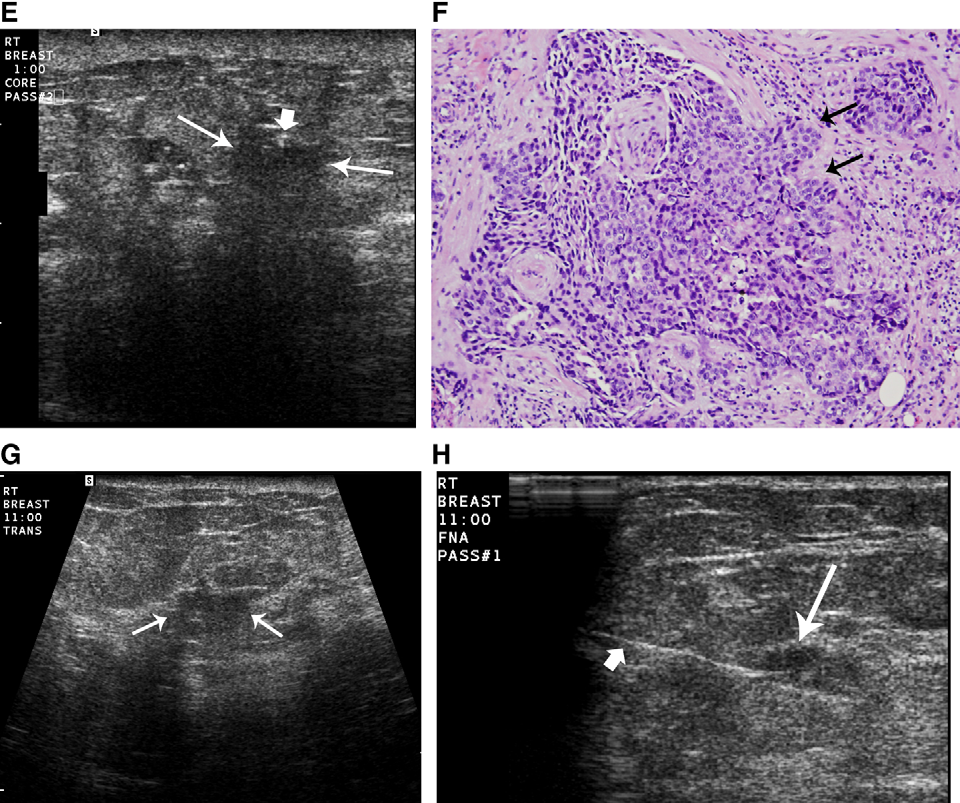
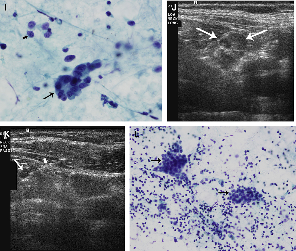
44 Ultrasound-Guided Breast Biopsies Fig. 4 (continued) 609
Breast Ultrasound: Benign vs. Malignant Lesions
 October 25-November 19, 2004 Breast Ultrasound: Benign vs. Malignant Lesions Jill Steinkeler,, Tufts University School of Medicine IV Breast Anatomy Case Presentation-Patient 1 62 year old woman with a
Breast Sonography general goal. Optimizing Breast Sonography. BUS indications -- all. Breast Sonography specific goals.
 Optimizing general goal Cindy Rapp BS, RDMS, FAIUM, FSDMS University of Colorado Hospital Denver, Colorado to make a more specific diagnosis than can be made with clinical and mammographic findings alone
D. FREQUENTLY ASKED QUESTIONS
 ACR BI-RADS ATLAS D. FREQUENTLY ASKED QUESTIONS 1. Under MQSA, is it necessary to include a numeric assessment code (i.e., 0, 1, 2, 3, 4, 5, or 6) in addition to the assessment category in all mammography
Nicole Kounalakis, MD
 Breast Disease: Diagnosis and Management Nicole Kounalakis, MD Assistant Professor of Surgery Goal of Breast Evaluation The goal of breast evaluation is to classify findings as: normal physiologic variations
MAMMOGRAPHY GOALS AND OBJECTIVES
 MAMMOGRAPHY GOALS AND OBJECTIVES GOALS: After completion of the mammography rotations, the resident will be able to: 1. Demonstrate learning of the knowledge-based objectives-(practice Base Learning) 2.
Mammography Education, Inc.
 Mammography Education, Inc. 2011 LÁSZLÓ TABÁR, M.D.,F.A.C.R (Hon) 3D image of a milk duct MULTIMODALITY DETECTION and DIAGNOSIS of BREAST DISEASES PRAGUE, Czech Republic Crown Plaza, Prague June 29 - July
Evaluation and Management of the Breast Mass. Gary Dunnington,, M.D. Department of Surgery Internal Medicine Ambulatory Conference December 4, 2003
 Evaluation and Management of the Breast Mass Gary Dunnington,, M.D. Department of Surgery Internal Medicine Ambulatory Conference December 4, 2003 Common Presentations of Breast Disease Breast Mass Abnormal
Guideline for the Imaging of Patients Presenting with Breast Symptoms incorporating the guideline for the use of MRI in breast cancer
 Guideline for the Imaging of Patients Presenting with Breast Symptoms incorporating the guideline for the use of MRI in breast cancer Version History Version Date Summary of Change/Process 0.1 09.01.11
VI. FREQUENTLY ASKED QUESTIONS CONCERNING BREAST IMAGING AUDITS
 ACR BI-RADS ATLAS VI. FREQUENTLY ASKED QUESTIONS CONCERNING BREAST IMAGING AUDITS American College of Radiology 55 ACR BI-RADS ATLAS A. All Breast Imaging Modalities 1. According to the BI-RADS Atlas,
III. REPORTING SYSTEM
 ACR BI-RADS ATLAS BREAST III. REPORTING SYSTEM American College of Radiology 121 2013 122 American College of Radiology ACR BI-RADS ATLAS BREAST A. REPORT ORGANIZATION The report should be concise and
OBJECTIVES By the end of this segment, the community participant will be able to:
 Cancer 101: Cancer Diagnosis and Staging Linda U. Krebs, RN, PhD, AOCN, FAAN OCEAN Native Navigators and the Cancer Continuum (NNACC) (NCMHD R24MD002811) Cancer 101: Diagnosis & Staging (Watanabe-Galloway
Breast MRI: Imaging and Intervention. Jaroslaw Nicholas Tkacz, M. D.
 Breast MRI: Imaging and Intervention Jaroslaw Nicholas Tkacz, M. D. Purpose To examine the typical morphologic, enhancement and kinetic features of breast lesions on MR Imaging and determine the role of
BREAST IMAGING. Developed by the Ad Hoc Committee on Resident and Fellow Education of the Society of Breast Imaging
 BREAST IMAGING Developed by the Ad Hoc Committee on Resident and Fellow Education of the Society of Breast Imaging Stephen A. Feig, M.D., Chair Ferris Hall, M.D. Debra Ikeda, M.D. Ellen Mendelson, M.D.
Breast Cancer: from bedside and grossing room to diagnoses and beyond. Adriana Corben, M.D.
 Breast Cancer: from bedside and grossing room to diagnoses and beyond Adriana Corben, M.D. About breast anatomy Breasts are special organs that develop in women during puberty when female hormones are
Breast cancer close to the nipple: Does this carry a higher risk ofaxillary node metastasesupon diagnosis?
 Breast cancer close to the nipple: Does this carry a higher risk ofaxillary node metastasesupon diagnosis? Erin I. Lewis, BUSM 2010 Cheri Nguyen, BUSM 2008 Priscilla Slanetz, M.D., MPH Al Ozonoff, Ph.d.
Breast Imaging Made Brief and Simple. Jane Clayton MD Associate Professor Department of Radiology LSUHSC New Orleans, LA
 Breast Imaging Made Brief and Simple Jane Clayton MD Associate Professor Department of Radiology LSUHSC New Orleans, LA What women are referred for breast imaging? Two groups of women are referred for
PROPERTY OF ELSEVIER SAMPLE CONTENT - NOT FINAL
 Oncoplastic breast conservation surgery Melvin J Silverstein C H A P T E R 5 Introduction Oncoplastic breast conservation surgery combines oncologic principles with plastic surgical techniques. But it
Table of Contents: Chapter 1: Screening for breast cancer in asymptomatic patients by Chris Comstock, MD and Marie Tartar, MD
 Table of Contents: Chapter 1: Screening for breast cancer in asymptomatic patients by Chris Comstock, MD and Marie Tartar, MD Case 1: Breast cancer presenting as a small new mass on Case 2: Breast cancer
Breast Cancer. Sometimes cells keep dividing and growing without normal controls, causing an abnormal growth called a tumor.
 Breast Cancer Introduction Cancer of the breast is the most common form of cancer that affects women but is no longer the leading cause of cancer deaths. About 1 out of 8 women are diagnosed with breast
The Male Breast: Masses, Malignancies and More
 The Male Breast: Masses, Malignancies and More Monique Marie Tyminski, DO, R Hultman, DO, J Watkins, MD, T Stockl, MD, E T Ghosh, MD, S A MacMaster, MD Teaching Points: Understand male breast anatomy and
ACR BI-RADS ATLAS MAMMOGRAPHY MAMMOGRAPHY II. REPORTING SYSTEM. American College of Radiology 121
 ACR BI-RADS ATLAS II. REPORTING SYSTEM American College of Radiology 121 2013 122 American College of Radiology ACR BI-RADS ATLAS A. REPORT ORGANIZATION (Guidance chapter, see page 147) The reporting system
Recommendations for cross-sectional imaging in cancer management, Second edition
 www.rcr.ac.uk Recommendations for cross-sectional imaging in cancer management, Second edition Breast cancer Faculty of Clinical Radiology www.rcr.ac.uk Contents Breast cancer 2 Clinical background 2 Who
Ductal carcinoma in situ (DCIS)
") DIAGNOSIS: DCIS Ductal carcinoma in situ (DCIS) This factsheet gives information on an early form of breast cancer called ductal carcinoma in situ (DCIS). It explains what it is, how your breast is made
DIAGNOSIS: DCIS Ductal carcinoma in situ (DCIS) This factsheet gives information on an early form of breast cancer called ductal carcinoma in situ (DCIS). It explains what it is, how your breast is made
Rotation Specific Goals & Objectives: University Health Network-Princess Margaret Hospital/ Sunnybrook Breast/Melanoma
 Rotation Specific Goals & Objectives: University Health Network-Princess Margaret Hospital/ Sunnybrook Breast/Melanoma Medical Expert: Breast Rotation Specific Competencies/Objectives 1.0 Medical History
Common Breast Complaints:
 : Palpable mass Abnormal mammogram with normal physical exam Vague thickening or nodularity Nipple Discharge Breast pain Breast infection or inflammation The physician s goal is to determine whether the
Introduction Breast cancer is cancer that starts in the cells of the breast. Breast cancer happens mainly in women. But men can get it too.
 Male Breast Cancer Introduction Breast cancer is cancer that starts in the cells of the breast. Breast cancer happens mainly in women. But men can get it too. Many people do not know that men can get breast
AJR:197, December 2011
 AJR:197, December 2011 William R. Geiser, Tamara Miner Haygood Lumarie Santiago, Tanya Stephens Debra Thames, Gary J. Whitman Presented by Chia-Ying WEN 2012/3/8 Early detection of breast cancer is directly
Benign Mimics of Malignancy on Breast Imaging. MM Tyminski, DO; JE Watkins, MD, ET Ghosh, MD; R Hultman, DO; T Stockl, MD; SA MacMaster, MD
 Benign Mimics of Malignancy on Breast Imaging MM Tyminski, DO; JE Watkins, MD, ET Ghosh, MD; R Hultman, DO; T Stockl, MD; SA MacMaster, MD Teaching Points: 1. Demonstrate benign entities of the female
INTERDISCIPLINARY CONFERENCE. Florence/Firenze, Italy Nov 27-29, 2012 Centro Congressi al Duomo, Firenze BREAST SEMINAR SERIES
 Since breast cancer is not a systemic disease from inception, when the imagers find in situ and 1-14 mm invasive breast cancer, it is the surgeon, specialized in the treatment of breast diseases, who should
Breast Implants: Local Complications and Adverse Outcomes
 Breast Implants: Local Complications and Adverse Outcomes This booklet highlights the most common problems associated with silicone gel-filled and saline-filled breast implants: those that occur in the
Mammography. What is Mammography?
 Scan for mobile link. Mammography Mammography is a specific type of breast imaging that uses low-dose x-rays to detect cancer early before women experience symptoms when it is most treatable. Tell your
Integumentary System Individual Exercises
 Integumentary System Individual Exercises 1. A physician performs an incision and drainage of a subcutaneous abscess in his office for a particularly uncooperative established patient. How should this
Infrared Thermography Not a Useful Breast Cancer Screening Tool
 Contact: Jeanne-Marie Phillips Sharon Grutman HealthFlash Marketing The American Society of Breast Surgeons 203-977-3333 877-992-5470 Infrared Thermography Not a Useful Breast Cancer Screening Tool Mammography
Q: What differentiates a diagnostic from a screening mammography procedure?
 The following Q&As address Medicare guidelines on the reporting of breast imaging procedures. Private payer guidelines may vary from Medicare guidelines and from payer to payer; therefore, please be sure
Ductal Carcinoma in Situ: A Case Report
 Ductal Carcinoma in Situ: A Case Report Abstract Breast ductal carcinoma in situ (DCIS) is a preinvasive form of breast cancer and is the most common type of in situ breast cancer found in women. There
Sonographic Evaluation of Isolated Abnormal Axillary Lymph Nodes Identified on Mammograms
 Article Sonographic Evaluation of Isolated Abnormal Axillary Lymph Nodes Identified on Mammograms Mahesh K. Shetty MD, FRCR, Wendy S. Carpenter, MD Objective. To evaluate the role of sonography in evaluation
Understanding your pathology report
 Understanding your pathology report 2 Contents Contents Introduction 3 What is a pathology report? 3 Waiting for your results 4 What s in a pathology report? 4 Information about your breast cancer 5 What
Objectives. Mylene T. Truong, MD. Malignant Pleural Mesothelioma Background
 Imaging of Pleural Tumors Mylene T. Truong, MD Imaging of Pleural Tumours Mylene T. Truong, M. D. University of Texas M.D. Anderson Cancer Center, Houston, TX Objectives To review tumors involving the
A Guide to Breast Imaging: The Latest Technology for Screening and Detecting Breast Cancer
 A Guide to Breast Imaging: The Latest Technology for Screening and Detecting Breast Cancer Sally Herschorn, MD Associate Professor of Radiology University of Vermont College of Medicine Medical Director
Breast Biphasic Compression versus Standard Monophasic Compression in X-ray Mammography 1
 Francesco Sardanelli, MD Franco Zandrino, MD Andrea Imperiale, MD Emma Bonaldo, MD Maria G. Quartini, RT Nadia Cogorno, RT Breast Biphasic Compression versus Standard Monophasic Compression in X-ray Mammography
Breast Density Legislation: Implications for primary care providers
 Breast Density Legislation: Implications for primary care providers Deborah J. Rhodes MD Associate Professor of Medicine 2012 MFMER slide-1 Disclosure Relevant financial relationship(s) None Off-label
Second look ultrasound examination for breast lesions: MRI and pathologic correlation
 Second look ultrasound examination for breast lesions: MRI and pathologic correlation Poster No.: C-0559 Congress: ECR 2015 Type: Scientific Exhibit Authors: E. Serrano Tamayo, E. López Soriano, M. Muñoz
Breast Implants and Reconstruction
 Last Review Date: October 9, 2015 Number: MG.MM.SU.fv2 Medical Guideline Disclaimer Property of EmblemHealth. All rights reserved. The treating physician or primary care provider must submit to EmblemHealth
Breast Health Starts Here
 Breast Health Starts Here k Breast Health Knowledge and self-awareness are powerful tools. Understanding and utilizing these tools starts with Lake Charles Memorial s Breast Health Program. Developing
PART TWO: DIAGNOSING BREAST CANCER
 PART TWO: DIAGNOSING BREAST CANCER What is Breast Cancer? Normal cells can turn into cancer because of genetic inheritance or repeated exposure to a cancer-related substance in the environment, such as
Sustaining a High-Quality Breast MRI Practice
 Sustaining a High-Quality Breast MRI Practice Christoph Lee, MD, MSHS Associate Professor of Radiology Adjunct Associate Professor, Health Services University of Washington September 11, 2015 Overview
Ultrasonography of the Adrenal Glands CVM 6105 Kari L. Anderson, DVM, Diplomate ACVR Associate Clinical Professor of Veterinary Radiology
 1: US of adrenal glands, KLA Ultrasonography of the Adrenal Glands CVM 6105 Kari L. Anderson, DVM, Diplomate ACVR Associate Clinical Professor of Veterinary Radiology Ultrasound has quickly become an important
Your Guide to the Breast Cancer Pathology Report
 Your Guide to the Breast Cancer Pathology Report Developed for you by Breastcancer.org is a nonprofit organization dedicated to providing education and information on breast health and breast cancer. The
Joelle M. Schoonjans, MD, Rachel F. Brem, MD
 Sonographic Appearance of Ductal Carcinoma In Situ Diagnosed with Ultrasonographically Guided Large Core Needle Biopsy: Correlation with Mammographic and Pathologic Findings Joelle M. Schoonjans, MD, Rachel
Medicare Part B. Mammograms - Updated Billing Guide for Screening and Diagnostic Tests
 Mammograms - Updated Billing Guide for Screening and Diagnostic Tests This article from Medicare B News Issue 223 dated October 21, 2005 is being updated and reprinted to ensure that the Noridian Administrative
Non-cancerous Breast Conditions
 Non-cancerous Breast Conditions Non-cancerous breast conditions are very common and most women have them. In fact, most breast changes that are tested turn out to be benign. Benign is another word for
Breast Cancer. The Pathology report gives an outline on direction of treatment. It tells multiple stories to help us understand the patient s cancer.
 Breast Cancer What Does the Pathology Report Say Normal Cells The Pathology report gives an outline on direction of treatment. It tells multiple stories to help us understand the patient s cancer. Non-Invasive
SUBJECT: MANAGEMENT OF BREAST EFFECTIVE DATE: 12/16/99 IMPLANTS REVISED DATE:
 MEDICAL POLICY SUBJECT: MANAGEMENT OF BREAST PAGE: 1 OF: 5 If the member's subscriber contract excludes coverage for a specific service it is not covered under that contract. In such cases, medical policy
Stereotactic Breast Biopsy
 Scan for mobile link. Stereotactic Breast Biopsy Stereotactic breast biopsy uses mammography a specific type of breast imaging that uses low-dose x-rays to help locate a breast lump or abnormality and
Michael A. Boss, M.D. FMH Plastic, Reconstructive und Aesthetic Surgery
 Michael A. Boss, M.D. FMH Plastic, Reconstructive und Aesthetic Surgery Boss Aesthetic Center Schauplatzgasse 23 CH-3011 Bern Switzerland +41 31 311 7691 www.aesthetic-center.com B r e a s t A u g m e
Understanding. Breast Changes. National Cancer Institute. A Health Guide for Women. National Institutes of Health
 National Cancer Institute Understanding Breast Changes A Health Guide for Women U.S. DEPARTMENT OF HEALTH AND HUMAN SERVICES National Institutes of Health It was easier to talk with my doctor after reading
Guide to Understanding Breast Cancer
 An estimated 220,000 women in the United States are diagnosed with breast cancer each year, and one in eight will be diagnosed during their lifetime. While breast cancer is a serious disease, most patients
Pathologic Assessment Of The Breast And Axilla After Preoperative Therapy
 Pathologic Assessment Of The Breast And Axilla After Preoperative Therapy W. Fraser Symmans, M.D. Associate Professor of Pathology UT M.D. Anderson Cancer Center Pathologic Complete Response (pcr) Proof
Breast Cancer. Presentation by Dr Mafunga
 Breast Cancer Presentation by Dr Mafunga Breast cancer in the UK Breast cancer is the second most common cancer in women. Around 1 in 9 women will develop breast cancer It most commonly affects women over
Current Status and Perspectives of Radiation Therapy for Breast Cancer
 Breast Cancer Current Status and Perspectives of Radiation Therapy for Breast Cancer JMAJ 45(10): 434 439, 2002 Masahiro HIRAOKA, Masaki KOKUBO, Chikako YAMAMOTO and Michihide MITSUMORI Department of Therapeutic
Lesions, and Masses, and Tumors Oh My!!
 Lesions, and Masses, and Tumors Oh My!! Presented by: Susan Ward, CPC, CPC-H, CPC-I, CPCD, CEMC, CPRC 1 1 CPT GUIDELINES Agenda CPT DEFINITIONS OP REPORT CASES 2 Definitions Cyst - a closed sac having
APGO Clinical Skills Curriculum. The Breast Exam
 APGO Clinical Skills Curriculum The Breast Exam Association of Professors of Gynecology and Obstetrics (APGO) Undergraduate Medical Education Committee 2008 The Breast Exam Table of Contents Intended Learning
Breast Cancer Awareness Month
 Aon Kenya Insurance Brokers Ltd Aon Hewitt Healthcare Division Breast Cancer Awareness Month Issue 12 October 2015 In this Issue 2 Cancer Statistics in Kenya 3 What is Breast Cancer? 4 Symptoms of Breast
It is important that you read this information carefully and completely.
 Placement of Permanent Breast Implant Following Tissue Expansion 1. I hereby authorize Dr. John P. Stratis and such assistants as may be selected to perform the following procedure or treatment INFORMED-
Provider Reimbursement for Women's Cancer Screening Program
 Reimbursement Schedule July 1, 2015 June 30, 2016 Office Visits - Established Patients Office Visit / Minimal / no physician 99211 $ 16.70 Office Visit / Problem focused History / exam 99212 $ 36.46 Preventive
Breast Reconstruction Surgery
 Breast Reconstruction Surgery I. Policy University Health Alliance (UHA) will reimburse for Breast Reconstruction Surgery when it is determined to be medically necessary and when it meets the medical criteria
dedicated to curing BREAST CANCER
 dedicated to curing BREAST CANCER When you are diagnosed with breast cancer, you need a team of specialists who will share their knowledge of breast disease and the latest treatments available. At Cancer
Cystic Neoplasms of the Pancreas: A multidisciplinary approach to the prevention and early detection of invasive pancreatic cancer.
 This lecture is drawn from the continuing medical education program Finding Hope: Prevention, Early Detection and Treatment of Pancreatic Cancer, Nov, 2011. Robert P. Jury, MD Cystic Neoplasms of the Pancreas:
Surgical guidelines for the management of breast cancer
 Available online at www.sciencedirect.com EJSO xx (2009) S1eS22 www.ejso.com Guidelines Surgical guidelines for the management of breast cancer Contents Association of Breast Surgery at BASO 2009 Introduction...
Variability and Accuracy in Mammographic Interpretation Using the American College of Radiology Breast Imaging Reporting and Data System
 Variability and Accuracy in Mammographic Interpretation Using the American College of Radiology Breast Imaging Reporting and Data System Karla Kerlikowske, Deborah Grady, John Barclay, Steven D. Frankel,
Invasive lobular breast cancer
 Invasive lobular breast cancer This booklet is about invasive lobular breast cancer. It describes what invasive lobular breast cancer is, the symptoms, how it s diagnosed and possible treatments. Diagnosed
What You Need to Know Before Treatment About: Breast Cancer
 What You Need to Know Before Treatment About: Breast Cancer Publication MDCH-1234 Revised: September 2008 Authority: Public Act 368 of 1978, as amended by Public Act 195 of 1986 and Public Act 15 of 1989.
Personalized Breast Screening Service
 Frequently Asked Questions WHAT IS BREAST DENSITY? Breasts are made up of a mixture of fibrous, glandular and fatty tissue. Your breasts are considered if you have predominantly fibrous or glandular tissue
Areola: The area of dark-colored skin on the breast that surrounds the nipple.
 Glossary of Terms Adenocarcinoma: Cancer arising in gland-forming tissue; the medical term meaning related to a gland is adeno ; breast lobules and ducts are types of glands. Adjuvant: Treatment given
The best treatment Your guide to breast cancer treatment in Scotland
 The best treatment Your guide to breast cancer treatment in Scotland If you are looking for information on secondary breast cancer treatment, please see Breast Cancer Now s website: breastcancernow.org/
Sonography of Wrist Ganglion Cysts
 CME Article Sonography of Wrist Ganglion Cysts Variable and Noncystic Appearances George Wang, MD, Jon A. Jacobson, MD, Felix Y. Feng, MD, Gandikota Girish, MBBS, FRCS, FRCR, Elaine M. Caoili, MD, Catherine
Surgery Choices. National Cancer Institute. For Women with DCIS or Breast Cancer. National Institutes of Health
 National Cancer Institute Surgery Choices For Women with DCIS or Breast Cancer U.S. DEPARTMENT OF HEALTH AND HUMAN SERVICES National Institutes of Health The National Cancer Institute is grateful for our
CMS Limitations Guide Mammograms and Bone Density Radiology Services
 CMS Limitations Guide Mammograms and Bone Density Radiology Services Starting July 1, 2008, CMS has placed numerous medical necessity limits on tests and procedures. This reference guide provides you with
NET della mammella: realtà o fantasia
 NET della mammella: realtà o fantasia Roberto BALDELLI M.D., Ph.D. Regina Elena, Italian National Cancer Institute, Rome Unit of Endocrinology baldelli@ifo.it Tumors arising from enterochromaffin cells
Wisconsin Cancer Data Bulletin Wisconsin Department of Health Services Division of Public Health Office of Health Informatics
 Wisconsin Cancer Data Bulletin Wisconsin Department of Health Services Division of Public Health Office of Health Informatics In Situ Breast Cancer in Wisconsin INTRODUCTION This bulletin provides information
Changes in Breast Cancer Reports After Second Opinion. Dr. Vicente Marco Department of Pathology Hospital Quiron Barcelona. Spain
 Changes in Breast Cancer Reports After Second Opinion Dr. Vicente Marco Department of Pathology Hospital Quiron Barcelona. Spain Second Opinion in Breast Pathology Usually requested when a patient is referred
Alaska Breast & Cervical Health Partnership. Healthy Women, Healthy Alaska Through Early Detection
 Alaska Breast & Cervical Health Partnership Healthy Women, Healthy Alaska Through Early Detection Southcentral Foundation (SCF) Breast and Cervical Health SouthEast Alaska Regional Health Consortium (SEARHC)
The best treatment Your guide to breast cancer treatment in England and Wales
 The best treatment Your guide to breast cancer treatment in England and Wales If you are looking for information on the treatment of secondary breast cancer (also known as advanced or metastatic breast
www.downstatesurgery.org
 Male Breast Cancer Rabih Nemr MD Kings County Hospital August 2008 ACGME Core Competencies 1 Patient t Care Medical Knowledge 2 g 3 4 Practice Based Learning/Improvement Interpersonal Communication Skills
Automated Breast Volume Scanning 3D Ultrasound of the Breast
 Automated Breast Volume Scanning 3D Ultrasound of the Breast Roel Mus, MD Radboud University Nijmegen Medical Centre (RUNMC), Nijmegen, The Netherlands Matthieu Rutten, MD, PhD Jeroen Bosch Ziekenhuis,
FRIEND TO FRIEND CPT CODES 2015 2016. Diagnostic digital breast tomosynthesis, unilateral (list separately in addition to code for primary procedure)
") FRIEND TO FRIEND CPT CODES 2015 2016 CPT CODE SERVICE DESCRIPTION FEE EFFECTIVE G0101 Screening pelvic examination $36.69 01 Jan 16 G0202 Mammography, screening, digital, bilateral (2 view film study of
FRIEND TO FRIEND CPT CODES 2015 2016 CPT CODE SERVICE DESCRIPTION FEE EFFECTIVE G0101 Screening pelvic examination $36.69 01 Jan 16 G0202 Mammography, screening, digital, bilateral (2 view film study of
Kidney Cancer OVERVIEW
 Kidney Cancer OVERVIEW Kidney cancer is the third most common genitourinary cancer in adults. There are approximately 54,000 new cancer cases each year in the United States, and the incidence of kidney
TOM J. POUSTI, MD, F.A.C.S. PLASTIC AND RECONSTRUCTION SURGERY
 TOM J. POUSTI, MD, F.A.C.S. PLASTIC AND RECONSTRUCTION SURGERY INFORMED CONSENT FOR BREAST AUGMENTATION SURGERY INSTRUCTIONS This is an informed consent document that has been prepared to help inform you
New Wireless Handheld Ultrasound-Guided Vacuum-Assisted Breast Biopsy (VABB) Devices: An Important Innovation in Breast Diagnosis
 Devices: An Important Innovation in Breast Diagnosis") Open Journal of Radiology, 2013, 3, 174-179 Published Online December 2013 (http://www.scirp.org/journal/ojrad) http://dx.doi.org/10.4236/ojrad.2013.34029 New Wireless Handheld Ultrasound-Guided Vacuum-Assisted
Open Journal of Radiology, 2013, 3, 174-179 Published Online December 2013 (http://www.scirp.org/journal/ojrad) http://dx.doi.org/10.4236/ojrad.2013.34029 New Wireless Handheld Ultrasound-Guided Vacuum-Assisted
Harlem Hospital Center Integrated Radiology Residency Program Mammography Educational goals and objectives
 Harlem Hospital Center Integrated Radiology Residency Program Mammography Educational goals and objectives Rotation 1 (Radiology year 1/2) Knowledge Based Objectives: At the end of the rotation, the resident
Gynecology Abnormal Pelvic Anatomy and Physiology: Cervix. Cervix. Nabothian cysts. cervical polyps. leiomyomas. Cervical stenosis
 Gynecology Abnormal Pelvic Anatomy and Physiology: (Effective February 2007) pediatric, reproductive, and perimenopausal/postmenopausal (24-28 %) Cervix Nabothian cysts result from chronic cervicitis most
Breast Augmentation. If you are dissatisfied with your breast size, augmentation surgery is a choice to consider. Breast augmentation can:
 Breast Augmentation What is Breast Augmentation? Also known as augmentation mammaplasty, breast augmentation involves using implants to fulfill your desire for fuller breasts or to restore breast volume
Technical Advices for Prostate Needle Biopsy Under Transrectal Ultrasound Guidance
 Technical Advices for Prostate Needle Biopsy Under Transrectal Ultrasound Guidance Makoto Ohori 1 and Ayako Miyakawa 2 1 Dept. of Urology, Tokyo Medical University 2 Dept. of Molecular Medicine and Surgery,
Spleen. Anatomy. (Effective February 2007) (1%-5%) Normal. Related Anatomy Anterior to spleen. Medial border. Posteriorly
 (1%-5%) Normal. Related Anatomy Anterior to spleen. Medial border. Posteriorly") Spleen (Effective February 2007) (1%-5%) Anatomy Normal Intraperitoneal, except hilum Left hypochondrium Left hemidiaphragm superior generally considered to be ovoid, with a convex superior and a concave
Spleen (Effective February 2007) (1%-5%) Anatomy Normal Intraperitoneal, except hilum Left hypochondrium Left hemidiaphragm superior generally considered to be ovoid, with a convex superior and a concave
Hereditary Multifocal Breast Cancer. Farin Amersi M.D., F.A.C.S Division of Surgical Oncology Department of Surgery Cedar Sinai Medical Center
 Hereditary Multifocal Breast Cancer Farin Amersi M.D., F.A.C.S Division of Surgical Oncology Department of Surgery Cedar Sinai Medical Center CASE STUDY 30 year old Ashkenazi Jewish woman Nulliparous Felt
MALE BREAST CANCER - CASE REPORT AND BRIEF REVIEW
 Middle East Journal of Family Medicine, 2004; Vol. 6 (6) MALE BREAST CANCER - CASE REPORT AND BRIEF REVIEW Elias A Sarru', MD, MS, AAFP, ABFP Family Physician, Al Hasa Primary Care Services Division Faysal
Florida Breast Health Specialists Breast Cancer Information and Facts
 Definition Breast cancer is a cancer that starts in the tissues of the breast. There are two main types of breast cancer: Ductal carcinoma starts in the tubes (ducts) that move milk from the breast to
Breast Augmentation Primer
 Breast Augmentation Primer Breast augmentation is typically a very rewarding procedure. In order to achieve the highest level of patient satisfaction, many decisions must be carefully made. We have put