WHAT IS THE REAL RISK OF RADIATION EXPOSURE FROM MEDICAL IMAGING? Stephanie Parks Taylor, MD Assistant Professor Division of Hospital Medicine
|
|
|
- Cornelius Blake
- 7 years ago
- Views:
Transcription
1 WHAT IS THE REAL RISK OF RADIATION EXPOSURE FROM MEDICAL IMAGING? Stephanie Parks Taylor, MD Assistant Professor Division of Hospital Medicine

2 WHAT IS THE REAL RISK OF RADIATION EXPOSURE FROM MEDICAL IMAGING? I have no disclosures, financial or otherwise, to report

3 OBJECTIVES Understand the reasons for concern about radiation exposure from medical imaging Learn basic radiobiology and radiation dosimetry Discuss evidence and consensus opinions on the risks of radiation Identify strategies to minimize radiation exposure for patients

4 CT scan!

5 QUESTION 1 CT scan of the abd/pelvis with and without contrast results in an effective radiation dose similar to: A: One transatlantic flight B: 10 chest radiographs C: Average exposure of Japanese atomic bomb survivors D: A block of kryptonite

6 QUESTION 2 According to modern risk models, what percentage of cancers in the US are attributable to radiation from CT scans? A: Definitely none, there is no risk of cancer B. Probably none, the risk is only theoretical C: 0.01% D: to 2.0% 20%

7 HOW OTHERS FARED Survey of radiologists and ER docs 75% significantly underestimated radiation dose from CT 53% of radiologists and 91% of ER docs did not think CT 53% of radiologists and 91% of ER docs did not think CT scans increase the risk of cancer

8 OBJECTIVES Understand the reasons for concern about radiation exposure from medical imaging Learn basic radiobiology and radiation dosimetry Discuss evidence and consensus opinions on the risks of radiation Identify strategies to minimize radiation exposure for patients

9 BENEFITS OF MEDICAL IMAGING Earlier and more accurate diagnosis of disease Non-invasive Screening tool Reassurance

10 HARMS ASSOCIATED WITH IMAGING Radiation exposure False positives: unnecessary follow-up testing, psychological py distress, resource utilization Incidental findings: cascade of testing to rule out disease Overdiagnosis: unnecessary treatment Contrast reactions: some major, most minor Healthcare costs

11 FEAR OF RADIATION: WHY NOW? Marked increase in radiation exposure from medical imaging Radiation overdoses (Mad River, Cedars-Sinai) leading to lay press activity Xrays classified as carcinogens by WHO, CDC, NIEHS

12 RADIATION EXPOSURE FROM CT 3 million CT scans in 1980 Almost 70 million in 2007 Imaging rate has tripled in last 10 years

13

14 UTILIZATION OF MEDICAL IMAGING: CT Brenner DJ, Hall EJ. N Engl J Med 2007;357:

15 RADIATION EXPOSURE OF US PUBLIC HAS DOUBLED DUE TO MEDICAL IMAGING 1985: total 3.7 msv 75% from natural sources 25% imaging 2006: total 6.2 msv 50% from natural sources 50% imaging

16 RADIATION FEAR: HISTORY Marie Curie Nobel prize for work in physics and chemistry Died of aplastic anemia Her papers from the 1890s are considered too dangerous to handle due to high radioactivity

17 RADIATION FEAR: CONTEMPORARY Arcata, CA 2008 CT head ordered for 2 ½ year old boy in ER T h i i l d 3 li 151 Technician erroneously scanned same 3mm slice 151 times

18

19 RADIATION FEAR: CONTEMPORARY Cedars-Sinai Medical Center, stroke patients receiving brain perfusion imaging S ft lf ti lt d i d i i t ti f 8 Software malfunction resulted in administration of 8 times the maximum dose

20
21 RADIATION FEAR: RECENT STUDIES Exposure to Low-Dose Ionizing Radiation from Medical Imaging g Procedures. NEJM, Number 9 Volume 361: Reza Fazel, M.D., et. Al Collected CT data from 5 Healthcare markets from Categorized effective radiation doses into 4 categories: Low (<3mSv): Moderate (>3-20mSv): 19.4% of enrollees High (>20-50mSv): 1.9% of enrollees Very high (>50mSv):.19% of enrollees
22
23 RADIATION DOSES FROM CT: HIGH AND VARIABLE Smith-Bindman et al., Arch Intern Med Study of 4 facilities in San Francisco Bay area Adults, median age 59 years January 1 May Dose from CT 1.5 to 5 times higher than cited Higher than necessary for diagnosis Wide variability in dose for same test and indication Vary times among facilities Even greater variation among patients Expect ~2 fold variation due to difference in body habitus
24 VARIATION ACROSS FACILITY AND PATIENT Head Site 1 Site 2 Site 3 Site 4 Range Across Patients Routine head Suspected stroke Chest Routine chest Suspected PE Coronary angiogram Abdomen-pelvis Routine Multiphase Smith-Bindman et al., Arch Intern Med, 2009
25 VARIATION ACROSS FACILITY AND PATIENT Smith-Bindman et al., Arch Intern Med, 2009
26 VARIATION ACROSS FACILITY AND PATIENT Average exposure among Japanese atomic bomb survivors Smith-Bindman et al., Arch Intern Med, 2009
27 RADIATION EXPOSURE Risks associated with radiation are not new Dramatic increase in exposure to ionizing radiation is the issue
28 OBJECTIVES Understand the reasons for concern about radiation exposure from medical imaging Learn basic radiobiology and radiation dosimetry Discuss evidence and consensus opinions on the risks of radiation Identify strategies to minimize radiation exposure for patients
29 RADIATION BIOLOGY
30 RADIATION BIOLOGY
31 BIOLOGIC EFFECTS OF RADIATION Stochastic effects Deterministic effects
32 STOCHASTIC EFFECTS All or none effect from exposure to low-dose radiation Severity independent of dose No safe threshold h dose (probability bilit of biological i l effect increases with dose) May take many years (or a lifetime) to manifest Carcinogenesis and genetic effects
33 DETERMINISTIC EFFECTS Result from high dose radiation exposure Severity is dose dependent Threshold h concept applies Ex: hair loss, cataracts, skin changes, GI effects, reproductive damage, death
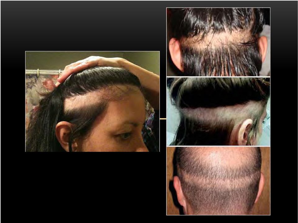
34 DETERMINISTIC EFFECTS: BAND ALOPECIA
35 RADIATION MEASUREMENTS Activity (Becquerel) Absorbed b dose (Gray = 100 rad) 1 joule of energy deposited per kg Effective dose (Sievert): takes into account type of radiation (gamma vs xrays) and tissue sensitivity Sievert = Gray * Q * N Q, quality (photons/electrons = 1, alpha particles = 20) N depends on body tissue Most tissues ~ Gonads ~ 0.2 Bone marrow, colon, lung, stomach = 0.12
36 TISSUE RADIOSENSITIVITY High Lymphoid Tissue Marrow Medium Skin Vascular endothelium Low Muscle Bone GI Epithelium Gonads Embryos Lung Kidney Liver Lens Connective tissue Cartilage
37 MODELS TO DETERMINE RADIATION RISK Linear no-threshold (LNT) Risk of stochastic health effects increases linearly with biologically effective absorbed dose Implies there is risk even to low levels of radiation Most widely accepted model Linear with threshold Risk increases linearly with exposure after exposure crosses a threshold level Implies that low levels of radiation does not have any risk Hormesis Low doses of radiation are beneficial whereas high doses are harmful Widely rejected
38
39 RADIATION MEASURES FOR CT CTDI VOL : volume CT dose index (mgy) DLP: Dose length product (mgy-cm)
40 CT RADIATION MEASUREMENTS: CTDI CTDI VOL (mgy) Represents radiation dose of a single CT slice using ]plastic phantoms either 16 or 32cm in diameter
41 CT RADIATION MEASUREMENTS: DLP DLP (mgy-cm) CTDI VOL x scan length Represents integrated dose across scan length Can be multiplied by conversion factor to yield estimate of effective dose
42 CT RADIATION MEASUREMENTS CTDI vol and DLP are shown on modern CT scanners Useful for comparing CT protocols between scanners Do NOT represent effective dose (msv) Can be used to estimate effective dose using tissue conversion factors
43
44
45
46 ESTIMATING EFFECTIVE DOSE FOR CT Effective dose estimates can be calculated by multiplying py DLP by tissue specific conversion factors Effective dose (msv) = DLP x k(e/dlp) Adult values for k(e/dlp) Head =.0021 Head/neck =.0031 Chest =.014 Abdomen/pelvis =.015 Trunk =.015
47
48 EXAMPLE: EFFECTIVE DOSE FROM ADULT CT HEAD CT abdomen/pelvis multiphase CTDI VOL = mgy DLP = mgy-cm Effective dose estimate: DLP x msv
49
50 OBJECTIVES Understand the reasons for concern about radiation exposure from medical imaging Learn basic radiobiology and radiation dosimetry Discuss evidence and consensus opinions on the risks of radiation Identify strategies to minimize radiation exposure for patients
51 RISKS OF RADIATION FROM MEDICAL IMAGING No data directly attributing cancer to CT scanning (yet ) Assumptions must be made based on other forms of radiation exposure with comparable doses Most commonly used source: Atomic bomb survivors Radiation used for medical purposes
52 ATOMIC BOMB LIFESPAN STUDY 120, survivors Median dose of survivors 40 msv 25,000 in range of 2-20mSV Followed incidence of cancer over 55 years Even at low dose (10 msv), significant increase in cancer risk
53 MEDICALLY IRRADIATED POPULATIONS Malignant disease Patients receiving XRT for malignant disease are at increased risk of secondary cancers In Hodgkin s survivors, radiation-induced malignancy is a leading cause of mortality
54 MEDICALLY IRRADIATED POPULATIONS Benign disease XRT commonly used for benign conditions Tinea capitis Enlarged tonsils Acne Breast conditions (postpartum mastitis) Peptic ulcer disease Increased risk of radiosensitive cancers Thyroid, salivary gland, CNS, skin ad breast
55 MEDICALLY IRRADIATED POPULATIONS Groups receiving repeated radiography Tuberculosis Scoliosis Children requiring cardiac catheterizations All significant ifi increased risk of developing cancer
56 THYROID CANCER AFTER CHILDHOOD XRT
57 BIER VII REPORT US National academies of Sciences Biological Effects of Radiation (BIER) Committee conducted comprehensive review of literature on health risks of low dose radiation exposure Members: leading scientists from broad range of disciplines Estimated cancer risk based on dose and age of g exposure using variety of studies
58 BIER VII REPORT The current scientific evidence is consistent with the hypothesis that there is a linear no-threshold dose response relationship between the exposure to ionizing radiation and the development of cancer in humans
59 BIER VII RISK ESTIMATES PER POPULATION OF 100,000 EXPOSED Solid Cancer Males Females Excess cases from exposure to 100mSv Cases in the absence of exposure 800 ( ) 1300 ( ) 45,500 36,900 Excess deaths from 410 ( ) 610 ( ) exposure to 100mSv Deaths in absence of exposure 22,100 17,500
60 ESTIMATING THE RISK Projected Cancer Risks From Computed Tomographic Scans Performed in the United States in Amy Berrington de González, Dphil et. al. Arch Intern Med. 2009;169(22): Sponsored by NIH and NCI Estimated 29,000 new cancers from CTs performed in 2007 Estimates based on BEIR VII risk modeling
61 CANCER RISKS ARE NOT NEGLIGIBLE Smith-Bindman et al., Arch Intern Med, 2009
62 CANCER RISKS ARE NOT NEGLIGIBLE If year old women undergo a CT abd and pelvis, 4 are estimated to develop cancer from the test (range in estimate 2 12) Smith-Bindman et al., Arch Intern Med, 2009
63 MORE THAN JUST CANCER Rate of Major Coronary Events According to Mean Radiation Dose to the Heart, as Compared with the Estimated Rate with No Radiation Exposure to the Heart.
64 RISKS OF RADIATION FROM MEDICAL IMAGING: SUMMARY Risks of CT scanning are NOT hypothetical or based on major extrapolations in dose Risks are based directly on measured radiation-related cancers in populations receiving i the same dose as CT Although the risk is small, it is cumulative Statistically significant increase in cancer risk above 50mSv Repeat exams are problematic The benefits of an indicated CT e am o t eigh the risks The benefits of an indicated CT exam outweigh the risks, but
65 OBJECTIVES Understand the reasons for concern about radiation exposure from medical imaging Learn basic radiobiology and radiation dosimetry Discuss evidence and consensus opinions on the risks of radiation Identify strategies to minimize radiation exposure for patients
66 RADIATION EXPOSURE FROM CT Collective dose to population is increasing Increasing dose per exam (prettier pictures) Increasing indications Increasing availability Quicker and easier to perform
67 CT DOSE REDUCTION Appropriate utilization + Optimize CT protocols
68 REASONS FOR OVER-UTILIZATION Defensive imaging Estimated that 30% of medical imaging is unnecessary, doesn t change management Patient demand Imaging the worried-well Perceived lack of disincentive Physician demand Easy Poor tolerance for ambiguity Physician self-referral (high profitability) Whole body CT screening
69 REASONS FOR OVER-UTILIZATION Patient expectation: More is better 2010 Archives of Internal Medicine Patients with nontraumatic abdominal pain were evaluated with or without CT Measured patient confidence in exam on 100-pt scale Without CT: 20 (95% CI, 16 to 25) With CT: 90 (85%CI, 88 to 91)
70 APPROPRIATE UTILIZATION Strategies to reduce radiation exposure from unnecessary CT scans Consider other imaging tests Avoid repeating studies (CTPA, CT A/P) Trust history and physical exam Tolerate some uncertainty
71 MGH RADIOLOGY ORDER ENTRY SYSTEM- DECISION SUPPORT (ROE-DS)
72 OPTIMIZE CT PROTOCOLS Ct technique should be individulaized to each patient and his/her body habitus Image gently campaign, ALARA policy
73 OPTIMIZE CT PROTOCOL Peak KvP optimization: BMI or weight based protocols Tube current adjustment (mas): AEC software Adjust pitch- increase pitch decreases dose Develop chart of tube-current settings based on patient weight or diameter and region of interest Avoid multi-phasic scans Limit scan range to necessary anatomic region
74 LIMIT MULTIPHASIC SCANS Almost never need with and without CT images Without contrast CT head for acute hemorrhage CT a/p for stone CT chest when interested in lung parenchyma With contrast CT neck CT a/p for abdominal pathology Metastatic disease evaluation
75 SURE EXPOSURE DOSE REDUCTION SYSTEM After the operator sets plan on scanogram, the scanner will calculate the absorption of patient body, and decide appropriate scan technique.during scanning, the scanner modulates ma with every gantry rotation. (right) 200mA 180mA 150mA 130mA 150mA 180mA 210mA 200mA 170mA A l d i i i d As a result, detector output is maintained. Therefore, the image noise of each slice is also maintained, providing the same Image Quality at a lower patient dose. (left)
76 FUTURE DIRECTIONS TO REDUCE CT DOSE Hardware improvements from vendors Move away from slice wars with emphasis on dose reduction Volume scanning: Aquillon One Dual energy- Siemens Definition Flash More efficient detectors: GEMS Software improvements: iterative reconstruction techniques ASIR: GEMS IRIS: Siemens
77 ASIR FOR CT DOSE REDUCTION
78 FUTURE DIRECTIONS TO REDUCE CT DOSE Requirements to display CTDI VOL and DLP with image data Programs to integrate patient dose profile at order entry level Requires provider to break the glass if patient has Requires provider to break the glass if patient has exceeded agreed upon cumulative dose thresholds
79 Dose index registry (DIR) FUTURE DIRECTIONS TO REDUCE CT DOSE part of National Radiology Data Registry (NRDR) Collect and provide feedback on dose estimate information from different modalities Will allow ao fine-tuning etu of protocols and increased awarenessa ess
80 FUTURE DIRECTIONS TO REDUCE CT DOSE Legislative and regulatory reform Congressional oversight and legislation to reduce medical radiation errors Goal to modify the currently fragmented oversight for medical use of radiation FDA regulations
81 WHAT CAN INTERNISTS DO? Become dose aware Check CTDI VOL or DLP on imaging i exams Websites Appropriate utilization ACR appropriateness criteria with RRL Utilize radiologists as a resource
82 INSERT appropriateness criteria here- headache
83 CONCLUSIONS Advances in CT technology have revolutionized the practice of medicine Increasing utilization has led to a marked increase in population radiation exposure Small but definite association between radiation exposure at CT doses and cancer Physicians have a role as primary gatekeepers Appropriateness criteria ALARA principle i Educate and counsel regarding radiation risks
Assessing Radiation Dose: How to Do It Right
 Assessing Radiation Dose: How to Do It Right Michael McNitt-Gray, PhD, DABR, FAAPM Professor, Department of Radiology Director, UCLA Biomedical Physics Graduate Program David Geffen School of Medicine
The Challenge of CT Dose Records
 The Challenge of CT Dose Records Kimberly Applegate, MD, MS, FACR Professor of Radiology and Pediatrics Emory University *financial disclosures: -Springer Textbook contracts -AIM advisory board for patient
Environmental Radiation Risk Assessment
 Environmental Radiation Risk Assessment Jerome Puskin, PhD Center for Science & Risk Assessment Radiation Protection Division Office of Radiation and Indoor Air (ORIA) 2 Outline 1. Ionizing radiation definitions,
Radiation Effects Modulating Factors and Risk Assessment An Overview
 Radiation Effects Modulating Factors and Risk Assessment An Overview Richard Wakeford Visiting Professor in Epidemiology, Dalton Nuclear Institute, The University of Manchester, UK (Richard.Wakeford@manchester.ac.uk)
University of Colorado Radiology Dose-Risk Smartcard
 University of Colorado Radiology Dose-Risk Smartcard Estimates of cancer risk from the low-dose radiation exposures in this card come from ICRP-103 and BEIR-VII (see references). These studies assume a
The effects of radiation on the body can be divided into Stochastic (random) effects and deterministic or Non-stochastic effects.
 effects and deterministic or Non-stochastic effects.") RADIATION SAFETY: HOW TO EDUCATE AND PROTECT YOURSELF AND YOUR STAFF John Farrelly, DVM, MS, ACVIM (Oncology), ACVR (Radiation Oncology) Cornell University Veterinary Specialists The Veterinary Cancer
RADIATION SAFETY: HOW TO EDUCATE AND PROTECT YOURSELF AND YOUR STAFF John Farrelly, DVM, MS, ACVIM (Oncology), ACVR (Radiation Oncology) Cornell University Veterinary Specialists The Veterinary Cancer
What Parents Should Know about the Safety of Dental Radiology.
 What Parents Should Know about the Safety of Dental Radiology. There are many different types of x-ray images (pictures) that can be taken of children in the dental office to assist in diagnosis. These
Low-dose CT Imaging. Edgar Fearnow, M.D. Section Chief, Computed Tomography, Lancaster General Hospital
 Lung Cancer Screening with Low-dose CT Imaging Edgar Fearnow, M.D. Section Chief, Computed Tomography, Lancaster General Hospital Despite recent declines in the incidence of lung cancer related to the
Multi-slice Helical CT Scanning of the Chest
 Multi-slice Helical CT Scanning of the Chest Comparison of different low-dose acquisitions Lung cancer is the main cause of deaths due to cancer in human males and the incidence is constantly increasing.
X-Rays Benefits and Risks. Techniques that use x-rays
 X-Rays Benefits and Risks X-rays are a form of electromagnetic radiation, just like light waves and radiowaves. Because X-rays have higher energy than light waves, they can pass through the body. X-rays
OPTIMIZING PATIENT EXPOSURE TO IONIZING RADIATION (OPEIR) MEASURES GROUP OVERVIEW 2015 PQRS OPTIONS FOR MEASURES GROUPS:
 MEASURES GROUP OVERVIEW 2015 PQRS OPTIONS FOR MEASURES GROUPS:") OPTIMIZING PATIENT EXPOSURE TO IONIZING RADIATION (OPEIR) MEASURES GROUP OVERVIEW 2015 PQRS OPTIONS F MEASURES GROUPS: 2015 PQRS MEASURES IN OPTIMIZING PATIENT EXPOSURE TO IONIZING RADIATION (OPEIR) MEASURES
OPTIMIZING PATIENT EXPOSURE TO IONIZING RADIATION (OPEIR) MEASURES GROUP OVERVIEW 2015 PQRS OPTIONS F MEASURES GROUPS: 2015 PQRS MEASURES IN OPTIMIZING PATIENT EXPOSURE TO IONIZING RADIATION (OPEIR) MEASURES
Pediatric Hospitals Bring Low-dose CT to the Middle East
 Pediatric Hospitals ring Low-dose CT to the Middle East For years, radiologists have been cognizant of the importance of limiting pediatric patients exposure to radiation dose. uilding on the LR principle,
Computed Tomography Radiation Safety Issues in Ontario
 Computed Tomography Radiation Safety Issues in Ontario Healthcare Human Factors Group Centre for Global ehealth Innovation University Health Network Toronto, ON, Canada June 16, 2006 Table of Contents
X-ray (Radiography) - Abdomen
 - Abdomen") Scan for mobile link. X-ray (Radiography) - Abdomen Abdominal x-ray uses a very small dose of ionizing radiation to produce pictures of the inside of the abdominal cavity. It is used to evaluate the stomach,
Scan for mobile link. X-ray (Radiography) - Abdomen Abdominal x-ray uses a very small dose of ionizing radiation to produce pictures of the inside of the abdominal cavity. It is used to evaluate the stomach,
Practical exercise: Effective dose estimate in CT
 Practical exercise: Effective dose estimate in CT TRAINING COURCE PROGRAM 19 20 May 2011, Sofia, Bulgaria Virginia Tsapaki Medical Physics Dpt Konstantopoulio General Hospital email: virginia@otenet.gr
Monitoring Patient Radiation Dose in VA. Charles M. Anderson MD, PhD Chief Consultant for Diagnostic Services Veterans Health Administration
 Monitoring Patient Radiation Dose in VA Charles M. Anderson MD, PhD Chief Consultant for Diagnostic Services Veterans Health Administration TWENTY ONE/NOVEMBER 2011 Computed Tomography (CT), Magnetic Resonance
Diagnostic Exposure Tracking in the Medical Record
 Diagnostic Exposure Tracking in the Medical Record J.A. Seibert, Ph.D. Department of Radiology Sacramento, California USA Vancouver. British Columbia Relevant disclosures None Learning objectives Understand
CT Dose to Patients. CT Dose Reporting Requirements of CA Senate Bill 1237. Sources of Ionizing Radiation Exposure (then) 5/3/2011
 5/3/2011") CT Dose to Patients CT Dose Reporting Requirements of CA Senate Bill 1237 In U.S., CT comprises only 11% of all exams but generates 67% of total diagnostic dose» Mettler 2000 Melissa C. Martin, M.S., FACMP,
CT Dose to Patients CT Dose Reporting Requirements of CA Senate Bill 1237 In U.S., CT comprises only 11% of all exams but generates 67% of total diagnostic dose» Mettler 2000 Melissa C. Martin, M.S., FACMP,
Patient sample criteria for the OPEIR Measures Group are all patients regardless of age, that have a specific CT procedure performed:
 OPTIMIZING PATIENT EXPOSURE TO IONIZING RADIATION (OPEIR) MEASURES GROUP OVERVIEW 2016 PQRS OPTIONS F MEASURES GROUPS: 2016 PQRS MEASURES IN OPTIMIZING PATIENT EXPOSURE TO IONIZING RADIATION (OPEIR) MEASURES
Radiation and Pregnancy
 Radiation and Pregnancy General Special attention must be paid to the pregnant, potentially pregnant, or breast feeding patient. The developing embryo or fetus is particularly sensitive to radiation. If
Suspected pulmonary embolism (PE) in pregnant women
 in pregnant women") Suspected pulmonary embolism (PE) in pregnant women What is a pulmonary embolus? A deep vein thrombosis (DVT) is a blood clot that forms in one of the deep veins of the leg. If the clot moves to the lung,
Suspected pulmonary embolism (PE) in pregnant women What is a pulmonary embolus? A deep vein thrombosis (DVT) is a blood clot that forms in one of the deep veins of the leg. If the clot moves to the lung,
Tracking Radiation Exposure From Medical Diagnostic Procedures: Siemens Perspectives
 Tracking Radiation Exposure From Medical Diagnostic Procedures: Siemens Perspectives Gilbert W. Beebe Symposium The National Academies Katharine Grant, PhD Staff Scientist 8 December 2011 For internal
First floor, Main Hospital North Services provided 24/7 365 days per year
 First floor, Main Hospital North Services provided 24/7 365 days per year General Radiology (X-ray) Fluoroscopy Ultrasound (Sonography) Nuclear Medicine P.E.T. imaging Computed Tomography (CT scan) Magnetic
Are children more sensitive to radiation than adults?
 Are children more sensitive to radiation than adults? By Madan M. Rehani Director of Radiation Protection, European Society of Radiology There is a commonly held belief that children may be two to three
Comparison of radiation dose from X-ray, CT, and PET/ CT in paediatric patients with neuroblastoma using a dose monitoring program
 Comparison of radiation dose from X-ray, CT, and PET/ CT in paediatric patients with neuroblastoma using a dose monitoring program Poster No.: C-0591 Congress: ECR 2015 Type: Authors: Keywords: DOI: Scientific
Design and Implementation of an Institution-Wide Patient-Specific Radiation Dose
 Design and Implementation of an Institution-Wide Patient-Specific Radiation Dose Monitoring Program for Computed Tomography, Digital Radiography, and Nuclear Medicine by Olav Christianson Medical Physics
Automated EMR Dose History Extraction and Monitoring
 Automated EMR Dose History Extraction and Monitoring Aaron Sodickson MD, PhD Section Chief, Emergency Radiology Medical Director of CT, Brigham Radiology Network Brigham and Women s Hospital Harvard Medical
Radiation dose from medical imaging has come under
 CONCISE REVIEW FOR CLINICIANS RADIATION RISK FROM MEDICAL IMAGING Radiation Risk From Medical Imaging Eugene C. Lin, MD This review provides a practical overview of the excess cancer risks related to radiation
Incidence of Incidental Thyroid Nodules on Computed Tomography (CT) Scan of the Chest Performed for Reasons Other than Thyroid Disease
 Scan of the Chest Performed for Reasons Other than Thyroid Disease") International Journal of Clinical Medicine, 2011, 2, 264-268 doi:10.4236/ijcm.2011.23042 Published Online July 2011 (http://www.scirp.org/journal/ijcm) Incidence of Incidental Thyroid Nodules on Computed
International Journal of Clinical Medicine, 2011, 2, 264-268 doi:10.4236/ijcm.2011.23042 Published Online July 2011 (http://www.scirp.org/journal/ijcm) Incidence of Incidental Thyroid Nodules on Computed
CT: Size Specific Dose Estimate (SSDE): Why We Need Another CT Dose Index. Acknowledgements
: Why We Need Another CT Dose Index. Acknowledgements") CT: Size Specific Dose Estimate (SSDE): Why We Need Another CT Dose Index Keith J. Strauss, MSc, FAAPM, FACR Clinical Imaging Physicist Cincinnati Children s Hospital University of Cincinnati College of
CT: Size Specific Dose Estimate (SSDE): Why We Need Another CT Dose Index Keith J. Strauss, MSc, FAAPM, FACR Clinical Imaging Physicist Cincinnati Children s Hospital University of Cincinnati College of
2/28/2011. MIPPA overview and CMS requirements. CT accreditation. Today s agenda. About MIPPA. Computed Tomography
 Today s agenda Computed Tomography Presented by: Dina Hernandez, BSRT, RT (R), CT, QM Krista Bush, RT, MBA Leonard Lucey, JD ACR Quality & Safety MIPPA overview and CMS requirements CT accreditation How
COMMUNICATING RADIATION RISKS IN PAEDIATRIC IMAGING
 COMMUNICATING RADIATION RISKS IN PAEDIATRIC IMAGING Information to support healthcare discussions about benefit and risk Executive summary Executive summary Advances in technologies using ionizing radiation
U.S. Bureau of Labor Statistics. Radiology Tech
 From the: U.S. Bureau of Labor Statistics Radiology Tech What They Do Radiologic technologists (RTs) perform diagnostic imaging examinations, such as x rays, on patients. Duties RTs typically do the following:
CONTENT SPECIFICATIONS FOR THE FLUOROSCOPY EXAMINATION
 CONTENT SPECIFICATIONS FOR THE FLUOROSCOPY EXAMINATION Publication Date: November 2010 Implementation Date: March 2011 The purpose of the American Registry of Radiologic Technologists Fluoroscopy Examination
Chest CT protocols. Mannudeep K. Kalra, MD, DNB. Dianna D. Cody, PhD. Massachusetts General Hospital Harvard Medical School
 Chest CT protocols Mannudeep K. Kalra, MD, DNB Dianna D. Cody, PhD Massachusetts General Hospital Harvard Medical School M.D. Anderson Cancer Center Specific principles Routine chest CT Lung nodule follow
MDCT Technology. Kalpana M. Kanal, Ph.D., DABR Assistant Professor Department of Radiology University of Washington Seattle, Washington
 MDCT Technology Kalpana M. Kanal, Ph.D., DABR Assistant Professor Department of Radiology University of Washington Seattle, Washington ACMP Annual Meeting 2008 - Seattle, WA Educational Objectives Historical
A Guide to Breast Imaging: The Latest Technology for Screening and Detecting Breast Cancer
 A Guide to Breast Imaging: The Latest Technology for Screening and Detecting Breast Cancer Sally Herschorn, MD Associate Professor of Radiology University of Vermont College of Medicine Medical Director
Required RS Training Info
 C-arm Radiation Safety at Tufts Required RS Training Info What are annual rad. dose limits? Who is our regulator? What should you do in an emergency? Are there health effects of radiation? C-arm beam awareness
Radiation Safety Issues for Radiologic Technologists
 Radiation Safety Issues for Radiologic Technologists Greg Sackett, M.S., CHP Medical Physicist Radiation Worker Risks? 1 Patient Risks? Acute Effects? Delayed Effects? Patient Questions? Radiation Dose
Patient Prep Information
 Stereotactic Breast Biopsy Patient Prep Information Imaging Services Cannon Memorial Hospital Watauga Medical Center Table Weight Limits for each facility Cannon Memorial Hospital Watauga Medical Center
SOURCES AND EFFECTS OF IONIZING RADIATION
 SOURCES AND EFFECTS OF IONIZING RADIATION United Nations Scientific Committee on the Effects of Atomic Radiation UNSCEAR 2008 Report to the General Assembly with Scientific Annexes VOLUME I UNITED NATIONS
Test Request Tip Sheet
 With/Without Contrast CT, MRI Studies should NOT be ordered simultaneously as dual studies (i.e., with and without contrast). Radiation exposure is doubled and both views are rarely necessary. The study
X-ray (Radiography) - Bone
 - Bone") Scan for mobile link. X-ray (Radiography) - Bone Bone x-ray uses a very small dose of ionizing radiation to produce pictures of any bone in the body. It is commonly used to diagnose fractured bones or
Scan for mobile link. X-ray (Radiography) - Bone Bone x-ray uses a very small dose of ionizing radiation to produce pictures of any bone in the body. It is commonly used to diagnose fractured bones or
CT RADIATION DOSE REPORT FROM DICOM. Frank Dong, PhD, DABR Diagnostic Physicist Imaging Institute Cleveland Clinic Foundation Cleveland, OH
 CT RADIATION DOSE REPORT FROM DICOM Frank Dong, PhD, DABR Diagnostic Physicist Imaging Institute Cleveland Clinic Foundation Cleveland, OH CT Patient comes out... Patient goes in... Big Black Box Radiology
Implementation of Cone-beam CT imaging for Radiotherapy treatment localisation.
 Implementation of Cone-beam CT imaging for Radiotherapy treatment localisation. Andrew Bridges Clinical Scientist Diagnostic Radiology & Radiation Protection Physics Overview What is CBCT? Use of CBCT
The disclaimer on page 1 is an integral part of this document. Copyright February 23, 2016 by AAPM. All rights reserved.
 DISCLAIMER: TO THE EXTENT ALLOWED BY LOCAL LAW, THIS INFORMATION IS PROVIDED TO YOU BY THE AMERICAN ASSOCIATION OF PHYSICISTS IN MEDICINE, A NON-PROFIT ORGANIZATION ORGANIZED TO PROMOTE THE APPLICATION
Comparison of Medical and DOE Health Physics Programs. Kevin Lee Radiation Safety Officer Palmetto Health
 Comparison of Medical and DOE Health Physics Programs Kevin Lee Radiation Safety Officer Palmetto Health Objectives To understand basic medical use of radiation producing equipment and radioactive materials.
Purchasing a cardiac CT scanner: What the radiologist needs to know
 Purchasing a cardiac CT scanner: What the radiologist needs to know Maria Lewis ImPACT St George s Hospital, London maria.lewis@stgeorges.nhs.uk CT scanner development Slice wars 1998 Increased z-coverage
Radiological protection education and training for healthcare staff and students
 1 2 ICRP ref 4811-3039-3350 May 5, 2010 3 4 5 6 Radiological protection education and training for healthcare staff and students 7 8 9 10 11 Chairman: Eliseo Vano Full Members (ICRP Committee 3 members):
Radiation Exposure in X-ray and CT Examinations
 Patient Safety-Xray: Radiation Exposure in X-ray and CT Examinations What are x-rays and what do they do? X-rays are forms of radiant energy, like light or radio waves. Unlike light, x-rays can penetrate
Patient-centered CT imaging: New methods for patient-specific optimization 1 of image quality and radiation dose
 Patient-centered CT imaging: New methods for patient-specific optimization 1 of image quality and radiation dose ipatient is an advanced platform that delivers focused innovations to facilitate patient-centered
Patient Dose Tracking for Imaging Studies. David E. Hintenlang, Ph.D., DABR University of Florida
 Patient Dose Tracking for Imaging Studies David E. Hintenlang, Ph.D., DABR University of Florida Conflict of Interest Statement No affiliation or financial interests in any of the commercial products or
Sophie ANCELET, IRSN/PRP-HOM/SRBE/LEPID
 Quantitative assessment of the childhood leukemia risks related to natural background radiation in France, exploring the impacts of uncertainty in risk models and parameters Sophie ANCELET, IRSN/PRP-HOM/SRBE/LEPID
Moving Forward What does this mean for the Medical Physicist and the Imaging Community?
 Moving Forward What does this mean for the Medical Physicist and the Imaging Community? John M. Boone, Ph.D., FAAPM, FACR Professor and Vice Chairman of Radiology University of California Davis Medical
Treating Thyroid Cancer using I-131 Maximum Tolerable Dose Method
 Treating Thyroid Cancer using I-131 Maximum Tolerable Dose Method Christopher Martel, M.Sc., CHP Lisa Thornhill,, NRRPT, RT(NM) Boston University Medical Center Thyroid Carcinoma New cases and deaths in
X-ray Radiation Safety Course. James Kane & Rob Deters Office of Radiological Control 545-7581
 X-ray Radiation Safety Course James Kane & Rob Deters Office of Radiological Control 545-7581 About the Course X-ray Radiation Safety X-ray radiation safety training is mandatory for radiation workers
Computed Tomography (CT) - Chest
 - Chest") Scan for mobile link. Computed Tomography (CT) - Chest Computed tomography (CT) of the chest uses special x-ray equipment to examine abnormalities found in other imaging tests and to help diagnose the
Scan for mobile link. Computed Tomography (CT) - Chest Computed tomography (CT) of the chest uses special x-ray equipment to examine abnormalities found in other imaging tests and to help diagnose the
ANALYSIS OF POTENTIAL RADIATION-INDUCED GENETIC AND SOMATIC EFFECTS TO HAN FROM MILLING OF URANIUM
 COl.'F-S.-i Michael H. Momeni* San Diego State University, San Diego, California,
REGULATION: QUALITY ASSURANCE PROGRAMS FOR MEDICAL DIAGNOSTIC X-RAY INSTALLATIONS N.J.A.C. 7:28-22
 REGULATION: QUALITY ASSURANCE PROGRAMS FOR MEDICAL DIAGNOSTIC X-RAY INSTALLATIONS N.J.A.C. 7:28-22 New Jersey Department of Environmental Protection Bureau of Radiological Health PO Box 415 Trenton NJ
Mammography. What is Mammography?
 Scan for mobile link. Mammography Mammography is a specific type of breast imaging that uses low-dose x-rays to detect cancer early before women experience symptoms when it is most treatable. Tell your
CT Protocol Optimization over the Range of CT Scanner Types: Recommendations & Misconceptions
 CT Protocol Optimization over the Range of CT Scanner Types: Recommendations & Misconceptions Frank N. Ranallo, Ph.D. Associate Professor of Medical Physics & Radiology University of Wisconsin School of
Exposure to Diagnostic Ionizing Radiation in Sports Medicine: Assessing and Monitoring the Risk
 Clinical Journal of Sport Medicine, 13:164 170 2003 Lippincott Williams & Wilkins, Inc., Philadelphia Exposure to Diagnostic Ionizing Radiation in Sports Medicine: Assessing and Monitoring the Risk Thomas
Patient Exposure Doses During Diagnostic Radiography
 Patient Exposure Doses During Diagnostic Radiography JMAJ 44(11): 473 479, 2001 Shoichi SUZUKI Associated Professor, Faculty of Radiological Technology, School of Health Sciences, Fujita Health University
MANCHESTER Lung Cancer Screening Program Dartmouth-Hitchcock Manchester 100 Hitchcock Way Manchester, NH 03104 (603) 695-2850
 695-2850") LEBANON Lung Cancer Screening Program One Medical Center Drive Lebanon, NH 03756 (603) 650-4400 (866) 966-1601 Toll-free cancer.dartmouth.edu/lungscreening MANCHESTER Lung Cancer Screening Program Dartmouth-Hitchcock
LEBANON Lung Cancer Screening Program One Medical Center Drive Lebanon, NH 03756 (603) 650-4400 (866) 966-1601 Toll-free cancer.dartmouth.edu/lungscreening MANCHESTER Lung Cancer Screening Program Dartmouth-Hitchcock
SUBCHAPTER 22 QUALITY ASSURANCE PROGRAMS FOR MEDICAL DIAGNOSTIC X-RAY INSTALLATIONS
 Note: This is a courtesy copy and is not the official version of this rule. The official, legally effective version of this rule is available through www.lexisnexic.com/bookstore (Phone: (800) 223-1940).
X-ray (Radiography) - Chest
 - Chest") Scan for mobile link. X-ray (Radiography) - Chest What is a Chest X-ray (Chest Radiography)? The chest x-ray is the most commonly performed diagnostic x-ray examination. A chest x-ray produces images of
Scan for mobile link. X-ray (Radiography) - Chest What is a Chest X-ray (Chest Radiography)? The chest x-ray is the most commonly performed diagnostic x-ray examination. A chest x-ray produces images of
How To Understand The Effects Of Radiation On A Cell
 Biological Effects of Radiation Whether the source of radiation is natural or man-made, whether it is a small dose of radiation or a large dose, there will be some biological effects. This chapter summarizes
RADIOLOGY SERVICES. By Dr Lim Eng Kok 1
 INTRODUCTION RADIOLOGY SERVICES By Dr Lim Eng Kok 1 Radiology is the branch of medicine that deals with the use of ionising (e.g. x- rays and radio-isotopes) and non-ionising radiation (e.g. ultrasound
Hodgkin Lymphoma Disease Specific Biology and Treatment Options. John Kuruvilla
 Hodgkin Lymphoma Disease Specific Biology and Treatment Options John Kuruvilla My Disclaimer This is where I work Objectives Pathobiology what makes HL different Diagnosis Staging Treatment Philosophy
Ionizing Radiation and Breast Cancer Risk
 Program on Breast Cancer Environmental Risk Factors Fact Sheet #52 October 2004 TOPICS What is ionizing radiation? Is ionizing radiation a cause of breast cancer? How high is the risk of radiation-induced
Procedures/risks: Radiology (CT, DXA, MRI, ultrasound, X-ray)
") Procedures/risks: Radiology (CT, DXA, MRI, ultrasound, X-ray) Computerized Axial Tomography (CT): Procedure: A Computerized Axial Tomography (CT) scan [of your heart] involves holding your breath for a
Procedures/risks: Radiology (CT, DXA, MRI, ultrasound, X-ray) Computerized Axial Tomography (CT): Procedure: A Computerized Axial Tomography (CT) scan [of your heart] involves holding your breath for a
SOP #: Revision #: Current Version Implementation Date: Page #: Page 1 of 10 Last Reviewed/Update Date: Expiration
 Implementation Page #: Page 1 of 10 Last Reviewed/Update 1. Purpose and Scope The purpose of this document is to describe the Medical Physics and Radiation Safety program at Boston University (BU) and
Gestión global de la dosis en TC. Sistema de registro y gestión
 Gestión global de la dosis en TC. Sistema de registro y gestión IV Jornada de Protección Radiológica Hospitalaria SARH FEA Radiofísica Hospital Virgen de las Nieves. Granada jalmansa.lopez@gmail.com 22
Guide to Low Dose. Answers for life.
 Guide to Low Dose Answers for life. Guide to Low Dose FOREWORD Foreword Dear reader, Radiation and radiation dose reduction are arguably the most controversial topics in medical imaging today, subjecting
An abdominal ultrasound produces a picture of the organs and other structures in the upper abdomen.
 Scan for mobile link. Ultrasound - Abdomen Ultrasound imaging of the abdomen uses sound waves to produce pictures of the structures within the upper abdomen. It is used to help diagnose pain or distention
 P R E S E N T S Dr. Mufa T. Ghadiali is skilled in all aspects of General Surgery. His General Surgery Services include: General Surgery Advanced Laparoscopic Surgery Surgical Oncology Gastrointestinal
GE Healthcare. Revolution EVO. More than just high tech. Higher purpose.
 GE Healthcare Revolution EVO More than just high tech. Higher purpose. Revolution EVO. Designed with purpose. Today s healthcare environment is about creating new solutions to pressing needs. It s about
Patient Radiation Overdose
 Patient Radiation Overdose Do You Know Your True Risk? Christopher T. Baird, MS, DABMP, LMP 2 August 2012 Evolving Technology, Regulatory and Safety Needs Technology Regulatory Safety Rapid evolution of
There must be an appropriate administrative structure for each residency program.
 Specific Standards of Accreditation for Residency Programs in Radiation Oncology 2015 VERSION 3.0 INTRODUCTION The purpose of this document is to provide program directors and surveyors with an interpretation
X-ray (Radiography), Chest
, Chest") X-ray (Radiography), Chest What is a Chest X-ray (Chest Radiography)? The chest x-ray is the most commonly performed diagnostic x-ray examination. A chest x-ray makes images of the heart, lungs, airways,
X-ray (Radiography), Chest What is a Chest X-ray (Chest Radiography)? The chest x-ray is the most commonly performed diagnostic x-ray examination. A chest x-ray makes images of the heart, lungs, airways,
ESTIMATING POPULATION DOSES FROM MEDICAL RADIOLOGY
 ESTIMATING POPULATION DOSES FROM MEDICAL RADIOLOGY B. Wall, (Co-ordinator) 1, D. Hart 1, H. Mol 2, A. Lecluyse 2, A. Aroua 3, P. Trueb 3, J. Griebel 4, E. Nekolla 4, P. Gron 5, H. Waltenburg 5, H. Beauvais-March
Prepublication Requirements
 Issued Prepublication Requirements The Joint Commission has approved the following revisions for prepublication. While revised requirements are published in the semiannual updates to the print manuals
R. Julian Preston NHEERL U.S. Environmental Protection Agency Research Triangle Park North Carolina. Ninth Beebe Symposium December 1, 2010
 Low Dose Risk Estimation: The Changing Face of Radiation Risk Assessment? R. Julian Preston NHEERL U.S. Environmental Protection Agency Research Triangle Park North Carolina Ninth Beebe Symposium December
Liver Transplantation for Hepatocellular Carcinoma. John P. Roberts, MD Chief, Division of Transplant Service University of California, San Francisco
 Liver Transplantation for Hepatocellular Carcinoma John P. Roberts, MD Chief, Division of Transplant Service University of California, San Francisco Hepatocellular Carcinoma HCC is the 5th most common
Marlene M Johnson, MEd, R.T.(R)
") Radiation Protection Education in Fluoroscopy Marlene M Johnson, MEd, R.T.(R) Potential biological damage from radiation received during fluoroscopy procedures is of particular concern because of the high
Radiation Protection Education in Fluoroscopy Marlene M Johnson, MEd, R.T.(R) Potential biological damage from radiation received during fluoroscopy procedures is of particular concern because of the high
I. GENERAL ISSUES IN ASSESSING DATA ON NON-CANCER DISEASES
 INTRODUCTION 1. The risks of cancer associated with exposure to ionizing radiation have been extensively studied and documented. Epidemiological data on the carcinogenic effects of exposure to ionizing
Prevention and Early Detection. Radiation Exposure and Cancer. Ionizing Radiation. print close
 Radiation Exposure and Cancer Prevention and Early Detection print close Radiation is the emission (sending out) of energy from any source. The light that comes from the sun is a source of radiation, as
2011 Radiology Diagnosis Coding Update Questions and Answers
 2011 Radiology Diagnosis Coding Update Questions and Answers How can we subscribe to the Coding Clinic for ICD-9 guidelines and updates? The American Hospital Association publishes this quarterly newsletter.
Recommendations for cross-sectional imaging in cancer management, Second edition
 www.rcr.ac.uk Recommendations for cross-sectional imaging in cancer management, Second edition Breast cancer Faculty of Clinical Radiology www.rcr.ac.uk Contents Breast cancer 2 Clinical background 2 Who
Radiology Workload and Follow-up Considerations
 Radiology Workload and Follow-up Considerations William C. Black, MD Department of Radiology Norris Cotton Cancer Center Dartmouth-Hitchcock Medical Center william.c.black@hitchcock.org No financial disclosures
Lung Cancer Screening
 Lung Cancer Screening Middlesex Hospital Total Lung Care Center Megin Iaccarino RN, BSN Lung Pathway Coordinator and Lung Nurse Navigator Middlesex Hospital Cancer Center and Surgical Alliance Lung Screening
American College of Radiology CT Accreditation Program. Testing Instructions
 American College of Radiology CT Accreditation Program Testing Instructions (Revised July 24, 2015) This guide provides all of the instructions necessary for clinical tests, phantom tests and general submission
Estimated New Cases of Leukemia, Lymphoma, Myeloma 2014
 ABOUT BLOOD CANCERS Leukemia, Hodgkin lymphoma (HL), non-hodgkin lymphoma (NHL), myeloma, myelodysplastic syndromes (MDS) and myeloproliferative neoplasms (MPNs) are types of cancer that can affect the
The AAPM does not endorse any products, manufacturers, or suppliers. Nothing in this publication should be interpreted as implying such endorsement.
 DISCLAIMER: This publication is based on sources and information believed to be reliable, but the AAPM, the editors, and the publisher disclaim any warranty or liability based on or relating to the contents
Nerve Blocks. What is a Nerve Block? What are some common uses of the procedure?
 Scan for mobile link. Nerve Blocks A nerve block is an injection to decrease inflammation or "turn off" a pain signal along a specific distribution of nerve. Imaging guidance may be used to place the needle
RADIATION AND HEALTH NOVEMBER 1996
 RADIATION AND HEALTH NOVEMBER 1996 Radiation and Health in Durham Region Durham Region Health Department November, 1996 Executive Summary All humans are exposed to ionizing radiation from the atmosphere,
Choosing Wisely Clinical Decision Support
 Choosing Wisely Clinical Decision Support Scott Weingarten, MD Senior VP and Chief Clinical Transformation Officer Cedars-Sinai Health System Scott.weingarten@cshs.org Point of Care Information 75% of
Justification of medical exposures Referral criteria and clinical decision support
 What do we need from ICRP in medicine? Justification of medical exposures Referral criteria and clinical decision support Dr Maria del Rosario Pérez Public Health and Environment 2 nd International Symposium
CHILDHOOD CANCER SURVIVOR STUDY Analysis Concept Proposal
 CHILDHOOD CANCER SURVIVOR STUDY Analysis Concept Proposal 1. STUDY TITLE: Longitudinal Assessment of Chronic Health Conditions: The Aging of Childhood Cancer Survivors 2. WORKING GROUP AND INVESTIGATORS: