MRI QA Technologist s Tests
|
|
|
- Gwendoline Black
- 7 years ago
- Views:
Transcription
1 AAPM 55th Annual Meeting MRI QA Technologist s Tests Guang Jia, PhD Associate Professor Department of Physics Louisiana State University

2 Background Outline QC importance Technologist s role QC test tips and problems Table OK? CF and TX Gain Phantom distance HR holes LCD spokes Additional QC tests Questions

3 Background Dr. Moriel S. NessAiver s experience 174 yearly performance tests (98 magnets over 3.5 years) 18 (10.3%) without deficiencies 19 (21.3%) with minor deficiencies, not affecting image quality 137 (78.7%) with deficiencies, directly affecting image quality 144 phased array coils (19.2%) with significant problems 22 systems (12.6%) with homogeneity problems 10-20% scanners: excessive RF noise, excessive ghosting, poor gradient calibration, poor hard copy (film), and soft copy performance 1 vendor s TSE with slice thickness 18-23% thicker than specified 1 vendor s TSE with slice thickness 20-25% thinner than specified Sobol WT, NessAiver MS, Orton CG, Med Phys 35(8)3419
 with homogeneity problems 10-20% scanners: excessive RF noise, excessive ghosting, poor gradient calibration, poor hard copy (film), and soft copy performance 1")
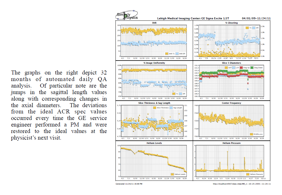
4 Daily/Weekly QA Program
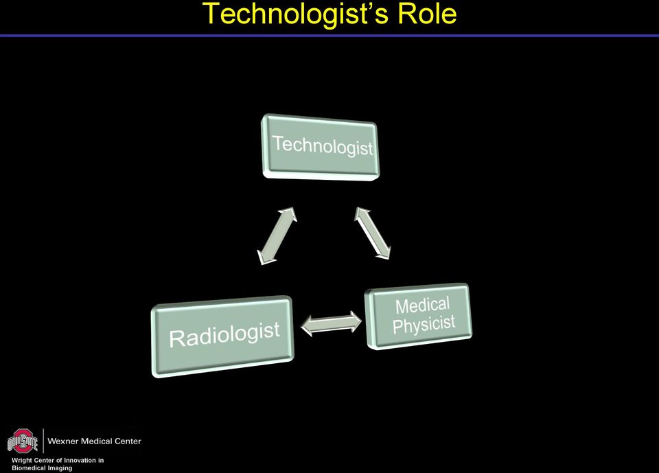
5 Technologist s Role

6 Record Book Examples Data form for weekly QC Template from ACR MRI QC Manual Examples


7 Record Book Examples QC visual checklist Template from ACR MRI QC Manual Examples

8 Table OK? Table OK? A test about the accuracy of the laser localizer Page 35 of ACR Manual 2004 version Frequently ignored by technologists Most technologists misunderstand that it is a check of whether the table move smoothly Laser Light

9 Table OK? on Siemens Scanner Procedure Open the sagittal localizer image User PixelLens to check the top edge of the vertical bars The location should be within ± 2mm < H(ead)2 or F(oot)2

10 Table OK? on GE Scanner Procedure Open the sagittal localizer image User Crosshair to check the top edge of the vertical bars The location should be within ± 2mm < S2 or I2

11 Table OK? Offline Measurement Philips EBW Open the sagittal localizer image User Crosshair to check the top edge of the vertical bars

12 Table OK? Offline Measurement Philips EBW Open the sagittal localizer image User Crosshair to check the top edge of the vertical bars

13 Magnetic Field Drift Definition Center frequency run down over time Drift reasons Windings are not perfect superconductors Eddy current interactions Drift rate < 0.01 ppm per hour < 107 Hz per week for 1.5T < 214 Hz per week for 3T Enshrined drift level 3 ppm per day (AAPM report 34, 1992) 1 ppm per day (AAPM report 100, 2010): 447 Hz/wk for 1.5T and 894 Hz/wk for 3T 1.5 ppm per day (ACR MRI QC guide, 2004) Corrective action Ask medical physicist or MRI scientist to re-check Ask service to monitor the units to ensure 1. B 0 field within RF transmit/receive frequency range 2. Draft rate does not increase

14 Central Frequency on GE scanner: 1 GE Signa Horizon 1.5T Place the phantom Click Auto Prescan before run the localizer sequence Record TG (transmit gain) and AX (central frequency)

15 Central Frequency on GE scanner: 2 GE Signa Horizon 1.5T Text page Series page Record central frequency and transmit gain

16 Central Frequency on Siemens scanner Procedure Before run a sequence, e.g. ACR T1 axial Options Adjustments Record central frequency and transmit gain

17 Distance Measurement: WW and WC Setting window and level/center To have the edges at the half-maximum value of the signal intensity Skipped by some Technologists First set window to 0 or 1 Adjust level/center about ½ white and ½ black, record level value width = recorded level value level/center = ½ recorded level value W = 606 C = 303 W = 1 C = 256 W = 256 C = 128 W = 768 C = 384 W = 1 C = 500 W = 500 C = 250

18 Distance Measurement: Sagittal Sagittal Image Avoid going through the black bar < 148 ± 2 mm

19 Distance Measurement: Problem Sagittal Loc Length mm (Stoneridge) mm (Morehouse) mm (Siemens SSCBC)
 147.")


20 Distance Measurement: Axial Axial slice #5 One vertical One horizontal < 190 ± 2 mm Examples:


21 Distance Measurement: Problem Vertical distance 194 mm (Siemens SSCBC 3T MRI) Out of spec Axial Curved border Axial Out of spec
22 Spokes GE Signa Horizon 1.5T Assign image 8 or 9? # of spokes on the image? Image 8 Image 9 Image 10 Image 11
 no columns or column 4 of the LR array could be resolved?")
23 Resolution GE Signa Horizon 1.5T (1.0) Rows 1 through 4 of the UL array are resolved (1.0) no columns or column 4 of the LR array could be resolved? ACR MRI Accreditation Program Phantom Test Guidance
24 Resolution: ZIP Zero filled interpolation (ZIP) ZIP512: zero fill up to 512x256 Better image resolution Cost 1. Slower reconstruction times 2. Longer to transfer 3. Takes up more disk space FT Unfiltered Image 256x256 k-space data 256x256 Courtesy of Dr. Moriel S. NessAiver
25 Resolution: ZIP Zero filled interpolation (ZIP) ZIP512: zero fill up to 512x256 Better image resolution Cost 1. Slower reconstruction times 2. Longer to transfer 3. Takes up more disk space FT Unfiltered Image 512x512 k-space data 512x512 Courtesy of Dr. Moriel S. NessAiver
26 Resolution: ZIP No ZIP UL: 1.0 LR: 1.1 Not passing ACR criteria with ZIP512 UL: 0.9 LR: 0.9 Passing ACR criteria
27 Common Problems Problems 1. Central frequency: 4 digits? (should be 9 at 3T) 2. Phantom distance: 3 digits? (should be 4) 3. LCD spokes: Always 10 spokes? (because the tech used slice #11, should use specified slice # 8) Slice 11 Slice
28 Additional Tests Advanced clinical MRI procedures MR spectroscopy Diffusion imaging Blood oxygen level dependent contrast (BOLD) imaging Angiographic and blood perfusion methods 1. DCE-MRI
29 Diffusion


30 DCE-MRI Purpose of the DCE-MRI Phantom Tests: Provide means for qualitative and quantitative evaluation, including T1 map error estimation, temporal resolution and image quality. Evaluate compliance with image acquisition protocols. ACRIN CQIE Body Phantom
31 20% 20% 20% 20% Question 1: Allowed weekly CF drift from ACR Manual? 20% ppm ppm ppm ppm ppm 10
32 20% 20% 20% 20% Question 2: Transaxial image measurements on slice #? 20% Any slice 10
33 20% 20% 20% 20% Question 3: Table OK is for checking? 20% 1. Bed position and lights 2. Laser light alignment 3. Patient monitors 4. Horizontal bed movement 5. Vertical motion smoothness 10
34 References 1. Sobol WT, NessAiver MS, Orton CG, Point/counterpoint. The physics components of the ACR MRI Accreditation Program are overly tedious and beyond what is needed to ensure good patient care.med Phys 35(8) ACR 2004 Magnetic Resonance Imaging Qaulity Control Manual 3. Ihalainen TM, Lönnroth NT, Peltonen JI, Uusi-Simola JK, Timonen MH, Kuusela LJ, Savolainen SE, Sipilä OE. MRI quality assurance using the ACR phantom in a multi-unit imaging center. Acta Oncol Aug;50(6): Chen CC, Wan YL, Wai YY, Liu HL. Quality assurance of clinical MRI scanners using ACR MRI phantom: preliminary results. J Digit Imaging Dec;17(4): NessAiver, Range of Results from over 534 ACR-mandated Annual MRI Performance Evaluations on over 204 Magnets from 8 Vendors Spanning a 10-year Period, MRI QA RSNA AAPM Report No. 110, Acceptance Testing and Quality Assurance Procedures for Magnetic Resonance Imaging Facilities, AAPM Report No. 34, Acceptance Testing of Magnetic Resonance Imaging Systems (Reprinted from Medical Physics, Vol. 19, Issue 1)


35 Acknowledgements Department of Radiology Rick Layman Brian Raterman Xiangyu Yang Georgeta Mihai Jun Zhang
MRI QA Technologist s Tests
 AAPM 55th Annual Meeting MRI QA Technologist s Tests Guang Jia, PhD, DABR Associate Professor Department of Physics, Louisiana State University Department of Radiology, The Ohio State University Outline
Breast MRI Quality Control
 Donna M. Reeve, MS, DABR, DABMP Department of Imaging Physics Educational Objectives Discuss the importance of breast MRI quality control (QC). Provide an overview of the new ACR Breast MRI Accreditation
Cirrus 0.2T. MRI for Everyone. North America, Asia, Europe. contact: kturek@mri-tech.pl
 Cirrus 0.2T MRI for Everyone North America, Asia, Europe contact: kturek@mri-tech.pl MRI-TECH inc. Cirrus MRI system for all your needs: Low costs Low maintenance High quality Open geometry Imaging of
SITE IMAGING MANUAL ACRIN 6698
 SITE IMAGING MANUAL ACRIN 6698 Diffusion Weighted MR Imaging Biomarkers for Assessment of Breast Cancer Response to Neoadjuvant Treatment: A sub-study of the I-SPY 2 TRIAL Version: 1.0 Date: May 28, 2012
GUIDE TO SETTING UP AN MRI RESEARCH PROJECT
 GUIDE TO SETTING UP AN MRI RESEARCH PROJECT Formal requirements and procedures OVERVIEW This document is intended to help a principle investigator set up a research project using magnetic resonance imaging
How To Improve Your Ct Image Quality
 Translating Protocols Between Scanner Manufacturer and Model Cynthia H. McCollough, PhD, FACR, FAAPM Professor of Radiologic Physics Director, CT Clinical Innovation Center Department of Radiology Mayo
ACR AAPM TECHNICAL STANDARD FOR DIAGNOSTIC MEDICAL PHYSICS PERFORMANCE MONITORING OF MAGNETIC RESONANCE IMAGING (MRI) EQUIPMENT
 EQUIPMENT") The American College of Radiology, with more than 30,000 members, is the principal organization of radiologists, radiation oncologists, and clinical medical physicists in the United States. The College
The American College of Radiology, with more than 30,000 members, is the principal organization of radiologists, radiation oncologists, and clinical medical physicists in the United States. The College
Phantom Test Guidance
 Phantom Test Guidance for the ACR MRI Accreditation Program The American College of Radiology 1891 Preston White Dr Reston, VA 20191-4397 www.acr.org Contents 0.0 INTRODUCTION............................................................................
Update on ACR Digital Mammography QC Manual
 Update on ACR Digital Mammography QC Manual Priscilla F. Butler, M.S. Medical Physicist and Senior Director, ACR, Reston, VA (with thanks to Eric Berns, Ph.D.) Overview Phantom Specifications QC Manual
QUANTITATIVE IMAGING IN MULTICENTER CLINICAL TRIALS: PET
 Centers for Quantitative Imaging Excellence (CQIE) LEARNING MODULE QUANTITATIVE IMAGING IN MULTICENTER CLINICAL TRIALS: PET American College of Radiology Clinical Research Center v.1 Centers for Quantitative
32-Channel Head Coil Imaging at 3T
 32-Channel Head Coil Imaging at 3T Thomas Benner Athinoula A. Martinos Center for Biomedical Imaging, Department of Radiology, Massachusetts General Hospital and Harvard Medical School, Boston, MA, USA
Moving Forward What does this mean for the Medical Physicist and the Imaging Community?
 Moving Forward What does this mean for the Medical Physicist and the Imaging Community? John M. Boone, Ph.D., FAAPM, FACR Professor and Vice Chairman of Radiology University of California Davis Medical
Quantitative Imaging In Clinical Trials Using PET/CT: Update
 Quantitative Imaging In Clinical Trials Using PET/CT: Update Paul Kinahan, Robert Doot Imaging Research Laboratory Department of Radiology University of Washington, Seattle, WA Supported by RSNA Quantitative
INTRODUCTION. A. Purpose
 New York State Department of Health Bureau of Environmental Radiation Protection Guide for Radiation Safety/Quality Assurance Programs Computed Radiography INTRODUCTION A. Purpose This guide describes
TISSUE MIMICKING GEL QUALITY LE PHANTOM SERIES DESIGN. performance the ultrasound labs ofand. icking material has the same attenuation mim-
 QUALITY Tissue Benefits Mimicking of s RMI recognized RMI as the ultrasound standard phantoms for quality are performance the ultrasound labs ofand hospitals, manufacturers. clinics Sophisticated and ultrasound
Quality Control and Maintenance Programs
 Quality Control and Maintenance Programs Cari Borrás, D.Sc., FACR, FAAPM Visiting Professor DOIN-DEN / UFPE Recife, Pernambuco, Brazil Co-Chair, IUPESM Health Technology Task Group 1 Medical Imaging Equipment
Toshiba Excelart Vantage 1.5T MRI Tech Specs (Technical Specifications)
") Toshiba Excelart Vantage 1.5T MRI Tech Specs (Technical Specifications) Excelart Vantage Magnet Configuration: Ultra-short-bore Strength (or W x H): 1.5 T Homogeneity, ppm V-RMS: Dimensions of maximum
Toshiba Excelart Vantage 1.5T MRI Tech Specs (Technical Specifications) Excelart Vantage Magnet Configuration: Ultra-short-bore Strength (or W x H): 1.5 T Homogeneity, ppm V-RMS: Dimensions of maximum
ParaVision 6. Innovation with Integrity. The Next Generation of MR Acquisition and Processing for Preclinical and Material Research.
 ParaVision 6 The Next Generation of MR Acquisition and Processing for Preclinical and Material Research Innovation with Integrity Preclinical MRI A new standard in Preclinical Imaging ParaVision sets a
Surveying and QC of Stereotactic Breast Biopsy Units for ACR Accreditation
 Surveying and QC of Stereotactic Breast Biopsy Units for ACR Accreditation LORAD Stereotactic Breast Biopsy System AAPM Spring Clinical Meeting Phoenix, AZ March 17, 2013 Melissa C. Martin, M.S., FACR,
5 Factors Affecting the Signal-to-Noise Ratio
 5 Factors Affecting the Signal-to-Noise Ratio 29 5 Factors Affecting the Signal-to-Noise Ratio In the preceding chapters we have learned how an MR signal is generated and how the collected signal is processed
Prepublication Requirements
 Issued Prepublication Requirements The Joint Commission has approved the following revisions for prepublication. While revised requirements are published in the semiannual updates to the print manuals
kv-& MV-CBCT Imaging for Daily Localization: Commissioning, QA, Clinical Use, & Limitations
 kv-& MV-CBCT Imaging for Daily Localization: Commissioning, QA, Clinical Use, & Limitations Moyed Miften, PhD Dept of Radiation Oncology University of Colorado Denver Questions Disease Stage (local, regional,
MDCT Technology. Kalpana M. Kanal, Ph.D., DABR Assistant Professor Department of Radiology University of Washington Seattle, Washington
 MDCT Technology Kalpana M. Kanal, Ph.D., DABR Assistant Professor Department of Radiology University of Washington Seattle, Washington ACMP Annual Meeting 2008 - Seattle, WA Educational Objectives Historical
Standardized MRI Protocol for Brain Tumor Clinical Trials. Benjamin M. Ellingson, Ph.D. Assistant Professor of Radiology at UCLA
 Standardized MRI Protocol for Brain Tumor Clinical Trials Benjamin M. Ellingson, Ph.D. Assistant Professor of Radiology at UCLA Standardized MRI Protocol for Therapeutic Studies FDA Meeting in January
Update on MQSA and ACR Mammography Accreditation
 MQSA - Who s Who Update on MQSA and ACR Mammography Accreditation The Law: Mammography Quality Standards Act (MQSA) The Regulator: US Food and Drug Administration (FDA) Pamela L. Platt, BSRT(R)(M)(CV)
User Manual Microsoft Dynamics AX Add-on LabAX Label Printing
 User Manual Microsoft Dynamics AX Add-on LabAX Label Printing Version 1.7 Last Update: 17.04.2011 User Manual Microsoft Dynamics AX Add-on LabAX Label Printing Page 2 / 23 Contents 1 Introduction... 3
CT RADIATION DOSE REPORT FROM DICOM. Frank Dong, PhD, DABR Diagnostic Physicist Imaging Institute Cleveland Clinic Foundation Cleveland, OH
 CT RADIATION DOSE REPORT FROM DICOM Frank Dong, PhD, DABR Diagnostic Physicist Imaging Institute Cleveland Clinic Foundation Cleveland, OH CT Patient comes out... Patient goes in... Big Black Box Radiology
Magnetic Resonance Imaging
 PRIMARY CERTIFICATION DIDACTIC AND CLINICAL COMPETENCY REQUIREMENTS EFFECTIVE JANUARY 2014 Magnetic Resonance Imaging 1. Introduction Candidates for certification and registration are required to meet
Quality Control of Full Field Digital Mammography Units
 Quality Control of Full Field Digital Mammography Units Melissa C. Martin, M.S., FACMP, FACR, FAAPM Melissa@TherapyPhysics.com 310-612-8127 ACMP Annual Meeting Virginia Beach, VA May 2, 2009 History of
GE Medical Systems Training in Partnership. Module 8: IQ: Acquisition Time
 Module 8: IQ: Acquisition Time IQ : Acquisition Time Objectives...Describe types of data acquisition modes....compute acquisition times for 2D and 3D scans. 2D Acquisitions The 2D mode acquires and reconstructs
Role of the Medical Physicist in Clinical Implementation of Breast Tomosynthesis
 Role of the Medical Physicist in Clinical Implementation of Breast Tomosynthesis Bob Liu, Ph.D. Department of Radiology Massachusetts General Hospital And Harvard Medical School Digital Breast Tomosynthesis
Current Industry Neuroimaging Experience in Clinical Trials Jerome Barakos, M.D.
 Current Industry Neuroimaging Experience in Clinical Trials Jerome Barakos, M.D. Melbourne Australia March 28, 2012 Synarc Experience and Expertise Largest imaging service provider dedicated to clinical
Routine Ultrasound Equipment Tests for Quality Assurance. Contents
 Routine Ultrasound Equipment Tests for Quality Assurance James A. Zagzebski, Ph.D. Nick Rupert, M.S. Ryan DeWall, M.S. Tomy Varghese, Ph.D. Dept. of Medical Physics University of Wisconsin, Madison, WI,
PET/CT QC/QA. Quality Control in PET. Magnus Dahlbom, Ph.D. Verify the operational integrity of the system. PET Detectors
 Quality Control in PET PET/CT QC/QA Magnus Dahlbom, Ph.D. Division of Nuclear Medicine Ahmanson Biochemical Imaging Clinic David Geffen School of Medicine at UCLA Los Angeles Verify the operational integrity
Applicable Models ME511L, ME551i2, MS31i2, MS33i2, MS51i2, MS53i2
 Version 3.1 Applicable Models ME511L, ME551i2, MS31i2, MS33i2, MS51i2, MS53i2 MQSA Quality Control Manual for Monochrome Monitors for Mammography Foreword Purpose of this Document This document is for
Q.A. Collectible. Sponsored by CRCPD s Committee on Quality Assurance in Diagnostic X-Ray (H-7)
") Q.A. Collectible Sponsored by CRCPD s Committee on Quality Assurance in Diagnostic X-Ray (H-7) Mammography Phantom Image Quality Evaluation (from the American College of Radiology 1999 Mammography Quality
Q.A. Collectible Sponsored by CRCPD s Committee on Quality Assurance in Diagnostic X-Ray (H-7) Mammography Phantom Image Quality Evaluation (from the American College of Radiology 1999 Mammography Quality
Performance testing for Precision 500D Classical R/F System
 Performance testing for Precision 500D Classical R/F System John M. Boudry, Ph.D. Image Quality Systems Engineer GE Healthcare Technologies Outline System background Image Quality Signature Test (IQST)
CT: Size Specific Dose Estimate (SSDE): Why We Need Another CT Dose Index. Acknowledgements
: Why We Need Another CT Dose Index. Acknowledgements") CT: Size Specific Dose Estimate (SSDE): Why We Need Another CT Dose Index Keith J. Strauss, MSc, FAAPM, FACR Clinical Imaging Physicist Cincinnati Children s Hospital University of Cincinnati College of
CT: Size Specific Dose Estimate (SSDE): Why We Need Another CT Dose Index Keith J. Strauss, MSc, FAAPM, FACR Clinical Imaging Physicist Cincinnati Children s Hospital University of Cincinnati College of
Tracking Radiation Exposure From Medical Diagnostic Procedures: Siemens Perspectives
 Tracking Radiation Exposure From Medical Diagnostic Procedures: Siemens Perspectives Gilbert W. Beebe Symposium The National Academies Katharine Grant, PhD Staff Scientist 8 December 2011 For internal
SUBCHAPTER 22 QUALITY ASSURANCE PROGRAMS FOR MEDICAL DIAGNOSTIC X-RAY INSTALLATIONS
 Note: This is a courtesy copy and is not the official version of this rule. The official, legally effective version of this rule is available through www.lexisnexic.com/bookstore (Phone: (800) 223-1940).
Purchasing a cardiac CT scanner: What the radiologist needs to know
 Purchasing a cardiac CT scanner: What the radiologist needs to know Maria Lewis ImPACT St George s Hospital, London maria.lewis@stgeorges.nhs.uk CT scanner development Slice wars 1998 Increased z-coverage
Assessing Radiation Dose: How to Do It Right
 Assessing Radiation Dose: How to Do It Right Michael McNitt-Gray, PhD, DABR, FAAPM Professor, Department of Radiology Director, UCLA Biomedical Physics Graduate Program David Geffen School of Medicine
Technical guidelines for magnetic resonance imaging for the surveillance of women at increased risk of developing breast cancer
 Technical guidelines for magnetic resonance imaging for the surveillance of women at increased risk of developing breast cancer NHSBSP PUBLICATION No 68 DECEMBER 2009 1 EDITORS Dr Barbara JG Dall Consultant
THEORY, SIMULATION, AND COMPENSATION OF PHYSIOLOGICAL MOTION ARTIFACTS IN FUNCTIONAL MRI. Douglas C. Noll* and Walter Schneider
 THEORY, SIMULATION, AND COMPENSATION OF PHYSIOLOGICAL MOTION ARTIFACTS IN FUNCTIONAL MRI Douglas C. Noll* and Walter Schneider Departments of *Radiology, *Electrical Engineering, and Psychology University
Atomic Force Microscope
 Atomic Force Microscope (Veeco Nanoman) User Manual Basic Operation 4 th Edition Aug 2012 NR System Startup If the system is currently ON To start the NanoScope software, double-click the NanoScope startup
Nuclear PACS: Problems, Solutions
 Nuclear PACS: Problems, Solutions by Jerold W. Wallis, MD Introduction Reprinted with permission: Wallis JW. Nuclear PACS: Problems, Solutions. Decisions in Imaging Economics. 2004;6;21-25. There is increasing
Advanced MRI methods in diagnostics of spinal cord pathology
 Advanced MRI methods in diagnostics of spinal cord pathology Stanisław Kwieciński Department of Magnetic Resonance MR IMAGING LAB MRI /MRS IN BIOMEDICAL RESEARCH ON HUMANS AND ANIMAL MODELS IN VIVO Equipment:
MGH Adult Diffusion Data Scanning Protocols
 MGH Adult Diffusion Data Scanning Protocols Structural scans SIEMENS MAGNETOM ConnectomA syngo MR D11 \\USER\INVESTIGATORS\Default\AAHScout_64 TA:0:14 PAT:3 Voxel size:1.6 1.6 1.6 mm Rel. SNR:1.00 :fl
Education. October, 1985 GE Medical Systems Milwaukee, WI Signa Operators Training Program
 William H. Faulkner, Jr. B.S.,R.T.(R)(MR)(CT), FSMRT 1554 Sedgefield Dr. Ooltewah, TN 37363 (423) 894-7214(W) (423) 893-9422 (H) Fax: (615) 290-5229 E-mail: faulkner@t2star.com Internet: http://www.t2star.com
Digital Mammography Update: Design and Characteristics of Current Systems
 : Design and Characteristics of Current Systems Kalpana M. Kanal, Ph.D., DABR Assistant Professor Department of Radiology University of Washington Seattle, Washington AAPM Annual Meeting 2009 Anaheim,
Proton magnetic resonance spectroscopy in the brain: Report of AAPM MR Task Group #9
 Proton magnetic resonance spectroscopy in the brain: Report of AAPM MR Task Group #9 Dick J. Drost a) Nuclear Medicine and MRI Department, St. Joseph s Health Centre, London, Ontario N6A 4L6, Canada William
Super-Resolution Reconstruction in MRI: Better Images Faster?
 Super-Resolution Reconstruction in MRI: Better Images Faster? Esben Plenge 1,, Dirk H. J. Poot 1, Monique Bernsen 2, Gyula Kotek 2, Gavin Houston 2, Piotr Wielopolski 2, Louise van der Weerd 3, Wiro J.
Imaging Informatics Ann Scherzinger, PhD, CIIP C-TRIC Lecture Series, May 18, 2011
 Imaging Informatics Ann Scherzinger, PhD, CIIP C-TRIC Lecture Series, May 18, 2011 Science of Information and Information Processing! Breadth, depth of current activities in human medical imaging informatics!
Correcting the Lateral Response Artifact in Radiochromic Film Images from Flatbed Scanners
 Correcting the Lateral Response Artifact in Radiochromic Film Images from Flatbed Scanners Background The lateral response artifact (LRA) in radiochromic film images from flatbed scanners was first pointed
MQSA Quality Control Manual for Monochrome Displays for Mammography
 MQSA Quality Control Manual for Monochrome Displays for Mammography Ver1.0 Applicable Models LMD-DM50, LMD-DM30 Sony Corporation - 1 - MQSA Quality Control Manual for Monochrome Displays for Mammography
Compliance Guidance for FLUOROSCOPIC QUALITY CONTROL
 Compliance Guidance for FLUOROSCOPIC QUALITY CONTROL New Jersey Department of Environmental Protection Bureau of Radiological Health PO Box 415 Trenton NJ 08625 FAX 609-984-5811 Website: http://www.state.nj.us/dep/rpp
The Dedicated MRI Factbook. Esaote MRI: A world of difference
 The Dedicated MRI Factbook Esaote MRI: A world of difference Esaote Dedicated MRI Measures of Excellence Creativity in Healthcare is Esaote s motto which represents the philosophy that has been adopted
CT QC Under the ACR QC Manual. What? There s a Manual?? Learning Objectives 8/6/12! ! YES!!! Almost. ! Doesn t have a pretty cover yet
 CT QC Under the ACR QC Manual Douglas Pfeiffer, MS, DABR Boulder Community Hospital What? There s a Manual??! YES!!! Almost! Doesn t have a pretty cover yet! Should be out by RSNA! Really seriously I mean
WHERE IN THE WORLD JILL LIPOTI?
 WHERE IN THE WORLD IS JILL LIPOTI? HELLO FROM NEW JERSEY CRCPD - National Symposium on Fusion Imaging and Multimodalities February 18-20, 2004 Kansas City, Missouri New Jersey s Requirements As They Pertain
Physics testing of image detectors
 Physics testing of image detectors Parameters to test Spatial resolution Contrast resolution Uniformity/geometric distortion Features and Weaknesses of Phantoms for CR/DR System Testing Dose response/signal
Whitepapers on Imaging Infrastructure for Research Paper 1. General Workflow Considerations
 Whitepapers on Imaging Infrastructure for Research Paper 1. General Workflow Considerations Bradley J Erickson, Tony Pan, Daniel J Marcus, CTSA Imaging Informatics Working Group Introduction The use of
Window Glass Design 5 According to ASTM E 1300
 A User s Guide to: Window Glass Design 5 According to ASTM E 1300 A product of: 1 Table of Contents Table of Contents List of Figures Chapter 1: Window Glass Design 5 1.1 Introduction 1.2 Features ii iv
Sample Table. Columns. Column 1 Column 2 Column 3 Row 1 Cell 1 Cell 2 Cell 3 Row 2 Cell 4 Cell 5 Cell 6 Row 3 Cell 7 Cell 8 Cell 9.
 Working with Tables in Microsoft Word The purpose of this document is to lead you through the steps of creating, editing and deleting tables and parts of tables. This document follows a tutorial format
Microsoft Excel 2010 Charts and Graphs
 Microsoft Excel 2010 Charts and Graphs Email: training@health.ufl.edu Web Page: http://training.health.ufl.edu Microsoft Excel 2010: Charts and Graphs 2.0 hours Topics include data groupings; creating
Medical Image Processing on the GPU. Past, Present and Future. Anders Eklund, PhD Virginia Tech Carilion Research Institute andek@vtc.vt.
 Medical Image Processing on the GPU Past, Present and Future Anders Eklund, PhD Virginia Tech Carilion Research Institute andek@vtc.vt.edu Outline Motivation why do we need GPUs? Past - how was GPU programming
Kap 8 Image quality, signal, contrast and noise
 4/5/ FYS-KJM 474 contrast SNR MR-teori og medisinsk diagnostikk Kap 8 Image qualit, signal, contrast and noise resolution vailable MRparameters speed Main source of noise in MRI: Noise generated within
Implementation of Cone-beam CT imaging for Radiotherapy treatment localisation.
 Implementation of Cone-beam CT imaging for Radiotherapy treatment localisation. Andrew Bridges Clinical Scientist Diagnostic Radiology & Radiation Protection Physics Overview What is CBCT? Use of CBCT
Elfring Fonts, Inc. PCL MICR Fonts
 Elfring Fonts, Inc. PCL MICR Fonts This package contains five MICR fonts (also known as E-13B), to print magnetic encoding on checks, and six Secure Number fonts, to print check amounts. These fonts come
REGULATION: QUALITY ASSURANCE PROGRAMS FOR MEDICAL DIAGNOSTIC X-RAY INSTALLATIONS N.J.A.C. 7:28-22
 REGULATION: QUALITY ASSURANCE PROGRAMS FOR MEDICAL DIAGNOSTIC X-RAY INSTALLATIONS N.J.A.C. 7:28-22 New Jersey Department of Environmental Protection Bureau of Radiological Health PO Box 415 Trenton NJ
High Spatial Resolution EPI Using an Odd Number of Interleaves
 Magnetic Resonance in Medicine 41:1199 1205 (1999) High Spatial Resolution EPI Using an Odd Number of Interleaves Michael H. Buonocore* and David C. Zhu Ghost artifacts in echoplanar imaging (EPI) arise
Automatic Wheel Profile Measuring Device for Tramway Vehicles
 Automatic Wheel Profile Measuring Device for Tramway Vehicles Technical description The automatic wheel profile measuring device is used to measure the wheel profile and diameter whilst a tramway vehicle
Technique and Safety of. by Pierluigi Castellone, Electronics Engineer Brain Products General Manager
 Technique and Safety of performing EEG/fMRI measurements by Pierluigi Castellone, Electronics Engineer Brain Products General Manager Contents of the presentation Why recording simultaneous EEG and fmri?
Wireless Radio Frequency Coil for Magnetic Resonance Image of Knee Joint
 Wireless Radio Frequency Coil for Magnetic Resonance Image of Knee Joint Tomohiro Sahara, Hiroshi Tsutsui, Shigehiro Hashimoto, Shuichi Mochizuki, Kenichi Yamasaki, Hideo Kondo, Teruo Miyazaki Department
Create Charts in Excel
 Create Charts in Excel Table of Contents OVERVIEW OF CHARTING... 1 AVAILABLE CHART TYPES... 2 PIE CHARTS... 2 BAR CHARTS... 3 CREATING CHARTS IN EXCEL... 3 CREATE A CHART... 3 HOW TO CHANGE THE LOCATION
The multitude of symptoms following a whiplash injury has given rise to much discussion because of the lack of objective radiological findings.
 HELPFUL PERSONAL INJURY INFORMATION COURTESY OF RIVERVIEW CHIROPRACTIC FROM ABSTRACT: Dynamic kine magnetic resonance imaging in whiplash patients Pain Research and Management 2009 Nov-Dec 2009;Vol. 14,
Three Dimensional Ultrasound Imaging
 Three Dimensional Ultrasound Imaging Hans Torp/ Sevald Berg/Kjell Kristoffersen m/flere Department of circulation and medical imaging NTNU Hans Torp NTNU, Norway Acquisition Reconstruction Filtering Collecting
MRI DATA PROCESSING. Compiled by: Nicolas F. Lori and Carlos Ferreira. Introduction
 MRI DATA PROCESSING Compiled by: Nicolas F. Lori and Carlos Ferreira Introduction Magnetic Resonance Imaging (MRI) is a clinical exam that is safe to the patient. Nevertheless, it s very important to attend
Chest CT protocols. Mannudeep K. Kalra, MD, DNB. Dianna D. Cody, PhD. Massachusetts General Hospital Harvard Medical School
 Chest CT protocols Mannudeep K. Kalra, MD, DNB Dianna D. Cody, PhD Massachusetts General Hospital Harvard Medical School M.D. Anderson Cancer Center Specific principles Routine chest CT Lung nodule follow
MRI SEQUENCES. 1 Gradient Echo Sequence
 5 An MRI sequence is an ordered combination of RF and gradient pulses designed to acquire the data to form the image. In this chapter I will describe the basic gradient echo, spin echo and inversion recovery
Quality control of CT systems by automated monitoring of key performance indicators: a two-year study
 JOURNAL OF APPLIED CLINICAL MEDICAL PHYSICS, VOLUME 16, NUMBER 4, 2015 Quality control of CT systems by automated monitoring of key performance indicators: a two-year study Patrik Nowik, a Robert Bujila,
Diffusione e perfusione in risonanza magnetica. E. Pagani, M. Filippi
 Diffusione e perfusione in risonanza magnetica E. Pagani, M. Filippi DW-MRI DIFFUSION-WEIGHTED MRI Principles Diffusion results from a microspic random motion known as Brownian motion THE RANDOM WALK How
CT Protocol Optimization over the Range of CT Scanner Types: Recommendations & Misconceptions
 CT Protocol Optimization over the Range of CT Scanner Types: Recommendations & Misconceptions Frank N. Ranallo, Ph.D. Associate Professor of Medical Physics & Radiology University of Wisconsin School of
Musculoskeletal MRI Technical Considerations
 Musculoskeletal MRI Technical Considerations Garry E. Gold, M.D. Professor of Radiology, Bioengineering and Orthopaedic Surgery Stanford University Outline Joint Structure Image Contrast Protocols: 3.0T
A software tool for Quality Assurance of Computed / Digital Radiography (CR/DR) systems
 systems") A software tool for Quality Assurance of Computed / Digital Radiography (CR/DR) systems Nikunj Desai a and Daniel J Valentino a,b a icr Company Inc, 2580 West 237th Street, Torrance, CA 90505, USA b Department
A software tool for Quality Assurance of Computed / Digital Radiography (CR/DR) systems Nikunj Desai a and Daniel J Valentino a,b a icr Company Inc, 2580 West 237th Street, Torrance, CA 90505, USA b Department
Basic Specifications. Electrical. EPSON Perfection 3170. Color EPSON MatrixCCD TM line sensor. device Effective pixels
 Scanner Parts Document table Power cord Document cover Transparency unit (TPU) under lid Web Transparency unit (TPU) connector TPU indicator light e-mail Carriage and lamp (in the home position) USB port
Annual MQSA Inspection Questions NMQAAC April 19, 2004
 1 1 1 1 1 1 1 1 0 1 0 1 0 1 0 1 Annual MQSA Inspection Questions NMQAAC April 1, 00 1.0 Inspection Information 1.1 Name and Address 1. Equipment Registration.0 Facility Inspection Download.0 Facility Inspections.0
2/28/2011. MIPPA overview and CMS requirements. CT accreditation. Today s agenda. About MIPPA. Computed Tomography
 Today s agenda Computed Tomography Presented by: Dina Hernandez, BSRT, RT (R), CT, QM Krista Bush, RT, MBA Leonard Lucey, JD ACR Quality & Safety MIPPA overview and CMS requirements CT accreditation How
Shorter Scanning Times Through Efficient Software. Annette Tuffs. Article from the customer magazine Medical Solutions, December 2010
 Shorter Scanning Times Through Efficient Software Annette Tuffs Article from the customer magazine Medical Solutions, December 2010 www.siemens.com/healthcare-magazine Highlight Topic Efficiency: MRI A
QAV-PET: A Free Software for Quantitative Analysis and Visualization of PET Images
 QAV-PET: A Free Software for Quantitative Analysis and Visualization of PET Images Brent Foster, Ulas Bagci, and Daniel J. Mollura 1 Getting Started 1.1 What is QAV-PET used for? Quantitative Analysis
Technical handbook Panel Anchoring System
 1 Basic principles of sandwich panels 3 Design conditions 4 Basic placement of anchors and pins 9 Large elements (muliple rows) 10 Small elements (two rows) 10 Turned elements 10 Slender elements 10 Cantilevering
Failure Modes and Effects Analysis (FMEA)
") Failure Modes and Effects Analysis (FMEA) Sasa Mutic Washington University School of Medicine St. Louis Missouri Failure Modes and Effects Analysis Objectives: To motivate the use of FMEA and to provide
Failure Modes and Effects Analysis (FMEA) Sasa Mutic Washington University School of Medicine St. Louis Missouri Failure Modes and Effects Analysis Objectives: To motivate the use of FMEA and to provide
Education, Training, and Certification of Medical Physicist in Korea
 Education, Training, and Certification of Medical Physicist in Korea Rena Lee, Education Committee, KSMP Tae-Suk Suh, President, KSMP The 7th Asia-Oceania Congress of Medical Physics IOMP Symposium on
Cynthia H. McCollough b) and Michael R. Bruesewitz Department of Radiology, Mayo Clinic College of Medicine, Rochester, Minnesota 55905
 and Michael R. Bruesewitz Department of Radiology, Mayo Clinic College of Medicine, Rochester, Minnesota 55905") The phantom portion of the American College of Radiology ACR Computed Tomography CT accreditation program: Practical tips, artifact examples, and pitfalls to avoid a Cynthia H. McCollough b) and Michael
The phantom portion of the American College of Radiology ACR Computed Tomography CT accreditation program: Practical tips, artifact examples, and pitfalls to avoid a Cynthia H. McCollough b) and Michael
DIRECTIONS FOR SETTING UP LABELS FOR MARCO S INSERT STOCK IN WORD PERFECT, MS WORD AND ACCESS
 DIRECTIONS FOR SETTING UP LABELS FOR MARCO S INSERT STOCK IN WORD PERFECT, MS WORD AND ACCESS WORD PERFECT FORMAT MARCO ITEM #A-3LI - 2.25 H x 3W Inserts First create a new document. From the main page
Scatter Plots with Error Bars
 Chapter 165 Scatter Plots with Error Bars Introduction The procedure extends the capability of the basic scatter plot by allowing you to plot the variability in Y and X corresponding to each point. Each
Unwarping Echo Planar Images Using CMTK 1 Release 1.2
 Unwarping Echo Planar Images Using CMTK 1 Release 1.2 Torsten Rohlfing October 1, 2012 Neuroscience Program, SRI International, Menlo Park, CA Abstract This document describes the workflow for unwarping
GE 3.0T NPW,TRF,FAST,F R NPW,TRF,FAST,F R
 GE 3.0T 3.0T WRIST Invivo 8CH Wrist Coil Sequence Ax T2 Cor PD Cor PDFS Cor T1 Cor PD (Small FOV) FOV (mm) 80 80 80 80 40 Matrix 384x224 384x256 320x256 384x320 320x192 Phase Direction RL RL RL RL RL #
Specifying Laser Scanning Services. A Quantapoint White Paper
 A Quantapoint White Paper ABSTRACT Specifying Laser Scanning Services to Help Ensure Trusted and Accurate Results As-built documentation created using laser scanning is rapidly being adopted as the tool
Merging Labels, Letters, and Envelopes Word 2013
 Merging Labels, Letters, and Envelopes Word 2013 Merging... 1 Types of Merges... 1 The Merging Process... 2 Labels - A Page of the Same... 2 Labels - A Blank Page... 3 Creating Custom Labels... 3 Merged