The Human Muscular System. Introduction Images Used Under Fair Use Copyright source:
|
|
|
- Avice Payne
- 7 years ago
- Views:
Transcription
1 The Human Muscular System Introduction Images Used Under Fair Use Copyright source:

2 REMEMBER!!!!!! 1. Muscles PULL!!!!! 2. Muscles do NOT push!!!!!! 3. Bones, ligaments, nerves and muscles work together as levers so that we have movement.

3 Why You Need to Know The Muscular System -- 1

4 Why You Need to Know The Muscular System Pull skin 2. Inject 3. Wait 10 seconds (or per manufacturer s label) 4. Release skin 5. E.g., Rhus toxin and Z- track
 4. Release skin 5. E.g.")
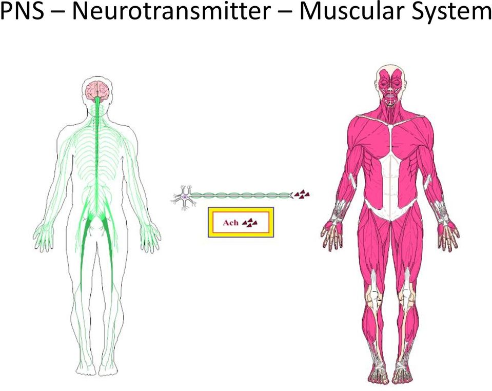
5 Introduction to Muscles Bones and articulations do not move themselves. In order to be moved, they require something to make the articulations bend or twist or turn and they require something to stimulate that which makes them move. The something that stimulates something else to make articulations move is the nervous system. 5

6 Nervous System Overview: FOCUS Peripheral Nervous System

7 PNS Neurotransmitter Muscular System
8 Ach Mechanism of Action

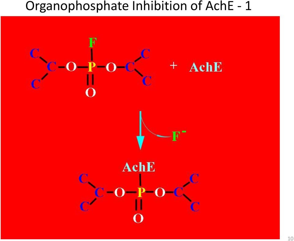
9 Ach Mechanism of Action -- Notes 1. Old data suggested simultaneous albeit disproportionate -- movement of Na + (ECF to ICF) and K + (ICF to ECF) 2. Ach alters the permeability of the cell membrane to Na + primarily K + secondarily 3. New data indicates that the movement of K + is not at the magnitude once believed 4. However, when extracellular K + is high enough, it will cause a Ca 2+ -dependent end plate current (aka spasm) 5. Mg 2+ blocks the end plate current by reducing Ca 2+ availability e.g., pre-term labor 9

10 Organophosphate Inhibition of AchE
11 S/S Organophosphate Poisoning 1. S 2. L 3. U 4. D 11
12 Organophosphate Inhibition of AchE

13 Un-Inhibition of AchE Due to Organophosphate Poisoning 1. 2-PAM pralidoxime -- reverses the inhibition in spite of the fact that this inhibition is uncompetitive 2. NO SLUD 3. 2-PAM must be given within a few hours of poisoning for maximal effect 4. Atropine used to ease breathing 5. Valium used for muscle spasms 13

14 Nicotinic Acetylcholine Receptors (NACHR s) in the CNS REM sleep (associated with high degree of dreaming) associated with NAchR activation? May explain why smokers don t awaken in the night to smoke? Dopaminergic/Nicotinic systems are close to each other. This suggests that smoking a cigarette provides a reward to the smoker neuroelectrically, similar to the self-medication undertaken by many drug addicts (includes alcohol and nicotine) and carbohydraholics. 14

15 N 1 receptors N 2 receptors At NMJ At ganglia Mixed cationic channels Na + /K + fast: NO 2d messenger Ca 2+ /Cl -?? Intermediate?? Ca 2+ /K + slow: requires 2d messenger Nicotine facilitates: Ach release 5-HT, NE and dopamine release PRL, AVP, ACTH, corticosterone release increased brain carbohydrate metabolism 15

16 Nicotine causes affects on behavior: Locomotor activity Schedule-controlled behaviors (OCD) Attention span Processing information Short-term memory Nicotine appears to enhance cognitive functioning and seems to increase attention to the task at hand which all lead to information storage, i.e., memory, BUT results on the effects of nicotine and memory, proper, are mixed. NAchR s mediate rapid excitatory responses 16

17 Nerve and Muscle Supply: the Neuro-Muscular Junction (NMJ) Term Description Acetylcholine (Ach) The neurotransmitter released into the synapse. Motor end plate Polarized Depolarized Troponin Myosin Actin The portion of the muscle membrane directly under the end of the axon. In a muscle at rest, there is an electrical potential difference (voltage) maintained between the inside of the fiber and the ECF's around it. This is due to active transport systems that keep Na+ outside the fiber and K+ inside the fiber. Also means relaxation. Is the permeability of that muscle membrane to Na+ and K+ and is followed by a loss of potential. Also means contraction. Binds actin with myosin; a contractile protein. ATP is bound to myosin cross-links (keeps muscle relaxed). Another contractile protein. In the presence of Ca2+ it acts as an enzyme to hydrolyze ATP to ADP and Pi. Another contractile protein. It cross-links with myosin after ATP is hydrolyzed and causes contraction. 17


18 The NMJ. The relationship of the axonal pad to the motor endplate and the synapse. 18


19 The best simple way to describe this graphic is that energy flows into the muscle cell, causing it to contract. 19

20 Excitation-Contraction Coupling Mechanism There are several theories "out there" that try to explain muscle contraction. As usual, I suspect that it's probably not one of the individual theories, but the sum of the theories that actually explains how muscle contracts. 20

21 The more scientific manner is as follows: when the Ach is released into the synaptic cleft, it then binds with an Ach receptor on the motor endplate. That causes the sarcolemma to depolarize. This depolarization alters the membrane's permeability to Na + and K +, causing the former to leak into the cell and the latter to leak out of the cell. This, in turn, causes the T tubule to depolarize which causes the sarcoplasmic reticulum (SR) to depolarize. When the SR is depolarized, it causes Ca 2+ to be released from the SR. The Ca 2+ binds to contractile proteins (troponin). The troponin changes shape and exposes the actin binding sites. Myosin heads attach to actin binding sites. The head tilts and pulls the actin molecules together. The tilt exposes ATP binding sites on the head and ATP binds to it. The head detaches and ATP is hydrolyzed to ADP, Pi and energy. The energy cocks the myosin and it attaches to a new site on actin. As long as the steps in red continue, this will cause the muscle to contract. Once the action potential (depolarization/repolarization cycle remember the "wave" some of you did in class during neuro?) stops, the Ca 2+ is released from the contractile proteins and pumped back into the SR. The muscle relaxes. When viewing the steps, above, think of this contraction mechanism as being very much like a very rapid rachet mechanism, similar to that on a wrench or on straps for tie down on a truck. 21
22 22
 that are separated by Z lines. There are thick and thin fibers (filaments) in each sarcomere.")
23 The Sliding Filament Theory of Muscle Contraction It seems to me that the previous mechanism just doesn't work without this mechanism. This mechanism says that relaxed muscles consist of sarcomeres (see graphic, next slide) that are separated by Z lines. There are thick and thin fibers (filaments) in each sarcomere. The thick fibers make up the A bands and the gaps between the thin filaments make up the H zone. As the muscle contracts, the thin filaments cock to a side to accommodate their sliding over each other until the A bands are in contact with the Z lines at maximal contraction. 23
24 Energy Sources from Intermediary Metabolism The first system is the phosphagen system. In this system, the source of the energy is ATP. Remember that during muscular contraction, ATP is hydrolyzed to ADP, P i and energy. When this happens, there is only enough energy for 5-6 seconds. So how do our cells get additional energy? Our cells get it via a compound called phosphocreatine (PCr). The concentration of PCr is about 2-3 times greater than the concentration of ATP. When PCr is available, it is hydrolyzed to Cr and P i and energy. The P i is used to re-phosphorylate ADP to make more ATP. This gives us about 15 seconds of maximal contractions and is used for short bursts. 24
25 PCr Shuttle Quick View To give you an idea of what is involved in PCr utilization and synthesis, let's examine the PCr shuttle in cardiac and skeletal muscle. This shuttle increases incredibly the movement/transport of high-energy phosphate (ATP) from the matrix of the mitochondrion to the cytosol of the cell. 25
26 In the first step (1), an ADP-ATP translocase re-phosphorylates ADP to form ATP in the mitochondrial matrix. This occurs via electron transport/oxidative phosphorylation (the "ETOP" in the graphic). When the ATP is "dumped" into the intermembrane space, it is reacted with creatine via a mitochondrial creatine kinase (6) to form PCr. The PCr is then transported via a creatine-creatine phosphate (C:PCr) transport pore (2) into the cytosol to "dump" into a cytosolic PCr store. 26
27 The cytosolic PCr store is derived from an EMP-linked (glycolysis-linked) creatine kinase (CK) (3) and another CK that maintains the equilibrium between C and PCr (or ATP and ADP, if you prefer)(4). The utilization of the PCr from the cytosolic store occurs via an ATP dependent CK that cranks out hugely elevated levels of ATP (5). The ATP is used by processes requiring high energy, e.g., contracting muscle. 2 7
28 Of significance is that there are three isozymes (enzymes with the same function that are found in different tissues and are slightly different, structurally) of CK: CK-BB CK-MB CK-MM = CK-1 = CK-2 = CK-3 Brain, uterus, prostate, lung Heart (and skeletal muscle) Skeletal muscle (and cardiac, too) 28
29 Creatine, Urea, TCA and Malate-Aspartate Shuttle Interconnections 1. Creatine: is a nitrogenous organic acid that occurs naturally in vertebrates and helps to supply energy to muscle and nerve cells. In humans and animals, approximately half of stored creatine originates from food (mainly from fresh meat). Ninety-five percent of creatine is later stored in the skeletal muscles. 2. Creatinine: is a break-down product of creatine phosphate in muscle, and is usually produced at a fairly constant rate by the body (depending on muscle mass). Creatinine is chiefly filtered by the kidney, though a small amount is actively secreted. There is little-to-no tubular reabsorption of creatinine. If the filtering of the kidney is deficient, blood levels rise. Men tend to have higher levels of creatinine because they have more skeletal muscle than women. Vegetarians tend to have lower creatinine levels, because vegetables contain no creatine. 29
30 Creatine/CreatinINe 1. Mitochondrial Arginine-glycine: amidinotransferase (E.C ) 2. ga Transporter (from mito to cytosol) 3. Cytosolic S-adenosylmethionine :guanidinoacetate-n-methyltransferase (SAM:gA NMT) 4. Cytosolic Creatine kinase 5. Cytosolic non-enzymatic cyclization 30
31 Inside black lines is mito matrix Outside black lines is cytosol N-acetylglutamate is an obligatory on switch for CPS-I. Note relationships between Urea cycle, TCA, Creatine Synthesis and Asp-Malate Shuttle. 31
32 ATP Fine Tune
33 NADH: through ETS ADP-ATP Translocase coupled with Dihydrogen phospate/proton Co-Transporter Complex 1 requires 4 protons to pass on electron to Complex III Complex III requires 2 protons to pass on electron to Complex IV Complex IV requires 4 protons to reduce molecular oxygen to water 10 protons exported per NADH to make an ATP
34 FADH 2 : through ETS ADP-ATP Translocase coupled with Dihydrogen phospate/proton Co-Transporter Complex III requires 2 protons to pass on electron to Complex IV Complex IV requires 4 protons to reduce molecular oxygen to water 6 protons exported per FADH 2 to make an ATP
35 Complex V and Co-Transporter 3 protons required to turn on F o 1 proton co-transported with P i 4 protons imported to synthesize 1 ATP molecule Currently Accepted Stoichiometry # ATP 1NADH 10 protons NADH 4 protons ATP 2.5 ATP NADH # ATP 1FADH 2 6 protons FADH 2 4 protons ATP 1.5 ATP FADH 2 Suggested Source:
36 ATP Fine Tune Part 2 Type I Muscle Fibers Red Fibers Slow Twitch For Endurance Small Diameter Aerobic Lots of Mitochondria Malate-Aspartate Shuttle Liver, Heart, Kidney Back Muscles, too Type II Muscle Fibers White Fibers Fast Twitch For Explosive Bursts of Power Large Diameter Anaerobic Fewer Mitochondria -Glycerol Phosphate Shunt Skeletal Muscle, Brain Digits and Extraocular Muscles Store Glycogen
37 Key to understanding muscles is that they ALWAYS PULL -- the NEVER push. Bones and joints and muscles are levers. Muscles function between agonists and antagonists, i.e., one muscle or set of muscles lets your body move one way (agonists) and another (set) acts in the exact opposite way (antagonists). 37
38 Bones, ligaments and muscles (and tendons -- coming later) make the body move under neurological stimulation. To move, our body is put together as a series of levers. There are 3 types of levers: LEVERS Class I Class II Class III In the case of a Class I lever, the fulcrum (F) is between the load (L) and the moving force (P). One example is a teeter totter; another is the articulation between the head and cervical spine that allows us to nod our heads. In the case of a Class II lever, the load (L) is between the fulcrum (F) and the moving force (P). One example is a wheel barrow; another is the articulation between our tibia and talus that allows us to plantarflex our foot as we step off. In the case of a Class III lever, the moving force (P) is between the fulcrum (F) and the load (L). One example is carrying a loaded shovel; another is the articulation between the humerus and ulna that allows us to bend our elbow when we have a drink of something. 38
39 Biological and Practical Levers Other Type I s: scissors, teeter totter, forearm extension Other Type II s: wheelbarrow Other Type III s: forceps, tweezers, carrying loaded shovel 39
40 Mechanical Advantage -- MA The less the MA, the faster the lever [horse foreleg = 0.08 (runner!)] The greater the MA, the stronger the lever [armadillo foreleg = 0.25 (digger!]) Type I is > OR < 1; Type II > 1 and Type III <1 40
41 Is the ability of a force to cause rotation If Force goes WITH or AGAINST the same direction as radius, there is no torque this is due to equation (trigonometric) requirements, as well as intuition. 41
42 Torque and Lifting 45# box Bad back: E. spinae tension = 608.7#; vertebral compression = 595.5# -- ~13 X > the original weight!!!!!!!! == back injury!!!!!!!! Good back: E. spinae tension = 45#; vertebral compression = 45# -- original weight!!!!!! == NO back injury!!!!!!!! 42
43 Perspective Even a 6# weight causes 325# of tension and 317# of compression when lifted incorrectly in a 150# person. 43
44 Pulleys Are combinations of levers and torques 1 pulley can change the direction of a force Multiple pulleys can reduce the Force needed to lift a heavy load Key: The MA equals the number of parallel ropes wrapped around the pulleys NOT including the anchoring rope That means that if there are 2 parallel ropes, it ll be like lifting half the weight of the object 44
45 MA = 2 Pulleys MA = 6 The force exerted is as follows: Wt F 2 And the MA is as follows: Wt MA F 2or 6 45
46 Traction Pulley Application 46
47 The Human Muscular System Introduction Images Used Under Fair Use Copyright source:
48 MUSCLE ORIGIN INSERTION ACTION Frontalis Orbicularis oculi Orbicularis oris Occipitalis Galea aponeurotica Frontal bone and maxilla Muscle fibers surrounding the mouth Galea aponeurotica Frontal and nasal bones The origin Skin at corners of mouth Occipital bone and mastoid process Raises eyebrows; draws scalp forward Closes eyelids gently as in sleep or blinking involuntarily and firmly, voluntarily Brings lips together, shapes lips during speech, kissing muscle Draws scalp backwards 48
49 MUSCLE ORIGIN INSERTION ACTION Attrahens auriculam Attolens auriculam Retrahens auriculam Galea aponeurotica Galea aponeurotica Mastoid portion of temporal bone Front of helix fold of ear Upper part of pinna (outer ear) Lower part of concha (opening into meatus) Superficial muscles Draws ear upward and forward Slightly raises ear Lower part of concha (opening into meatus) 49
50 MUSCLE ORIGIN INSERTION ACTION Temporalis Frontal, parietal, temporal and sphenoid bones Coronoid process of mandible Assists in jaw closure Deep Muscle 50
51 MUSCLE ORIGIN INSERTIO N Corrugator supercilii Levator labii superioris et alaquae nasi Depressor alae nasi not shown, here Frontal bone and maxilla Maxilla and above infraorbital foramen Incisive fossa of maxilla (over central/ lateral incisors) Deep skin, superiorly and laterally Ala of nose and upper lip Nasal septum and posterior ala ACTION Passes between two fascicles of the O. oculi frowning muscle Sneer muscle -- Elvis Constricts the nares 51
52 MUSCLE ORIGIN INSERTION ACTION Zygomaticus minor Malar bone, posterior to maxillary suture Angle of mouth Moves mouth backwards, upward and out as in sadness Zygomaticus major Malar bone, anterior to zygomatic suture Angle of mouth Moves mouth backward and upward as in laughing Levator anguli oris K-9 fossa below infraorbital foramen Angle of mouth Raises angle of mouth 52
53 MUSCLE ORIGIN INSERTION ACTION Levator menti (Mentalis) Incisive fossa lateral to mandibular symphysis Skin of chin Protrudes lower lip forward; doubt/disdain Depressor labii inferioris (quadratus menti) Between mandibular symphysis and mental foramen Skin of lower lip Draws lip down and out; irony Depressor anguli oris (triangularis menti) Same as Q. menti Angle of mouth Depresses angle of mouth 53
54 MUSCLE ORIGIN INSERTION ACTION Buccinator 1 st, 2 d and 3 d molar alveolar processes upper & lower Angle of mouth Compresses cheeks; sucking muscle; Dizzy Gillespie Risorius Fascia over the masseter Skin at the angle of the mouth Retracts the angles of the mouth; produces the sardonic expression Masseter Malar process of maxilla; zygomatic arch Angle/ramus of jaw and coronoid process Elevates the mandible as in closing the mouth; walnut cracking muscle 54
55 MUSCLE ORIGIN INSERTION ACTION Sternocleidomastodeus (SCM) Sternum (manubrium) and clavicel) Outer surface of mastoid process and occipital bone Rotates head to opposite side; draws head to same side Platysma Fascia covering upper chest and shoulders Skin of face and about angle of mouth Draws down lower lip; expression of melancholy Trapezius Inner third of occipital bone and protuberance; thoracic vertebra Posterior border of clavicle; inner margin of acromion; crest of scapular spine Retracts scapula; braces shoulder; rotates scapula; draws head backward 55
56 MUSCLE ORIGIN INSERTION ACTION Pterygoideus externus great wing of the sphenoid neck of the condyle of the mandible Protrudes mandible Pterygoideus internus lateral pterygoid plate and the pyramidal process of the palatine bone ramus and angle of the mandible, as high as the mandibular foramen Raises mandible with masseter and temporalis 56
57 MUSCLE ORIGIN INSERTION ACTION Posterior digastricus belly Inner mastoid Hyoid bone Prevents return of morsel into mouth after swallowed Stylohyoid Styloid process Body of hyoid As with above Anterior digastricus belly Mandibular symphysis Hyoid bone Pushes tongue against roof of mouth in swallowing 57
58 MUSCLE ORIGIN INSERTION ACTION Mylohyoid Levator anguli scapula Mandibular symphysis to last molar Transverse processes of C1-4 Body of hyoid Posteromedial border of scapula Pushes tongue against roof of mouth in swallowing like ant. digastricus Shrugs shoulders 58
59 MUSCLE ORIGIN INSERTION ACTION Styloglossus Styloid process Anterior/tip of tongue Hyoglossus Hyoid bone Middle of tongue Geniohyoid Inner mandibular symphysis Body of hyoid Curls tongue backwards Rolls tongue As with ant. Digastricus: tongue against roof of mouth 59
60 MUSCLE ORIGIN INSERTION ACTION Scalenus anticus Scalenus medius Scalenus posticus Tranverse processes of C vertebra Transverse processes of C2-7 Transverse processes of C5-7 Inner and upper surface of rib 1 Upper surface of rib 1 Outer surface of rib 2 Cone shaped; elevates first two ribs: inspiration Largest of the three; elevates first two ribs: inspiration Deepest of the three; elevates first two ribs: inspiration Bend vertebral column laterally and medially; slight flexion; when acting antagonistically, there is NO lateral movement of the vertebral column 60
61 MUSCLE ORIGIN INSERTION ACTION Trapezius Inner third of occipital bone and protuberance; thoracic vertebra Posterior border of clavicle; inner margin of acromion; crest of scapular spine Retracts scapula; braces shoulder; rotates scapula, draws head backward Latissimus dorsi T6-S5; iliac crests; ribs 9-12 Lower half of intertubercular groove Twisted upon itself; depresses humerus; draws humerus backwards; adducts humerus; rotates humerus inward; involved primarily in downward blow of arm 61
62 MUSCLE ORIGIN INSERTION ACTION Levator anguli scapuli Transverse processes of C1-4 Posteromed ial border of scapula Shrugs shoulders Rhomboideus minor C7 and T1 Root of scapular spine Rotates scapula Rhomboideus major T1-4 or 5 Lower root of scapular spine and inferior angle Rotates scapula backward and upward 62
63 MUSCLE ORIGIN INSERTION ACTION Serratus posticus superioris C7-T3 spinous processes Upper borders of ribs 2-4 Elevates the ribs on inspiration; beneath Rhomboids Serratus posticus inferioris T11-L2 or3 Lower borders of ribs 9-12 Draws lower ribs downward in inspiration 63
64 MUSCLE ORIGIN INSERTION ACTION Erector spinae (sacrospinalis) superficial layer of the deep muscles Mid-sacral crest; spinous processes of L1-5 and T 11-12; to rough ridge on inner lip of iliac crests superior to auricular surface 9-way split Extends vertebral column and bends vertebral column to one side. 64
65 MUSCLE ORIGIN INSERTION ACTION Multifidus spinae Rotatores spinae S4 foramen; medial PSIS; all mammillary processes in T spine; articular processes of C4-7 Transverse process of one vertebra Obliquely ascend to spinous processes from C5-L5 Base of the spinous process of the vertebra above it Extends vertebral column and rotates to opposite side Extends vertebral column ands rotates to opposite side; longi: crosses obliquely breves: crosses horizontally 65
66 Chest and Shoulder MUSCLE ORIGIN INSERTION ACTION Pectoralis major Sternal half of clavicle; anterior half of sternum down to rib 6 or 7; cartilages of all true ribs Outer bicipital ridge Adducts arm; brings arm in front of chest; rotates arm inward Deltoideus Outer third of anterosupero surface of clavicle Deltoid tuberosity Abducts arm; draws the arm forward/ backward 66
67 MUSCLE ORIGIN INSERTION ACTION Pectoralis major Sternal half of clavicle; anterior half of sternum down to rib 6 or 7; cartilages of all true ribs Outer bicipital ridge Adducts arm; brings arm in front of chest; rotates arm inward Deltoideus Outer third of anterosupero surface of clavicle Deltoid tuberosity Abducts arm; draws the arm forward/ backward Biceps brachii Apex of coracoid process; upper glenoid fossa Radial tuberosity Flexes forearm; supinator Brachialis anticus Lower half of humeral shaft Anterior ulnar coronoid process Flexes forearm; protects elbow joint 67
68 MUSCLE ORIGIN INSERTION ACTION Pectoralis minor Superolateral surface of ribs 3-5 Coracoid process of scapula Beneath major; depresses shoulder; forced inspiration Serratus magnus Ribs 1-8 Vertebral border of scapula Carries scapula forward; raises arm External intercostals 11 on each side; lower border of each rib Upper border of rib below Enlarge thoracic cavity on inspiration; fibers directed obliquely medially Internal intercostals 11 on each side; inner surface of each rib Upper border of rib below Under externals; fibers directed obliquely laterally; decrease thoracic cavity on expiration 68
69 MUSCLE ORIGIN INSERTION ACTION Coracobrachialis Apex of coracoid process Midhumeral shaft Draws humerus forward and in; elevates humerus towards scapula 69
70 Rotator Cuff Muscles MUSCLE ORIGIN INSERTION ACTION Supraspinatus Supraspinous fossa Superior greater tuberosity Abducts arm; prevents superior dislocation Infraspinatus Infraspinous fossa Mid-greater tuberosity Rotate humeral head outward; carries arm backwards; prevents posterior dislocation Teres minor Axillary border of scapula Lower greater tuberosity Same as infraspinatus SITTS Teres major Subscapularis Inferior angle of scapula Subscapular fossa Inner bicipital ridge Lesser tubercle Splits long head of triceps and latissimus dorsi; draws humerus down and back Medial arm rotation 70
71 MUSCLE ORIGIN INSERTION ACTION Triceps brachii Below the glenoid fossa and humerus Common tendon of triceps: back part of upper olecranon process Extends forearm 71
72 Fascia Description Linea alba Linea semilunares Poupart s ligament Extends from xiphoid process to symphysis pubis; formed by aponeurosis of Obliquii and Transversus muscles Outer border of rectus abdominis; cartilage of 9 th rib to pubic spine ASIS to pubic spine; from aponeurosis of external oblique Linea transversae Intersects recti muscles; connect semilunareas to alba 72
73 MUSCLE ORIGIN INSERTION ACTION External oblique Lower outer surface of ribs 5-12 Iliac crest and ASIS Compresses abdominal viscera; flexes chest on thighs; outermost layer 73
74 MUSCLE ORIGIN INSERTION ACTION Internal oblique Anterior 2/3 of iliac crest and lumbar fascia Crest of os pubis Compresses abdominal viscera; flexes chest on thighs; 2d layer muscle Cremaster Poupart s ligament and forms loops Loop ends insert into crest of os pubis Lower testes when hot; raises testes when cool or excited 74
75 MUSCLE ORIGIN INSERTION ACTION Transversalis Poupart s ligament; inner lip of iliac crest; inner surfaces of cartilages of ribs 7-12 Linea alba; crest of os pubis Compresses abdominal viscera; flexes chest on thighs; innermost (4 th deepest) layer Rectus abdominis Crest of os pubis Cartilage of ribs 5-7 As with other abdominal muscles; third layer 75
76 1. First layer = external oblique 2. Second layer = internal oblique 3. Third layer = rectus 4. Fourth layer = transversalis 76
77 MUSCLE ORIGIN INSERTION ACTION Internal sphincter ani External sphincter ani Transversus perinaei Levator ani Surrounds the lower extremity of the rectum for about an inch Tip and back of coccyx Ischial tuberosity Posterior os pubis Perineal raphe Perineal raphe (male); central point of perineum; external sphincter ani and sphincter vaginae (female) Apex of coccyx, rectum, side of prostate Occludes the anal aperature Keeps anal orifice closed; may be tightened voluntarily in expiration On contraction, fixes the raphe (male); fixes central part of the perineum (female) Supports lower end of rectum and vagina; supports bladder during expulsion 77
78 MUSCLE ORIGIN INSERTION ACTION Accelerator urinae (bulbospongiosus) Perineal raphe Corpora cavernosa, corpus spongiosum and os pubis To empty urethral canal after micturition; erection of penis (compresses corpus spongiosum) Erector penis (ischiocavernosus) Ischial tuberosity, crus, ischial ramus Sides and the under surface of the crus penis Maintains erection due to compressing the crus, retarding the return of blood Compressor urethrae Pubic and ischial rami; about ½ to ¾ of an inch Prostate gland to surround urethra Ejects last few drops of fluid like accelerator urinae 78
79 MUSCLE ORIGIN INSERTION ACTION Internal sphincter ani External sphincter ani Transversus perinaei Levator ani Surrounds the lower extremity of the rectum for about an inch Tip and back of coccyx Ischial tuberosity Posterior os pubis Perineal raphe Perineal raphe (male); central point of perineum; external sphincter ani and sphincter vaginae (female) Apex of coccyx, rectum, side of prostate Occludes the anal aperature Keeps anal orifice closed; may be tightened voluntarily in expiration On contraction, fixes the raphe (male); fixes central part of the perineum (female) Supports lower end of rectum and vagina; supports bladder during expulsion 79
80 MUSCLE ORIGIN INSERTION ACTION Sphincter vaginae (bulbospongiosus) External sphincter ani Corpora cavernosa of clitoris to compress dorsal vein Analogous to accelerator urinae in male; decreases size of vaginal orifice; clitoral erection Erector clitoridis (ischiocavernosus) Ischial tuberosity, clitoral crus, ischial ramus Crus clitoris Maintains erection due to compressing the crus, retarding the return of blood Compressor urethrae Pubic rami In front of urethra and wall of vagina Ejects last few drops of fluid like accelerator urinae 80
81 MUSCLE ORIGIN INSERTION ACTION Supinator longus (brachioradialis) Lateral supracondylar ridge of humerus Base of styloid of radius Most superficial muscle on radial side of forearm; flexes elbow joint and will supinate and pronate hand; moves arm across chest Pronator radii teres Superior medial condyle of humerus; medial cortonoid process of ulna Mid-lateral surface of radial shaft Rotates radius on ulna; pronation 81
82 MUSCLE ORIGIN INSERTION ACTION Flexor carpi radialis Medial condyle Base of metacarpal bones on index/society fingers Medial to previous muscle; flexes wrist; assists in pronation Palmaris longus Medial condyle Palmar fascia to all 5 digits Medial to previous muscle; flexes wrist/elbow; tenses palmar fascia Flexor sublimis digitorum Medial condyle; medial side of coronoid process; oblique line of radius Digits 2-4 Beneath the preceding two muscles and P.r.t. and F.c.u.; flexes first the middle phalanx, then the proximal phalanx Flexor carpi ulnaris Medial condyle; medial olecraonon process Pisiform bone; 5 th metacarpal; hamate bone Lies along ulnar side of forearm; flexes wrist; bends elbow 82
83 MUSCLE ORIGIN INSERTION ACTION Extensor carpi radialis Lateral supracondylar ridge of humerus; lateral condyle of humerus Base of metacarpals of index and society fingers Extends wrist; abducts hand; steadies elbow Extensor communis digitorum Lateral condyle of humerus 2d and 3d phalanges of fingers Extends phalanges, then wrist, then elbow Extensor carpi ulnaris Lateral condyle of humerus Base of metacarpal bone of pinky Extend hand and elbow joint 83
84 MUSCLE ORIGIN INSERTION ACTION Anconeus Lateral condyle of humerus Side of olecranon process Extension of triceps; extends elbow Extensor minimi digiti Lateral condyle of humerus 2d and 3d phalanges of fingers Extends little finger and wrist Extensor longus pollicis Posterolateral ulnar shaft Base of distal phalanx of thumb Extends distal phalanx of thumb, first; extends/abducts wrist, second 84
85 MUSCLE ORIGIN INSERTION ACTION Opponens pollicis Trapezium and annular ligament Whole length of metacarpal bone on radial side Draws thumb inward over palm Abductor pollicis Annular ligament; scaphoid and trapezium Radial side 1 st phalanx of thumb Moves metacarpal bone outward away from index finger Adductor transversus pollicis Lower 2/3 of metacarpal bone of society finger With inner part of flexor brevis pollicis into ulnar side of base of proximal phalanx of the first digit Move metacarpal bone of thumb towards index finger Flexor brevis pollicis Outer 2/3 of annular ligament Outer side of the base of 1 st phalanx of thumb Flexes/adducts proximal phalanx of thumb 85
86 MUSCLE ORIGIN INSERTION ACTION Abductor minimi digiti Pisiform bone and tendon of flexor carpi ulnaris Ulnarside of base of proximal phalanx of 5 th digit (superficial) abducts little finger; flexes proximal phalanx Flexor brevis minimi digiti From hook of hamate bone Inner side of base of proximal phalanx of 5 th digit Ibid Palmaris brevis Annular ligament and palmar fascia Skin of medial border of palm Corrugates the skin of inner side of palm Lumbricales Flexor profundus digitorum tendon Radial side of corresponding fingers 2d layer; flexes 1 st phalanx of digits 2,3,4,5 86
87 MUSCLE ORIGIN INSERTION ACTION Quadratus lumborum Transverse process of L5 to sacral base; iliac crest just ventral to the SI articulation Rib 12; transverse processes of L 1-4 Draws rib 12 to pelvis; flexes L spine laterally towards the flexed muscle Psoas minor Sides of T12 and L1 bodies Percineal line and iliopectineal emininence and iliac fossa Ventral to major; flexes pelvis and L spine Iliacus Superior 2/3 of iliac fossa and inner lip of iliac crest; dorsal sacral base and ASIS and AIIS Lateral side of psoas major Flex thigh Psoas major Transverse processes of L1-5 and vertebral bodies and disks of T12-L5 Lesser trochanter Flex thigh; flex L spine; bends L- spine laterally 87
88 MUSCLE ORIGIN INSERTION ACTION Tensor fasciae latae Anterior iliac crest ITB Abducts, flexes and medially rotates thigh Sartorius ASIS Medial surface of tibial shaft to crest Pectineus Gracilis Ischial ramus; pubic spine Lower margin of pubic symphysis and pubic arch From lesser trochanter to linea aspera Superomedial surface of tibial shaft Rotates tibia inward; flexes pelvis on thigh; rotates pelvis Behind pectineus and adductor longus; adducts thigh; flexes thigh on pelvis Flexes leg, rotates leg inward; adducts thigh 88
89 MUSCLE ORIGIN INSERTION ACTION Rectus femoris AIIS and brim above acetabulum Into patella Supports pelvis and trunk on femur; flexes thigh on pelvis Vastus (externus) lateralis Upper ½ of anterior intertrochanteric line; root of greater trochanter; upper ½ of linea aspera Into patella Extends leg on thigh Vastus (internus) medialis Lower half of anterior intertrochanteric line; linea aspera Into patella Draws patella inward as well as upward Vastus intermedius Anterolateral femur Into patella Extends leg at knee 89
90 MUSCLE ORIGIN INSERTION ACTION Gluteus maximus Curved line of ilium including crest; sacrum and coccyx Greater trochanter to linea aspera Extends femur; brings bent thigh into a line with the body; regain erect position after stooping Gluteus medius Ilium; outer lip of crest Outer surface of greater trochanter Abduct extended limb; supports body on one limb; rotate thigh inward Gluteus minimus Outer surface of ilium; greater sciatic notch Anterior border of greater trochanter Immediately beneath the preceding; actions as with other 2 glutei 90
91 MUSCLE ORIGIN INSERTION ACTION Biceps femoris Posterior ischial tuberosity; lateral lip of linea aspera Outer side of fibular head and lateral surface of tibia Flexes lower leg on thigh; rotates leg slightly outward Semitendinosus Ischial tuberosity common to long head of biceps tendon Superior medial surface of tibial shaft as far as anterior border Rotate leg inward; flexes lower leg on thigh; sets on top of semimembranosus Semimembranosus Ischial tuberosity common to the long head of the biceps tendon Superomedioposterior aspect of tibia Rotate leg inward; flexes lower leg on thigh; also medialmost posterior thigh muscle 91
92 MUSCLE ORIGIN INSERTION ACTION Tibialis anticus Lateral surface of tibia Posteromedial surface of 2d cuneiform bone and base of 1 st metatarsal Flexor of foot and ankle joint; inverts foot Extensor proprius hallucis Anterior surface of fibula Base of distal phalanx of great toe Extend phalanges of toes; flex foot on leg Extensor longus digitorum Lateral surface of tibia; anterior fibular shaft 2d and 3d phalanges of the 4 lesser toes Extends phalanges of toes; flex foot on leg 92
93 MUSCLE ORIGIN INSERTION ACTION Peroneus longus Head and upper 2/3 of lateral fibular shaft Base of metatarsal bone of great toe and 3d cuneiform Extends foot on one leg; everts foot Peroneus brevis Lower 2/3 lateral surface of fibular shaft Base of metatarsal bone of little toe Lies beneath P. longus; extends foot on one leg; everts foot Peroneus tertius Anterior lower fibula Posterior surface of base of 5 th metatarsal A part of E.l. digitorum and P. brevis; everts foot when present 93
94 MUSCLE ORIGIN INSERTION ACTION Gastrocnemius Both femoral condyles Posterior os calcis as Achilles tendon Most superficial; extends foot at ankle; used in walking, standing, dancing, leaping 94
95 MUSCLE ORIGIN INSERTION ACTION Soleus Posterior head of fibular shaft and oblique line of tibia (a posterior line from lateral to medial) Posterior os calcis as Achille s tendon Second layer muscle; extends foot at ankle; used in walking, standing, dancing, leaping; steadies leg on foot; prevents body from falling forward 95
96 MUSCLE ORIGIN INSERTION ACTION Plantaris Popliteus Inferior linea aspera and posterior ligament of knee joint Lateral side of lateral femoral condyle and posterior ligament of knee Posterior os calcis Above oblique line in triangle shape Extremely small; accessory to gastroc extending foot if ankle is free or bending the knee if the foot is fixed Flexes leg on thigh; rotates tibia inward as when knee is bent 96
97 MUSCLE ORIGIN INSERTION ACTION Tibialis posticus Posterolateral tibial surface between oblique line and 2/3 down shaft; upper 2/3 of fibula Navicular and 2d cuneiform bones Lies between the two preceding muscles; extends foot at ankle; with anticus, inverts foot; with peronei, everts foot; maintains foot arch Flexor longus digitorum Below oblique line to 3 short of the distal extremity Divides into 4 tendons into bases of last phalanges of 4 lesser toes Fibular side of leg; flexes phalanges; extends foot on leg; plantarflexion Flexor longus hallucis Lower 2/3 of fibula Base of last phalanx of great toe Fibular side of leg; flexes phalanges; extends foot on leg; plantarflexion 97
98 MUSCLE ORIGIN INSERTION ACTION Abductor hallucis Under surface of os calcis Inner side of phalanx 1, base of digit 1 Like abductor pollicis with thumb Flexor brevis digitorum Under surface of os calcis and center of plantar fascia Digits 2-5; phalanges 1 and 2 Like tendons in palm of hand Abductor minimi digiti Outer and under portion of calcaneus Outer portion of phalanx 1 of digit 5 Like abductor minimi digiti with pinky 98
99 MUSCLE ORIGIN INSERTION ACTION Lumbricales Flexor longus digitorum tendon 1 st phalanx of digits 2-5; medial aspect Same as lumbricales in hand Flexor accessorius Inner and outer edges of calcaneus Tendon of flexor longus digitorum Fixes and assists Flex. long. digit. 99
100 MUSCLE ORIGIN INSERTION ACTION Flexor brevis minimi digiti Base of metatarsal 5 Base of phalanx 1 in digit 5 on lateral aspect Same as Flex. brevis minimi digiti in hand 100
Skin of eyebrows galea aponeurotica. Muscle and skin of mouth
 : SEE ALSO THE AP SITE FOR OTHER TABLES GROSS ANATOMY OF THE MUSCULAR SYSTEMM Muscles of the Head and Neck: Occipitofrontalis Frontalis Occipitalis Orbicularis oculi Orbicularis oris Buccinator Masseter
Anatomy and Physiology 121: Muscles of the Human Body
 Epicranius Anatomy and Physiology 121: Muscles of the Human Body Covers upper cranium Raises eyebrows, surprise, headaches Parts Frontalis Occipitalis Epicranial aponeurosis Orbicularis oculi Ring (sphincter)
Muscles of the Neck and Vertebral Column Sternocleidomastoid (anterior neck) Origin Insertion Action
 Origin Insertion Action") Muscular movements of the head (at the cervical spine/neck) and of the torso (thoracic and lumbar spine/upper, middle, and lower back): flexion, extension, lateral flexion, rotation. Muscles of the Neck
Muscular movements of the head (at the cervical spine/neck) and of the torso (thoracic and lumbar spine/upper, middle, and lower back): flexion, extension, lateral flexion, rotation. Muscles of the Neck
Muscles of Mastication
 Muscles of Mastication Masseter Zygomatic Arch Mandibular angle Elevates mandible Mandibular ramus Temporalis Temporal fossa of the temporal bone Coronoid process of the mandible Elevates mandible Retracts
Buccinator Presses cheek against molar teeth Facial (CNVII) wrinkles forehead
 wrinkles forehead") Muscles to Identify on the Cadaver and/or Models You are required to identify each of the following muscles or associated structures on the cadavers and/or models in lab. If the box is shaded in a particular
Muscles to Identify on the Cadaver and/or Models You are required to identify each of the following muscles or associated structures on the cadavers and/or models in lab. If the box is shaded in a particular
Muscle Name Origin Insertion Action Innervation Muscles of Upper Extremity Pectoralis Major Medial half of clavicle, front of sternum, costal
 Muscle Name Origin Insertion Action Innervation Muscles of Upper Extremity Pectoralis Major Medial half of clavicle, front of sternum, costal Crest of greater tubercle (Lateral lip of bicipital groove)
Anatomy of Human Muscles
 Anatomy of Human Muscles PURPOSE: To develop skill in identifying muscle names and locations relative to other regional structures. To determine origin, insertion and principle action of muscles through
Chapter 8. Muscular System: Skeletal Muscles of the Body
 Chapter 8 Muscular System: Skeletal Muscles of the Body INTRODUCTION This chapter continues our study of the muscular system by examining the distribution of muscles throughout the body. We learned in
Muscular System. Student Learning Objectives: Identify the major muscles of the body Identify the action of major muscles of the body
 Muscular System Student Learning Objectives: Identify the major muscles of the body Identify the action of major muscles of the body Structures to be identified: Muscle actions: Extension Flexion Abduction
Name the muscle, A: (Action), O: (Origin), and I: (Insertion)
, O: (Origin), and I: (Insertion)") FRONTALIS - A: (Action) Elevates eyebrows in glancing upward and expressions of surprise or fright; draws scalp forward and wrinkles skin of forehead; O: (Origin) Galea aponeurotica; I: Subcutaneous tissue
FRONTALIS - A: (Action) Elevates eyebrows in glancing upward and expressions of surprise or fright; draws scalp forward and wrinkles skin of forehead; O: (Origin) Galea aponeurotica; I: Subcutaneous tissue
Chapter 10: The Muscular System
 Chapter 10: The Muscular System Objectives: 1. Describe the function of prime movers, antagonists, synergists, and fixators. 2. List the criteria used in naming muscles. Provide an example to illustrate
Anatomy & Physiology 120. Lab #7 Muscle Tissue and Skeletal Muscles
 Anatomy & Physiology 120 Lab #7 Muscle Tissue and Skeletal Muscles What you Need to Know Look briefly at the Structure of: 1) Skeletal, 2) Smooth & 3) Cardiac Muscle Naming, Identification, Functions You
Chapter 11 The Muscular System. Muscle Attachment Sites: Origin and Insertion
 Chapter 11 The Muscular System Skeletal muscle major groupings How movements occur at specific joints Learn the origin, insertion, function and innervation of all major muscles Important to allied health
Chapter 8 - Muscular System 8.1 Introduction (p. 178 ) A. The three types of muscle in the body are skeletal, smooth, and cardiac muscle. B.
 A. The three types of muscle in the body are skeletal, smooth, and cardiac muscle. B.") Chapter 8 - Muscular System 8.1 Introduction (p. 178 ) A. The three types of muscle in the body are skeletal, smooth, and cardiac muscle. B. This chapter focuses on skeletal muscle. 8.2 Structure of a
Chapter 8 - Muscular System 8.1 Introduction (p. 178 ) A. The three types of muscle in the body are skeletal, smooth, and cardiac muscle. B. This chapter focuses on skeletal muscle. 8.2 Structure of a
Chapter 6: The Muscular System
 Chapter 6: The Muscular System I. Overview of Muscle Tissues Objectives: Describe the similarities and differences in the structure and function of the three types of muscle tissue, and indicate where
The Muscular System. PowerPoint Lecture Presentations prepared by Jason LaPres. Lone Star College North Harris. 2012 Pearson Education, Inc.
 11 The Muscular System PowerPoint Lecture Presentations prepared by Jason LaPres Lone Star College North Harris An Introduction to the Muscular System Learning Outcomes 11-1 Describe the arrangement of
Anatomy and Pathomechanics of the Sacrum and Pelvis. Charles R. Thompson Head Athletic Trainer Princeton University
 Anatomy and Pathomechanics of the Sacrum and Pelvis Charles R. Thompson Head Athletic Trainer Princeton University Simplify Everything There are actually only three bones: Two innominates, one sacrum.
Practice Chapter 6. Figure 6.3. Multiple Choice Identify the choice that best completes the statement or answers the question.
 Practice Chapter 6 Multiple Choice Identify the choice that best completes the statement or answers the question. 1. Voluntary muscle tissue is; a. smooth muscle b. skeletal muscle c. dense regular d.
Chapter 9 The Hip Joint and Pelvic Girdle
 Copyright The McGraw-Hill Companies, Inc. Reprinted by permission. The Hip Joint and Pelvic Girdle Chapter 9 The Hip Joint and Pelvic Girdle Structural Kinesiology R.T. Floyd, Ed.D, ATC, CSCS Hip joint
International Standards for the Classification of Spinal Cord Injury Motor Exam Guide
 C5 Elbow Flexors Biceps Brachii, Brachialis Patient Position: The shoulder is in neutral rotation, neutral flexion/extension, and adducted. The elbow is fully extended, with the forearm in full supination.
II. Axial Skeleton (Skull, Thoracic Cage, and Vertebral Column)
") THE SKELETAL SYSTEM Lab Objectives Students should be able to: 1. Recognize bones and bone markings for the axial and appendicular skeleton 2. Recognize bones disarticulated and/or articulated 3. Identify
THE SKELETAL SYSTEM Lab Objectives Students should be able to: 1. Recognize bones and bone markings for the axial and appendicular skeleton 2. Recognize bones disarticulated and/or articulated 3. Identify
Upper Limb QUESTIONS UPPER LIMB: QUESTIONS
 1 Upper Limb QUESTIONS 1.1 Which of the following statements best describes the scapula? a. It usually overlies the 2nd to 9th ribs. b. The spine continues laterally as the coracoid process. c. The suprascapular
LABORATORY EXERCISE 12 BONE STRUCTURE AND CLASSIFICATION
 LABORATORY EXERCISE 12 BONE STRUCTURE AND CLASSIFICATION FIG. 12.1 1. Articular cartilage (hyaline cartilage) 6. Periosteum 2. Spongy bone (red marrow) 7. Proximal epiphysis 3. Medullary cavity 8. Diaphysis
The Muscular System General & Anatomy
 The Muscular System General & Anatomy General Functions: 1. movement voluntary skeletal muscles 2. internal movement of substances through various tubes and passageways eg blood, food, urine heart pumps
THE SHOULDER JOINT T H E G L E N O H U M E R A L ( G H ) J O I N T
 J O I N T") THE SHOULDER JOINT T H E G L E N O H U M E R A L ( G H ) J O I N T CLARIFICATION OF TERMS Shoulder girdle = scapula and clavicle Shoulder joint (glenohumeral joint) = scapula and humerus Lippert, p115
THE SHOULDER JOINT T H E G L E N O H U M E R A L ( G H ) J O I N T CLARIFICATION OF TERMS Shoulder girdle = scapula and clavicle Shoulder joint (glenohumeral joint) = scapula and humerus Lippert, p115
Ken Ross BSc ST, Nat Dip ST
 Ken Ross BSc ST, Nat Dip ST Trunk Most people will suffer from back pain at some point in their lives. Good spinal posture places minimal strain on the muscles which maintain the natural curve of the spine
Laerdal' Human Anatomy Manual The Skeleton
 Human Anatomy Manual The Skeleton Laerdal Texas P.O. Box 38.226 EM. 116 Gatesville,Texas U.S.A.76528 U.S.A.1-800-433-5539 IntemationaI1-254-865-7221 24 Hour Fax 254-865-8011 ~ Laerdal' TABLE OF CONTENTS
Anterior Superior Iliac Spine. Anterior Inferior Iliac Spine. head neck greater trochanter intertrochanteric line lesser trochanter
 Ilium Bones The Skeleton Ischium Pubis Sacro-iliac Joint Iliac Crest Anterior Superior Superior Pubic Ramus Anterior Inferior Acetabulum Obturator Foramen Ischio-pubic ramus Ischial tuberosity Pubic Crest
SPORT AND PHYSICAL ACTIVITY
 2016 Suite Cambridge TECHNICALS LEVEL 3 SPORT AND PHYSICAL ACTIVITY Unit 1 Body systems and the effects of physical activity K/507/4452 Guided learning hours: 90 Version 2 - Revised content - March 2016
Flexibility Assessment and Improvement Compiled and Adapted by Josh Thompson
 Flexibility Assessment and Improvement Compiled and Adapted by Josh Thompson Muscles must have a full and normal range of motion in order for joints and skeletal structure to function properly. Flexibility
Organization of the Muscular System. Introduction
 Organization of the Muscular System Introduction There are over 600 muscles in the human body. In BIOL 223, you are expected to learn upwards of 350 of these muscles. In BIOL 204, we'll gloss over about
Deltoid Trapezius. Identify the muscle pair(s) that work together to produce the movements listed above.
 that work together to produce the movements listed above.") Shoulder- the major muscles in this group are the infraspinatus, subscapularis, terems major, teres minor deltoid, and trapezius. These muscles work together to move the shoulder area, allowing you, for
Shoulder- the major muscles in this group are the infraspinatus, subscapularis, terems major, teres minor deltoid, and trapezius. These muscles work together to move the shoulder area, allowing you, for
Objectives continued- Answer each of the objectives on a separate sheet of paper to demonstrate content mastery. Attach answers to back of packet.
 Anatomy and Physiology Chapter 6: The Muscular System Name: Objectives- By the end of this chapter I will be able to: 1. Describe similarities and differences in the structure and function of the three
Structure & Function of the Ankle and Foot. A complicated model of simplicity that you really think little about until you have a problem with one.
 Structure & Function of the Ankle and Foot A complicated model of simplicity that you really think little about until you have a problem with one. The Foot and Ankle Terminology Plantar flexion Dorsi flexion
Muscle Movements, Types, and Names
 Muscle Movements, Types, and Names A. Gross Skeletal Muscle Activity 1. With a few exceptions, all muscles cross at least one joint 2. Typically, the bulk of the muscle lies proximal to the joint it crossed
Diagnostic MSK Case Submission Requirements
 Diagnostic MSK Case Submission Requirements Note: MSK Ultrasound-Guided Interventional Procedures (USGIP) is considered a separate specialty. Corresponds with 4/21/16 Accred Newsletter* From the main site:
GROSS ANATOMY. Unit #4: Upper and Lower Limbs. Lecture Syllabus 2008
 GROSS ANATOMY Lecture Syllabus 2008 Unit #4: Upper and Lower Limbs ANAT 6010 - Gross Anatomy Department of Neurobiology and Anatomy University of Utah School of Medicine G24- Upper Limb Overview, Shoulder,
EXERCISE MANUAL PERSONALITY GYM
 EXERCISE MANUAL PERSONALITY GYM EXERCISE MANUAL PERSONALITY GYM legs. 1 calves raise Stand with the wide part of one foot on the seated row foot support. Start in a position with your calves stretched.
The Muscular System. Appendicular Musculature
 11 The Muscular System Appendicular Musculature CHAPTER OBJECTIVES 1. Describe the functions of the appendicular musculature. 2. Identify and locate the principal appendicular muscles of the body, together
UNIT 2 - CHAPTER 9: MUSCULAR SYSTEM
 LEARNING OUTCOMES: 9.1 Introduction UNIT 2 - CHAPTER 9: MUSCULAR SYSTEM 1. List various outcomes of muscle actions. 9.2 Structure of a Skeletal Muscle 2. Describe the structure of a skeletal muscle. 3.
MUSCULAR SYSTEM REVIEW. 1. Identify the general functions of the muscular system
 MUSCULAR SYSTEM REVIEW 1. Identify the general functions of the muscular system 2. Define the four characteristics of muscular tissue a. irritability (excitability) - b. extensibility- c. contractibility
Surgical Art. Formulaic Drawing Method. DRAWING WORKSHOP Learning to sketch for patient notes
 DRAWING WORKSHOP Learning to sketch for patient notes Surgical Art Formulaic Drawing Method Formulaic figure drawing systems involve using abstract rhythms and interlocking shapes to construct the human
Stretching the Major Muscle Groups of the Lower Limb
 2 Stretching the Major Muscle Groups of the Lower Limb In this chapter, we present appropriate stretching exercises for the major muscle groups of the lower limb. All four methods (3S, yoga, slow/static,
An overview of the anatomy of the canine hindlimb
 An overview of the anatomy of the canine hindlimb Darren Kelly Artwork by Paddy Lennon Original photos courtesy of Mary Ferguson Students at University College Dublin, School of Veterinary Medicine. Video
Clarification of Terms
 Shoulder Girdle Clarification of Terms Shoulder girdle = scapula and clavicle Shoulder joint (glenohumeral joint) = scapula and humerus What is the purpose (or function) of the shoulder and entire upper
DSM Spine+Sport - Mobility
 To set yourself up for success, practice keeping a neutral spine throughout all of these movements. This will ensure the tissue mobilization is being applied to the correct area, and make the techniques
CHAPTER 9: THE MUSCULAR SYSTEM. 2. Describe three similarities among the three muscle tissues.
 OBJECTIVES: 1. Compare and contrast the types of muscle tissues in terms of structure, control, location, and type of contraction, and function. 2. Describe three similarities among the three muscle tissues.
Thank You for Your Support!
 Thank You for Your Support! This PDF document has been placed on the Internet with the goal of providing quality learning material at a low price to cover web operating expenses. This document is shareware,
Figure 6.1. 2) The A band within a skeletal muscle fiber is indicated by letter. Answer: A Diff: 2 Page Ref: 188
 The A band within a skeletal muscle fiber is indicated by letter. Answer: A Diff: 2 Page Ref: 188") Essentials of Anatomy and Physiology, 9e (Marieb) Chapter 6 The Muscular System Short Answer Figure 6.1 Using Figure 6.1, match the following: 1) The I band within a skeletal muscle fiber is indicated
Essentials of Anatomy and Physiology, 9e (Marieb) Chapter 6 The Muscular System Short Answer Figure 6.1 Using Figure 6.1, match the following: 1) The I band within a skeletal muscle fiber is indicated
The Pilates Studio of Los Angeles / PilatesCertificationOnline.com
 Anatomy Review Part I Anatomical Terminology and Review Questions (through pg. 80) Define the following: 1. Sagittal Plane 2. Frontal or Coronal Plane 3. Horizontal Plane 4. Superior 5. Inferior 6. Anterior
Muscular System. Principles of Health Science Dr. Wood
 Muscular System Principles of Health Science Dr. Wood Characteristics of muscles Excitability: : irritability or ability to respond to stimulus Contractibility: : ability to contract (become short and
Structure & Function of the Knee. One of the most complex simple structures in the human body. The middle child of the lower extremity.
 Structure & Function of the Knee One of the most complex simple structures in the human body. The middle child of the lower extremity. Osteology of the Knee Distal femur (ADDuctor tubercle) Right Femur
Muscle Organization and Function. Chapter 10: The Muscular System. Parallel (Fusiform) Muscles. Organization of Skeletal Muscle Fibers
 Muscles. Organization of Skeletal Muscle Fibers") Muscle Organization and Function Chapter 10: The Muscular System Muscle organization affects power, range, and speed of muscle movement Muscle cells (fibers) are organized in bundles (fascicles) Fibers
Muscle Organization and Function Chapter 10: The Muscular System Muscle organization affects power, range, and speed of muscle movement Muscle cells (fibers) are organized in bundles (fascicles) Fibers
PRIMARY HUMAN ANATOMY: BIOL20600 SPRING 2014
 PRIMARY HUMAN ANATOMY: BIOL20600 SPRING 2014 Instructors: Kit Muma, Rm. 158 CNS, (607) 274-3610, muma@ithaca.edu Michelle Bamberger, Rm. 118A Williams Hall, mbamberger@ithaca.edu Mark Baustian, Rm. 118A
Biology 105 Human Biology PRACTICE MIDTERM EXAM 1. Essentials of Anatomy and Physiology, 5e (Martini/Nath) Chapter 7 The Muscular System
 Chapter 7 The Muscular System") Essentials of Anatomy and Physiology, 5e (Martini/Nath) Chapter 7 The Muscular System Multiple-Choice Questions 1) Which of the following is (are) a function of skeletal muscle? A) produce movement B)
Essentials of Anatomy and Physiology, 5e (Martini/Nath) Chapter 7 The Muscular System Multiple-Choice Questions 1) Which of the following is (are) a function of skeletal muscle? A) produce movement B)
EXTENSOR POLLICIS TENDONITIS SYNDROME
 EXTENSOR POLLICIS TENDONITIS SYNDROME The extensor pollicis longus muscle has its origin on the lateral part of the middle third of the ulnar shaft on the dorsal border below the abductor pollicis longus
MET: Posterior (backward) Rotation of the Innominate Bone.
 Rotation of the Innominate Bone.") MET: Posterior (backward) Rotation of the Innominate Bone. Purpose: To reduce an anterior rotation of the innominate bone at the SI joint. To increase posterior (backward) rotation of the SI joint. Precautions:
MET: Posterior (backward) Rotation of the Innominate Bone. Purpose: To reduce an anterior rotation of the innominate bone at the SI joint. To increase posterior (backward) rotation of the SI joint. Precautions:
Muscles of the Spinal Column. Chapter 12
 Muscles of the Spinal Column Chapter 12 Cervical Muscles Splenius Splenius (capitis and cervicis) Origin: Cervicis spinous process of T3-T6 Capitis - lower half of ligmentum nuchea & spinous process of
Chapter 5. The Shoulder Joint. The Shoulder Joint. Bones. Bones. Bones
 Copyright The McGraw-Hill Companies, Inc. Reprinted by permission. Chapter 5 The Shoulder Joint Structural Kinesiology R.T. Floyd, Ed.D, ATC, CSCS Structural Kinesiology The Shoulder Joint 5-1 The Shoulder
Detailed Knowledge of Anatomy, Physiology, and Kinesiology
 Detailed Knowledge of Anatomy, Physiology, and Kinesiology 2 chapter CHAPTER OUTLINE Areas of Competence Anatomical Position Planes of Motion Cavities of the Body Body Movements Types of Contractions Muscle
ANATOMY 1 LEARNING TARGETS
 ANATOMY 1 LEARNING TARGETS ORGANIZATION OF THE BODY 1. Define "anatomy" and "physiology." 2. Describe homeostasis. 3. Identify examples of homeostasis 4. Describe the organization of the body according
TOTAL BODY: POWER/EXPLOSIVE EXERCISES
 Referring to Chapters 12-14 TOTAL BODY: POWER/EXPLOSIVE EXERCISES Power Snatch hip extension Muscle group/ gluteals gluteus maximis hamstrings semimembranosus semitendinosus biceps femoris knee extension
Objectives AXIAL SKELETON. 1. Frontal Bone. 2. Parietal Bones. 3. Temporal Bones. CRANIAL BONES (8 total flat bones w/ 2 paired)
") Objectives AXIAL SKELETON SKULL 1. On a skull or diagram, identify and name the bones of the skull 2. Identify the structure and function of the bones of the skull 3. Describe how a fetal skull differs
Objectives AXIAL SKELETON SKULL 1. On a skull or diagram, identify and name the bones of the skull 2. Identify the structure and function of the bones of the skull 3. Describe how a fetal skull differs
Classification of bones Any bone may be classified into one of the following groups:
 Skeletal system This system is made up of hard tissues like bone and cartilages. This system gives form and shape to animal body The skeleton of a living animal is made up living structures of bones. The
Muscle Tissue. Muscle Physiology. Skeletal Muscle. Types of Muscle. Skeletal Muscle Organization. Myofibril Structure
 Muscle Tissue Muscle Physiology Chapter 12 Specially designed to contract Generates mechanical force Functions locomotion and external movements internal movement (circulation, digestion) heat generation
Elbow & Forearm H O W V I T A L I S T H E E L B O W T O O U R D A I L Y L I V E S?
 Elbow & Forearm H O W V I T A L I S T H E E L B O W T O O U R D A I L Y L I V E S? Clarification of Terms The elbow includes: 3 bones (humerus, radius, and ulna) 2 joints (humeroulnar and humeroradial)
Anatomy of Skeletal System
 Anatomy of Skeletal System two main subdivisions of skeletal system: axial : skull, vertebral column, rib cage appendicular: arms and legs and girdles Bone Markings: Foramen: opening in bone passageway
Pilates to correct overactive upper trapezius muscles and prevent scapular elevation.
 Pilates to correct overactive upper trapezius muscles and prevent scapular elevation. Stephanie Blum July 13 th, 2014 Flow Studios, Chicago, IL Page 1 of 19 Abstract It s important to recognize where your
SECTION II General Osteopathic Techniques
 SECTION II General Osteopathic Techniques Chapter Four The Lower Extremities 40 Ligamentous Articular Strain The lower extremities are among the most important structures of the body and yet are often
UNIT 5 - MUSCULAR SYSTEM LECTURE NOTES
 UNIT 5 - MUSCULAR SYSTEM LECTURE NOTES 5.0I MUSCLE TISSUE FUNCTIONS A. Motion by moving the skeletal levers of the body B. Posture - stabilizing body positions C. Regulation of organ volume D. Thermogenesis
Muscles How muscles contract - The Sliding Filament Theory
 Muscles How muscles contract - The Sliding Filament Theory A muscle contains many muscle fibers A muscle fiber is a series of fused cells Each fiber contains a bundle of 4-20 myofibrils Myofibrils are
Hemiplegic shoulder pain/shoulder subluxation
 UPPER LIMB NEUROMUSCULAR ELECTRICAL STIMULATION: Electrode positions Please note that the polarity (red and black leads) can be altered according to your clinical reasoning. The area in which you want
Massage and Movement
 Massage and Movement Incorporating Movement into Massage Part One: Theory and Technique in Prone With Lee Stang, LMT NCBTMB #450217-06 1850 West Street Southington, CT 06489 860.747.6388 www.bridgestohealthseminars.com
Musculoskeletal Ultrasound Technical Guidelines. IV. Hip
 European Society of MusculoSkeletal Radiology Musculoskeletal Ultrasound Technical Guidelines IV. Ian Beggs, UK Stefano Bianchi, Switzerland Angel Bueno, Spain Michel Cohen, France Michel Court-Payen,
Chapter 4 The Shoulder Girdle
 Chapter 4 The Shoulder Girdle Key Manubrium Clavicle Coracoidprocess Acromionprocess bony landmarks Glenoid fossa Bones Lateral Inferior Medial border angle McGraw-Hill Higher Education. All rights reserved.
Chapter 9: Muscular System
 Shier, Butler, and Lewis: Hole s Human Anatomy and Physiology, 10 th ed. Chapter 9: Muscular System Chapter 9: Muscular System I. Structure of a Skeletal Muscle A. Introduction 1. A skeletal muscle is
Principles of Functional Exercise
 Principles of Functional Exercise FOR PROFESSIONAL FITNESS TRAINERS FIRST EDITION Charles DeFrancesco, NASM, NFPT Dr. Robert Inesta, DC, CCSp, CSCS For more information vist us online at: www.nfpt.com
THE SKELETAL SYSTEM FUNCTIONS OF THE SKELETAL SYSTEM
 THE SKELETAL SYSTEM The skeleton is the body s bony framework which consists of 206 bones. The bones are made up of water(45%), calcium and phosphorous(35%) and other organic materials(20%). The calcium
Human Anatomy & Physiology
 PowerPoint Lecture Slides prepared by Barbara Heard, Atlantic Cape Community College Ninth Edition Human Anatomy & Physiology C H A P T E R 7 The Skeleton: Part B Annie Leibovitz/Contact Press Images Vertebral
Human Body Vocabulary Words Week 1
 Vocabulary Words Week 1 1. arteries Any of the blood vessels that carry blood away from the heart to all parts of the body 2. heart The muscular organ inside the chest that pumps blood through the body
NETWORK FITNESS FACTS THE HIP
 NETWORK FITNESS FACTS THE HIP The Hip Joint ANATOMY OF THE HIP The hip bones are divided into 5 areas, which are: Image: www.health.com/health/static/hw/media/medical/hw/ hwkb17_042.jpg The hip joint is
Medical Terminology, Anatompy & Physiology
 1. Which of the following BEST describes the anatomical position? a. Supine with arms crossed over the chest and knees slightly bent b. Standing, facing forward, with arms raised above the head c. Standing,
Muscles of the Forearm and Hand
 8 Muscles of the Forearm and Hand 132 PRONATOR TERES Strengthening exercises Pronation with strength bar Self stretches Weight of stick increases supination via gravity PRONATOR TERES 133 Latin, pronate,
Chapter 7 The Wrist and Hand Joints
 Chapter 7 The Wrist and Hand Manual of Structural Kinesiology R.T. Floyd, EdD, ATC, CSCS Many Archery, Relate wrist require sports require precise functioning of flexion, & hand & hand functional combined
Spinal Exercise Program/Core Stabilization Program Adapted from The Spine in Sports: Robert G. Watkins
 Spinal Exercise Program/Core Stabilization Program Adapted from The Spine in Sports: Robert G. Watkins Below is a description of a Core Stability Program, designed to improve the strength and coordination
CHAPTER 9 BODY ORGANIZATION
 CHAPTER 9 BODY ORGANIZATION Objectives Identify the meaning of 10 or more terms relating to the organization of the body Describe the properties of life Describe the function for the structures of the
Rehabilitation with Pilates
 Rehabilitation with Pilates Flat Feet Pes Planus Naiké Durel January 2016 Montreal, Canada Table of contents Title Table of contents Abstract Anatomy of the foot p.1 p.2 p.3 p.4-5- 6-7 Case of study BASI
A proper warm-up is important before any athletic performance with the goal of preparing the athlete both mentally and physically for exercise and
 A proper warm-up is important before any athletic performance with the goal of preparing the athlete both mentally and physically for exercise and competition. A warm-up is designed to prepare an athlete
Unit 4: Skeletal System Test Review Test Review
 Name: Period: Unit 4: Skeletal System Test Review Test Review 1. List four functions of the skeletal system: a. b. c. d. 2. Define ossification and identify the roles of the osteoblasts, osteocytes, and
THE BENJAMIN INSTITUTE PRESENTS. Excerpt from Listen To Your Pain. Assessment & Treatment of. Low Back Pain. Ben E. Benjamin, Ph.D.
 THE BENJAMIN INSTITUTE PRESENTS Excerpt from Listen To Your Pain Assessment & Treatment of Low Back Pain A B E N J A M I N I N S T I T U T E E B O O K Ben E. Benjamin, Ph.D. 2 THERAPIST/CLIENT MANUAL The
EHFA Assessment Strategy (EAD 03) Name of Training Organisation:
 Name of Training Organisation:") EHFA Assessment Strategy (EAD 03) Name of Training Organisation: Introduction European Health and Fitness Association (EHFA) Standards The following EHFA standards are currently available: Fitness Instructor
EHFA Assessment Strategy (EAD 03) Name of Training Organisation: Introduction European Health and Fitness Association (EHFA) Standards The following EHFA standards are currently available: Fitness Instructor
Skeletal system. 2012 Pearson Education, Inc.
 NURS1004 Week 6 Part I Prepared by Didy Button Skeletal system An Introduction to the Skeletal System The Skeletal System Includes: Bones of the skeleton Cartilages, ligaments, and connective tissues 6-1
Definition: A joint or articulation is a place in the body where two bones come together.
 Definition: A joint or articulation is a place in the body where two bones come together. CLASSES OF JOINTS. 1. Joints are classified according to how the bones are held together. 2. The three types of
Trigger Point Master Course. Chapter 12. Muscles of the Leg and Foot
 Trigger Point Master Course Chapter 12 Muscles of the Leg and Foot 12 Muscles of the Leg and Foot Regional Trigger Points for Lower Leg, Ankle, and Foot Pain MUSCLE PAGE REFERENCE Abductor digiti minimi...
Sheet 1A. Treating short/tight muscles using MET. Pectorals. Upper trapezius. Levator scapula
 Sheet 1A Treating short/tight muscles using MET Pectorals Once daily lie at edge of bed holding a half-kilo can, arm out sideways. Raise arm and hold for 10 seconds, then allow arm to hang down, stretching
Strength Training for the Runner
 Strength Training for the Runner Strength Training for the Runner What? The goal of resistance training for runners is not necessarily adding muscle mass but 1. improving muscular strength, 2. improving
THE SKELETAL AND MUSCULAR SYSTEMS
 SECTION A A general overview of the skeletal system is required, but will not be directly examined, so we include a general overview here. Introduction The skeletal system The appendicular skeletal system
McMaster Spikeyball Therapy Drills
 BODY BLOCKS In sequencing Breathing and Tempo Flexibility / Mobility and Proprioception (feel) Upper body segment Middle body segment Lower body segment Extension / Static Posture Office / Computer Travel
Biology 2401 Anatomy and Physiology I Exam 3 Notes- Muscular System Ch. 8
 Biology 2401 Anatomy and Physiology I Exam 3 Notes- Muscular System Ch. 8 Functions of the muscular system: movement of body or body parts and materials within the body maintain posture and body position