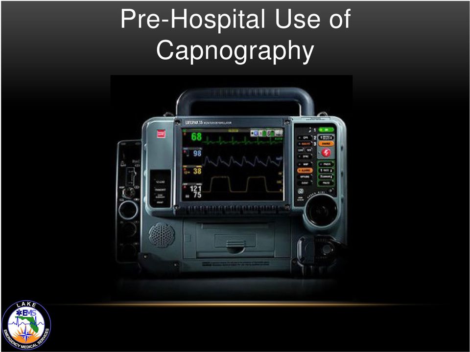
CAPNOGRAPHY IN EMS. Scott Temple Lake EMS February 2012
|
|
|
- Scot Lindsey
- 7 years ago
- Views:
Transcription
1 CAPNOGRAPHY IN EMS Scott Temple Lake EMS February 2012

2 What is the difference Oxygenation vs. Ventilation

3 Oxygenation vs. Ventilation Oxygenation - the ability of the red blood cells to pick up oxygen and move them to the tissues of the body Ventilation - the exchange of air between the lungs and the environment, including inhalation and exhalation

4 Cellular Metabolism Oxygen must first be picked up by red blood cells, then dumped off at the tissue level The body must then take the O 2 and glucose to create energy through cellular metabolism This leaves the waste product (CO 2 ) which needs to be discarded through exhalation
 which needs to be discarded through")
5 PULSE OXIMETRY AND CAPNOGRAPHY Pulse oximetry measures oxygenation and can be provided in a waveform Capnography measures ventilation and can also provide a graphical waveform for interpretation

6 Oxygenation Measured with pulse oximetry (SpO 2 ) Noninvasive measurement Expresses the percentage of oxygen in red blood cells Changes in oxygenation may take minutes to be detected (up to 5 minutes at times) Affected by motion artifact, poor perfusion nail polish, and some dysrhythmias
 Affected by motion artifact, poor perfusion nail polish, and")
7 Pulse Oximetry Waveforms

8 Ventilation Measured by End-Tidal CO 2 Partial pressure (mmhg) or volume (% vol.) of CO 2 in the airway at the end of exhalation Breath-to-breath measurement provides information within seconds Not affected by motion artifact, poor perfusion or dysrhythmias

9 Capnography Waveforms

10 Oxygenation and Ventilation Oxygenation Oxygen for metabolism SpO 2 measures % of O 2 in RBC Reflects change in oxygenation within 5 minutes Ventilation Carbon dioxide from metabolism EtCO 2 measures exhaled CO 2 at the point of exit Reflects change in ventilation within 10 seconds

11 Don t believe me about the time Try this: Turn on the monitor and put an EtCO 2 filter line on and the SpO 2 sensor Now hold your breath You will see that the capnography will show apnea almost immediately The SpO 2 will continue to show a high saturation Because it does not register for several minutes

12 Pre-Hospital Use of Capnography
13 Utilizing Capnography Immediate information via breath-to-breath monitoring Information on the ABCs Airway Breathing Circulation Documentation purposes

14 Capnography Uses - Airway Airway Verification of ET tube placement Continuous monitoring of ET tube position

15 Capnography Uses - Breathing Breathing Hyperventilation Hypoventilation Asthma COPD

16 Capnography Uses - Circulation Circulation Checking the effectiveness of compressions during cardiac arrest First indicator of ROSC Monitors low perfusion states

17 Documentation Purposes As with all documentation, capnography can be utilized as legal evidence pertaining to the care that a patient received during a call The following are examples of the information that capnography provides Baseline capnography assessments Waveform appearance Changes in the waveform with treatment (TRENDING) EtCO 2 values Documenting the actual numbers shown on the monitor how they change during transport
 EtCO 2 values Documenting the actual numbers shown on the monitor how they change")
18 Capnography and Intubated Patients Verifies and documents ET tube placement Immediately detects changes in ET tube position Assesses effectiveness of chest compressions Earliest indication of ROSC Indicator of probability of successful resuscitation Allows crews to adjust manual ventilations in accordance with the patient s needs

19 Research and capnography A 2005 study compared field intubations that used capnography to confirm ETT placement vs. intubations that did not use capnography to confirm placement. The study showed: That 0% of intubations went unrecognized if misplaced when ETCO 2 monitoring was used 23% went unrecognized in the non-etco 2 monitored group That s why we confirm intubations with waveform capnography Silverstir, Annals of Emergency Medicine, May 2005

20 Capnography and Non-Intubated Patients Assists in the assessment of acute respiratory disorders Asthma COPD Among many others Helps in determining a patient s response to our treatment(s) Contributes in the formation of a field diagnosis
 Contributes in the formation of a field")
21 Hypoventilating Non-intubated Patients Capnography can estimate the severity of the condition that leads to hypoventilation Stroke Head injury ETOH intoxication Drug overdose DKA patients Congestive heart failure Analgesia and sedation medications Great for assessing perfusion status of these patients
22 End-tidal CO 2 (EtCO 2 ) Reflects changes in Ventilation - movement of air in and out of the lungs Diffusion - exchange of gases between the air-filled alveoli and the pulmonary circulation Perfusion - circulation of blood
23 What s occurring in the Lungs Ventilation Right Ventricle Artery Alveoli Vein Left Atrium CO2 O 2 O 2 Perfusion Pulmonary Blood Flow
24 Anatomy and Physiology The Right Ventricle of the Heart pumps blood into the Pulmonary Artery Remember the Pulmonary Artery (P.A.) is the only artery that carries deoxygenated blood When in the lungs, the blood will dump CO 2 off where it will be diffused into the alveoli In return the alveoli will push O 2 into the blood, oxygenating the blood The Pulmonary Vein (P.V.), the only vein that carries oxygenated blood, then takes the newly oxygenated blood back into the heart, specifically into the Left Atrium Look at the diagram again P.A P.V. This is occurring in the alveoli
25 How CO 2 gets to the Alveoli Ventilation Right Ventricle Artery Alveoli Vein Left Atrium See how the pulmonary artery dumps CO 2 off CO2 O 2 O 2 Deoxygenated blood Perfusion Oxygenated blood Pulmonary Blood Flow
26 Oxygenated blood Ventilation Right Ventricle Artery Alveoli Vein Left Atrium CO2 O 2 Perfusion O 2 The pulmonary vein then picks up oxygen from the alveoli and carries it back to the heart
27 Ventilation Artery Alveoli Vein Now carbon dioxide (CO 2 ) is exhaled and we capture it via the capnography filter line CO2 O 2 O 2
28 Changes in EtCO 2 Levels
29 Why ETCO 2 levels decrease Decreased cardiac output Cardiac arrest Pulmonary embolism Bronchospasm Decreased muscular activity Hypothermia
30 Why ETCO 2 levels increase Increased cardiac output Good compressions during cardiac arrest Increased muscular activity shivering Effective therapy for bronchospasms
31 Anatomy of a waveform TIME The height shows amount of carbon dioxide exhaled The length shows the time This represents the normal waveform of one respiratory cycle
32 Waveform breakdown Phase 1 Baseline of waveform Initial upstroke of the waveform Represents exhaled gas from upper airways a.k.a. anatomical dead space Exhalation begins many reverse this and believes that it begins inhalation, it does not
33 PHASE 1 Dead Space Ventilation Dead space area is shaded orange Begins exhalation CO 2 is absent Air is coming from the mouth, nose, and pharynx Gas exchange does not occur here
34 Define Dead Space Ventilated areas that do not take part in the exchanging of gases: Airways leading to the alveoli and Ventilated areas in the lungs without blood flow
35 Waveform breakdown Phase 2 Transitions from upper to lower airway ventilation Shows changes in the perfusion status of a patient CO 2 present and increasing in exhaled air
36 CO 2 from the alveoli reaches the upper airway and mixes with the dead space air Why you see a rise in CO 2 CO 2 is now detectable in exhaled air via capnography PHASE 2 Ascending Phase
37 Waveform breakdown Phase 3 Shows alveolar gas exchange Indicates changes in gas distribution Increases in this slope indicates problems with distribution of gas delivery CO 2 exhalation - the wave plateaus
38 CO 2 now makes up the majority of the exhaled air The concentration of the CO 2 remains fairly constant from alveoli to the nose/mouth at this phase Phase 3 Alveolar Plateau
39 PHASE 3 END-TIDAL The end of exhalation contains the highest concentration of CO 2 End-Tidal CO 2 The star marks the end of the wave of exhalation End-tidal
40 Waveform breakdown Phase 4 Beginning of inhalation CO 2 levels drop to baseline (zero) Oxygen is now in the airway Descending Phase Inhalation Inspiratory down stroke returns to zero or baseline 0
41 What is the normal range for a capnography waveform 45 0
42 Answer 45 0 Normal range = 35-45mmHg
43 ABNORMAL VALUES ETCO 2 less than 35 mmhg Hyperventilation / Hypocapnia hyperventilation Hypocapnia low CO 2 ETCO 2 greater than 45 mmhg Hypoventilation / Hypercapnia hypoventilation Hypercapnia high CO 2
44 Question How would your waveform change if you intentionally started to breathe at a rate of 40? Frequency of waveforms Duration of waveforms Height of waveforms
45 Hyperventilation 1) Note how the respirations increase seen in the increased number of waves in the bottom capnogram, the duration of each wave is also shorter 2) Notice how the pink waves are under the 45mmHg line this shows that the ETCO 2 levels have dropped (the patient is blowing CO 2 off) 45 Normal 0 45 Hyperventilation 0
46 Hyperventilation Waveform Indicates CO 2 deficiency Hyperventilation Decreased pulmonary perfusion Hypothermia Decreased metabolism Interventions Adjust ventilation rate Evaluate for adequate sedation Evaluate anxiety Conserve body heat Regular Shape, Plateau Below Normal
47 Question How would your waveform change if you intentionally decreased your respiratory rate to 10? Frequency Duration Height
48 45 Hypoventilation 1) Note how the respirations decrease seen in the decreased number of waves in the bottom capnogram, the duration of each wave is also longer 2) Notice how the green waves are over the 45mmHg line this shows that the ETCO 2 levels have increased (the patient is retaining CO 2 ) Normal 0 Hypoventilation 45 0
49 Hypoventilation Waveform Indicates increase in ETCO 2 Hypoventilation Respiratory depressant drugs Increased metabolism Interventions Adjust ventilation rate Decrease respiratory depressant drug dosages Maintain normal body temperature Regular Shape, Plateau Above Normal
50 Waveform patterns 45 Normal 0 45 Hyperventilation 0 Hypoventilation 45 0
51 What is happening here? 45 0
52 Bronchospasm Waveform Bronchospasm restrict proper ventilation Alveoli unevenly fill during inspiration They also empty unevenly during expiration Uneven air flow during exhalation dilutes exhaled CO 2 This alters the ascending phase and plateau Slower rise in CO 2 concentration Characteristic pattern for bronchospasm Shark Fin shape to waveform
53 Capnography waveforms 45 Normal Hyperventilation Hypoventilation Bronchospasm 0
54 Capnography and Advanced Airways In-line capnography
55 Confirmation of the tube Utilized to confirm that your tube is in place Remember improper placement of the tube will result in the lack of a waveform This tube is confirmed!
56 Med-Legal Capnography provides Documentation of correct placement Ongoing documentation over time through the trending printout Documentation of correct position at ED arrival
57 Dislodgement of tube Tube dislodgment is immediately detected when a waveform drops suddenly Initially this tube was confirmed, then displaced
58 Detect ET Tube Displacement This is only provided through the use of capnography A continuous numerical value of EtCO 2 with apnea alarm after 30 seconds (if you have the alarm set) and A continuous graphic waveform for immediate visual recognition 45 0 Source: Linko K. et. al Capnography for detection of accidental oesophageal intubation. Acta Anesthesiol Scand 27:
59 Can Capnography Assist With CPR?
60 Absolutely! Determination of how effective your chest compressions are Helps to show early detection of ROSC Provides data for the decision to cease resuscitation i.e. the EtCO 2 reading remains under 10mmHg for over 20 minutes of working a code
61 Capnography and CPR Assessment When a patient is in cardiac arrest, remember the EtCO 2 is going to be low the patient is not perfusing enough therefore gas exchange is not occurring appropriately In cardiac arrest we are hoping to keep the EtCO 2 around 10 to 15mmHg. Without a number this high the chances of getting ROSC are very low You can increase the EtCO 2 by providing better compressions (thus providing better perfusion to the alveoli) during CPR How does providing better compressions help to raise EtCO 2 numbers?
62 REMEMBER THIS? Ventilation Right Ventricle Arter y Vein Left Atrium O CO2 2 O2 Perfusion Better compressions simply means we are moving blood better A patient in cardiac arrest is not circulating blood to the lungs and therefore limits the amount of CO 2 being diffused into the alveoli When you provide quality compressions it increases the movement of blood to the lungs where CO 2 can diffuse into the alveoli and O 2 can diffuse into the pulmonary vein If you notice your capnogram wave is low during the next code, just compress harder and faster and watch the boxes increase in height
63 In 1978 A scientist, Kalenda, reported a decrease in ETCO 2 as the person performing CPR fatigued, followed by an increase in ETCO 2 as a new rescuer took over, presumably providing better chest compressions. Gravenstein, Capnography: Clinical Aspects, Cambridge Press, 2004
64 Another study suggests Reductions in ETCO 2 during CPR are associated with comparable reductions in cardiac output...the extent to which resuscitation maneuvers, especially precordial compression, maintain cardiac output may be more readily assessed by measurements of ETCO 2 than palpation of arterial pulses. Yeah it s old but it still applies today! -Max Weil, M.D., Cardiac Output and End-Tidal carbon dioxide, Critical Care Medicine, November 1985
65 New Guidelines With the new American Heart Association Guidelines calling for quality compressions ("push hard, push fast, push deep"), rescuers should switch places every two minutes You should be setting the monitor up so the compressors can view the ETCO 2 readings as well as the EKG waveform generated by their compressions We encourage you to keep the ETCO 2 number up as high as possible during CPR
66 What is this a sign of?
67 What is this a sign of? This capnogram is a sign that your patient has just had a return of spontaneous circulation (ROSC) Note how high the wave is why does it look this way?
68 Retention of CO 2 The wave is large because the patient in cardiac arrest has been retaining CO 2 When ROSC is achieved the patient, whose heart is now working, is able to perfuse better getting rid of the retained CO 2 This exhalation of retained CO 2 is what gives the box such a high appearance Remember the box is a representation of an actual figure. In this case the EtCO 2 number is about 60mmHg
69 ROSC and ETCO 2 Survived to discharge from hospital Average EtCO 2 : 30mmHg Non-survivor Average EtCO 2 : 4-10mmHg
70 INCREASED INTRACRANIAL PRESSURE (ICP) AND CAPNOGRAPHY
71 ICP and EtCO 2 Levels Capnography should also be utilized in patients with ICP issues The following ICP patients are very sensitive to fluctuations in EtCO 2 levels: Stroke patients (possible bleeds) Trauma to the head Neoplasms (brain tumors) Brain infections
72 Question What do you think will occur to cerebral vasculature if the patient has high CO 2 levels?
73 Cerebral Vasodilation High CO 2 levels cause cerebral vasodilatation The Good: It increases cerebral blood flow (CBF) to counter cerebral hypoxia The Bad: Increased CBF increases ICP and may increase brain edema
74 Question What do you think will occur to cerebral vasculature if the patient has low CO 2 levels?
75 Cerebral Vasoconstriction Low CO 2 levels lead to cerebral vasoconstriction The Good: It lowers CO 2 levels that can cause mild cerebral vasoconstriction which may decrease ICP The Bad: Although low CO 2 levels decrease ICP it may cause or increase cerebral hypoxia
76 Pearls Hypoventilation increases CO 2 levels Not blowing off the CO 2 Results in cerebral vasodilation Hyperventilation decreases CO 2 levels Blowing off too much CO 2 Results in cerebral vasoconstriction
77 Treatment Goals Avoid cerebral hypoxia Maintain adequate cerebral blood flow Monitor oxygen saturation with pulse oximetry Utilize the capnography in conjunction with pulse oximetry Your target should be an EtCO 2 of 35mmHg
78 Warning when hyperventilating patients Recent evidence suggests hyperventilation leads to ischemia almost immediately...current models of both ischemic and Traumatic Brain Injury (TBI) suggest an immediate period during which the brain is especially vulnerable to secondary insults. This underscores the importance of avoiding hyperventilation in the pre-hospital environment. --Capnography as a Guide to Ventilation in the Field, D.P. Davis, Gravenstein, Capnography: Clinical Perspectives, Cambridge Press, 2004
79 The Dyspnea Patient
80 Other Patients Needing CO 2 Monitoring COPD Emphysema Chronic Bronchitis Asthma CHF/Pulmonary Edema Pneumonia Possible Pulmonary Embolism Cardiac Ischemia
81 Why you should use capnography for these patients To assess and trend your patients Low-perfusion status Hyperventilation status Hypoventilation status To identify changes in capnography waveforms i.e. bronchospasms in COPD and Asthma patients
82 Capnography and Bronchospasms Produces changes in ascending phase (2) with loss of the sharp upslope A B C 2 D E Alters alveolar plateau (3) producing a shark fin 3
83 Normal vs. Asthma Normal Bronchospasm Source: Krauss B., et al FEV1 in Restrictive Lung Disease Does Not Predict the Shape of the Capnogram. Oral presentation. Annual Meeting, American Thoracic Society, May, Seattle, WA
84 What you should see after treatment Prior to therapy After therapy
85 Role of capnography with COPD In COPD patients the PaCO 2 or the partial pressure of carbon dioxide in the arterial blood INCREASES This is the waveform that you will probably see Notice how the ascending phase and plateau look abnormal This is because there is an uneven emptying of gases Normal patient presentation Abnormal patient presentation Do you see the COPD patient is retaining CO 2?
86 70 year old female Scenario C.C.: Dyspnea, with moderate chest pain History: MI, CABG X 3, on oxygen at 2 l/m Pulse 60, BP 144/70, RR 38 labored and shallow, skin cool and diaphoretic, some pedal edema Initial SpO 2 79%; EtCO 2 20mmHG
87 You. Place the patient on NRBM 15L (100% O 2 ) Administer 1 inch nitro paste During transport you trend her vitals, this is what you see: SpO 2 99% EtCO 2 35mmHg In order to see documented improvement in your patients you must TREND!
88 Pulmonary Embolism and Capnography
89 Ventilation/Perfusion Mismatch (V/Q Mismatch) This is when the amount of ventilation or perfusion is altered In pulmonary embolism patients there is a decrease in perfusion when compared to ventilation This increases deadspace Remember deadspace is the parts of the airway that are unable to take part in gas exchange due to the lack of perfusion
90 INCREASED DEADSPACE P.E. patients therefore show a reduction in exhaled CO 2 on your capnography due to the increased deadspace The green line represents the normal level of CO 2. See how this PE patient is way under it? Low EtCO 2 due to increased deadspace
91 Why use capnography in P.E. We should utilize capnography in these patients because it can assist us in forming a field diagnosis Most of us know what to look for in these patients. Give me some examples of how P.E. patients present
92 Presentation of pulmonary embolism patients Sudden dyspnea Tachycardia RR > 20 Chest pain Hypoxia Anxiety Feeling of doom Hemoptysis Syncope And if you were paying attention A sudden decrease in EtCO 2
93 25 year old female Scenario C.C. shortness of breath and sharp pain in chest History: healthy no issues Meds: Chantix (smoking cessation), Yasmin (birth control) Sound like a possible P.E. candidate? YUP!
94 Patient begins to hyperventilate You place her on capnography and see this Without warning you see a marked decrease in EtCO2 and patient mental status
95 So what caused the drop in EtCO 2 Typically hyperventilation alone does not cause a sudden rapid drop in EtCO 2 Pulmonary Embolisms however DO! Remember a P.E. will increase the deadspace of the lung The increased deadspace will produce a decrease in exhaled CO 2 Now put the P.E. signs and symptoms together with the EtCO 2 information and you have just made an excellent field diagnosis Having the tools are nice, but knowing when to use them is key!
96 Using Capnography for Hypoventilatory States
97 First of all, think of some differential diagnosis that can cause a patient s breathing to be slow
98 Differentials Patients with altered mental status Head injury CVA Drug overdose Heavily sedated ETOH Sepsis/Infection (specifically CNS in nature)
99 Which of these is a patient hypoventilating A 45 0 B 45 0 C 45 0
100 Answer C 45 0 In this case the patient is retaining too much CO 2 Note: the EtCO 2 is above 45mmHg
101 What is this patient doing Hint: he is still hypoventilating This patient is actually breathing very shallow which causes most of the waveforms to be small and uneven. However when the patient lets out a larger exhale you note the height increase of one of the boxes. This shows he was breathing shallow then exhaled a high amount of retained CO 2
102 WHAT IS OCCURRING HERE? You are called to a nursing home. As you approach your patient you note that the patient is on a O 2 mask and lying semi fowlers (his chin is nearly touching his chest) You place him on capnography and this is what you see Tell me what is going on based on this waveform? Observe this one carefully, something is not right
103 DID YOU SEE IT? Note that the baseline is elevated This is a situation that many patients experience and we often don t recognize Patient Rebreathing 45 0 The baseline never comes back to zero because of the poor positioning of the patient. Also because he is wearing a mask that allows him to rebreath his own CO 2 He is not clearing the dead space that would allow a return to 0
104 DKA PATIENTS
105 What is occurring in DKA Diabetic Ketoacidosis (DKA) The issue with a Insulin Dependent Diabetic Mellitus (IDDM) and some Non-Insulin Dependent Diabetic Mellitus (NIDDM) patients is that they do not have insulin available to breakdown glucose (a.k.a. sugar) for energy creation Instead the IDDM (type 1) diabetic and in some case NIDDM (type 2) patients will sometimes have no choice but to utilize fat stores for energy production (termed Gluconeogenesis)
106 Fat for energy When fat stores are used for energy, acids called Ketones begin to build up in the blood and urine Ketones become toxic in high levels this is what we call ketoacidosis In many cases people find out that the have diabetes because they initially had a DKA experience These are the patients with very high blood sugar levels and whose breath smells sweet or some say like acetone
107 Buffering Eventually the body turns to the bicarbonate buffering system in order to balance the acid build up bicarbonate is a base that opposes acid, decreasing acid levels The problem is this system is quickly overwhelmed and other mechanisms must work to compensate for the acidosis So what does the body do? It triggers hyperventilation to lower the blood CO 2 levels
108 So how do DKA patients breathe? Remember what is occurring in these patients Ketones are building up (causing acidosis) This means that there is a high level of CO 2 in the blood Therefore what type of waveform will you see?
109 DKA and Capnography When patients are in (DKA) the body attempts to correct acidosis High levels of CO 2 in the body creates an acidotic environment 45 0 In this waveform you are seeing a patient whose body is attempting to correct the acidosis Note how the EtCO 2 is above 45mmHg showing an acidotic state This is why a patient breathes fast in DKA initially, they are trying to blow off the CO 2 and therefore lessen the acidosis that may kill them Be sure to utilize end-tidal with suspected DKA patients
110 However! Just like in the case of fast heart rates, a patient breathing this fast cannot keep it up, so what occurs? The patient begins a pattern of labored and deep breathing Remember in metabolic acidosis (in this case DKA) breathing is first rapid and shallow BUT As acidosis worsens breathing becomes deep, labored and gasping (Kussmaul respirations)
111 Shock Patients
112 Perfusing warning! Capnography can provide an early warning sign of shock A patient with a sudden drop in cardiac output will show a drop in ETCO 2 numbers that may be regardless of any change in breathing This has implications for trauma patients, cardiac patients any patient at risk for shock
113 Another Pig Study 5 pigs had hemorrhagic shock induced by bleeding, 5 pigs had septic shock induced by infusion of e-coli, and 6 pigs had cardiogenic shock induced by repeated episodes of v-fib. The pigs' cardiac output was continuously measured as well as their PETCO 2.
114 Results A patient with low cardiac output caused by cardiogenic shock or hypovolemia resulting from hemorrhage won t carry as much CO 2 per minute back to the lungs to be exhaled Therefore this patient s ETCO 2 will be reduced This doesn t necessarily mean the patient is hyperventilating or that their arterial CO 2 level will be reduced. Reduced perfusion to the lungs alone causes this phenomenon. The patient s lung function may be perfectly normal Baruch Krauss, M.D, JEMS, November 2003
115 When to use capnography Capnography is not cheap yet the benefits for the medic and the patient outweigh the cost However we still want to be selective when utilizing the filter lines
116 Utilize capnography for these patients STEMIs Cardiac arrest Respiratory patients Patients on sedation and analgesic meds Altered mental states CNS infections Sepsis Drug/ETOH intoxication Hyperglycemia DKA Patients with potential increased ICP issues
117 Capnography is just another tool in your arsenal of equipment that can be utilized to further confirm your field diagnosis. Remember that the your field diagnosis assists to lead you in your treatments. As stated previously the placement of EtCO 2 filter lines are not intended for every patient. Instead it should be used only for those patients who can benefit from it s information. If utilized please trend your findings after every treatment and document accordingly. Thank you for you attention!
118 Post-Test Please complete the post test and return to administration via interoffice mail or fax to: attention Scott Temple This program is worth 3 CEUs in the Airway, Medical, Operations, or Electives categories Please specify on your post test where you would like the CEUs to be inserted.
119 References D.P. Davis, Gravenstein Capnography as a Guide to Ventilation in the Field,, Capnography: Clinical Perspectives, Cambridge Press, 2004 Krauss B., et al FEV1 in Restrictive Lung Disease Does Not Predict the Shape of the Capnogram. Oral presentation. Annual Meeting, American Thoracic Society, May, Seattle, WA Max Weil, M.D., Cardiac Output and End-Tidal carbon dioxide, Critical Care Linko K. et. al. Capnography for detection of accidental oesophageal intubation. Acta Anesthesiol Scand 27: Medicine Silverstir, Annals of Emergency Medicine, May 2005
10 Things Every Paramedic Should Know About Capnography
 10 Things Every Paramedic Should Know About Capnography Capnography is the vital sign of ventilation. By tracking the carbon dioxide in a patient s exhaled breath, capnography enables paramedics to objectively
Capnography. McHenry Western Lake County EMS
 Capnography McHenry Western Lake County EMS What is Capnography? Capnography is an objective measurement of exhaled CO2 levels. Capnography measures ventilation. It can be used to: Assist in confirmation
Ventilation Perfusion Relationships
 Ventilation Perfusion Relationships VENTILATION PERFUSION RATIO Ideally, each alveolus in the lungs would receive the same amount of ventilation and pulmonary capillary blood flow (perfusion). In reality,
Oxygen Therapy. Oxygen therapy quick guide V3 July 2012.
 PRESENTATION Oxygen (O 2 ) is a gas provided in a compressed form in a cylinder. It is also available in a liquid form. It is fed via a regulator and flow meter to the patient by means of plastic tubing
Oxygen - update April 2009 OXG
 PRESENTATION Oxygen (O 2 ) is a gas provided in compressed form in a cylinder. It is also available in liquid form, in a system adapted for ambulance use. It is fed via a regulator and flow meter to the
Oxygenation. Chapter 21. Anatomy and Physiology of Breathing. Anatomy and Physiology of Breathing*
 Oxygenation Chapter 21 Anatomy and Physiology of Breathing Inspiration ~ breathing in Expiration ~ breathing out Ventilation ~ Movement of air in & out of the lungs Respiration ~ exchange of O2 & carbon
Understanding Hypoventilation and Its Treatment by Susan Agrawal
 www.complexchild.com Understanding Hypoventilation and Its Treatment by Susan Agrawal Most of us have a general understanding of what the term hyperventilation means, since hyperventilation, also called
Homeostasis. The body must maintain a delicate balance of acids and bases.
 Homeostasis The body must maintain a delicate balance of acids and bases. Metabolic and respiratory processes must work together to keep hydrogen ion (H+) levels normal and stable. ph of Blood The ph of
CHAPTER 1: THE LUNGS AND RESPIRATORY SYSTEM
 CHAPTER 1: THE LUNGS AND RESPIRATORY SYSTEM INTRODUCTION Lung cancer affects a life-sustaining system of the body, the respiratory system. The respiratory system is responsible for one of the essential
Question-and-Answer Document 2010 AHA Guidelines for CPR & ECC As of October 18, 2010
 Question-and-Answer Document 2010 AHA Guidelines for CPR & ECC As of October 18, 2010 Q: What are the most significant changes in the 2010 AHA Guidelines for CPR & ECC? A: Major changes for all rescuers,
Diabetic Emergencies. David Hill, D.O.
 Diabetic Emergencies David Hill, D.O. Class Outline Diabetic emergency/glucometer training Identify the different signs of insulin shock Diabetic coma, and HHNK Participants will understand the treatment
Anatomy and Physiology: Understanding the Importance of CPR
 Anatomy and Physiology: Understanding the Importance of CPR Overview This document gives you more information about the body s structure (anatomy) and function (physiology). This information will help
Oxygenation and Oxygen Therapy Michael Billow, D.O.
 Oxygenation and Oxygen Therapy Michael Billow, D.O. The delivery of oxygen to all body tissues is the essence of critical care. Patients in respiratory distress/failure come easily to mind as the ones
Diabetic Ketoacidosis: When Sugar Isn t Sweet!!!
 Diabetic Ketoacidosis: When Sugar Isn t Sweet!!! W Ricks Hanna Jr MD Assistant Professor of Pediatrics University of Tennessee Health Science Center LeBonheur Children s Hospital Introduction Diabetes
ACLS Provider Manual Comparison Sheet Based on 2010 AHA Guidelines for CPR and ECC. BLS Changes
 ACLS Provider Manual Comparison Sheet Based on 2010 AHA Guidelines for CPR and ECC CPR Chest compressions, Airway, Breathing (C-A-B) BLS Changes New Old Rationale New science indicates the following order:
Michigan Adult Cardiac Protocols CARDIAC ARREST GENERAL. Date: May 31, 2012 Page 1 of 5
 Date: May 31, 2012 Page 1 of 5 Cardiac Arrest General This protocol should be followed for all adult cardiac arrests. Medical cardiac arrest patients undergoing attempted resuscitation should not be transported
Medical Direction and Practices Board WHITE PAPER
 Medical Direction and Practices Board WHITE PAPER Use of Pressors in Pre-Hospital Medicine: Proper Indication and State of the Science Regarding Proper Choice of Pressor BACKGROUND Shock is caused by a
ETCO2 Monitoring: Riding the Wave! Disclosure 4/11/2013
 ETCO2 Monitoring: Riding the Wave! Debbie Fox, MBA, RRT-NPS, FAARC Director, Respiratory Care Wesley Medical Center Disclosure I have no financial conflicts to disclose. I have participated in focus groups
Tests. Pulmonary Functions
 Pulmonary Functions Tests Static lung functions volumes Dynamic lung functions volume and velocity Dynamic Tests Velocity dependent on Airway resistance Resistance of lung tissue to change in shape Dynamic
MECHINICAL VENTILATION S. Kache, MD
 MECHINICAL VENTILATION S. Kache, MD Spontaneous respiration vs. Mechanical ventilation Natural spontaneous ventilation occurs when the respiratory muscles, diaphragm and intercostal muscles pull on the
Pulmonary Diseases. Lung Disease: Pathophysiology, Medical and Exercise Programming. Overview of Pathophysiology
 Lung Disease: Pathophysiology, Medical and Exercise Programming Overview of Pathophysiology Ventilatory Impairments Increased airway resistance Reduced compliance Increased work of breathing Ventilatory
Chapter 26. Assisting With Oxygen Needs. Elsevier items and derived items 2014, 2010 by Mosby, an imprint of Elsevier Inc. All rights reserved.
 Chapter 26 Assisting With Oxygen Needs Oxygen (O 2 ) is a gas. Oxygen It has no taste, odor, or color. It is a basic need required for life. Death occurs within minutes if breathing stops. Brain damage
PULMONARY PHYSIOLOGY
 I. Lung volumes PULMONARY PHYSIOLOGY American College of Surgeons SCC Review Course Christopher P. Michetti, MD, FACS and Forrest O. Moore, MD, FACS A. Tidal volume (TV) is the volume of air entering and
The Sepsis Puzzle: Identification, Monitoring and Early Goal Directed Therapy
 The Sepsis Puzzle: Identification, Monitoring and Early Goal Directed Therapy Cindy Goodrich RN, MS, CCRN Content Description Sepsis is caused by widespread tissue injury and systemic inflammation resulting
Introduction to Cardiopulmonary Exercise Testing
 Introduction to Cardiopulmonary Exercise Testing 2 nd Edition Andrew M. Luks, MD Robb Glenny, MD H. Thomas Robertson, MD Division of Pulmonary and Critical Care Medicine University of Washington Section
Emphysema. Introduction Emphysema is a type of chronic obstructive pulmonary disease, or COPD. COPD affects about 64 million people worldwide.
 Emphysema Introduction Emphysema is a type of chronic obstructive pulmonary disease, or COPD. COPD affects about 64 million people worldwide. Emphysema involves damage to the air sacs in the lungs. This
AMERICAN HEART ASSOCIATION 2010 ACLS GUIDELINES: WHAT EVERY CLINICIAN NEEDS TO KNOW
 AMERICAN HEART ASSOCIATION 2010 ACLS GUIDELINES: WHAT EVERY CLINICIAN NEEDS TO KNOW Joseph Heidenreich, MD Texas A&M Health Science Center Scott & White Memorial Hospital Scott & White Memorial Hospital
Objectives COPD. Chronic Obstructive Pulmonary Disease (COPD) 4/19/2011
 4/19/2011") Objectives Discuss assessment findings and treatment for: Chronic Obstructive Pulmonary Disease Bronchitis Emphysema Asthma Anaphylaxis Other respiratory issues Provide some definitions Chronic Obstructive
Objectives Discuss assessment findings and treatment for: Chronic Obstructive Pulmonary Disease Bronchitis Emphysema Asthma Anaphylaxis Other respiratory issues Provide some definitions Chronic Obstructive
Common Ventilator Management Issues
 Common Ventilator Management Issues William Janssen, M.D. Assistant Professor of Medicine National Jewish Health University of Colorado Denver Health Sciences Center You have just admitted a 28 year-old
Pathophysiology of hypercapnic and hypoxic respiratory failure and V/Q relationships. Dr.Alok Nath Department of Pulmonary Medicine PGIMER Chandigarh
 Pathophysiology of hypercapnic and hypoxic respiratory failure and V/Q relationships Dr.Alok Nath Department of Pulmonary Medicine PGIMER Chandigarh Jan 2006 Respiratory Failure inadequate blood oxygenation
Diabetic Ketoacidosis
 Princess Margaret Hospital for Children PAEDIATRIC ACUTE CARE GUIDELINE Diabetic Ketoacidosis Scope (Staff): Scope (Area): All Emergency Department Clinicians Emergency Department This document should
Acid-Base Balance and the Anion Gap
 Acid-Base Balance and the Anion Gap 1. The body strives for electrical neutrality. a. Cations = Anions b. One of the cations is very special, H +, and its concentration is monitored and regulated very
Southern Stone County Fire Protection District Emergency Medical Protocols
 TITLE Pediatric Medical Assessment PM 2.4 Confirm scene safety Appropriate body substance isolation procedures Number of patients Nature of illness Evaluate the need for assistance B.L.S ABC s & LOC Focused
Vtial sign #1: PULSE. Vital Signs: Assessment and Interpretation. Factors that influence pulse rate: Importance of Vital Signs
 Vital Signs: Assessment and Interpretation Elma I. LeDoux, MD, FACP, FACC Associate Professor of Medicine Vtial sign #1: PULSE Reflects heart rate (resting 60-90/min) Should be strong and regular Use 2
Respiratory failure and Oxygen Therapy
 Respiratory failure and Oxygen Therapy A patient with Hb 15 G % will carry 3X more O2 in his blood than someone with Hb 5G % Give Controlled O2 treatment in acute pulmonary oedema to avoid CO2 retention
Update on Small Animal Cardiopulmonary Resuscitation (CPR)- is anything new?
- is anything new?") Update on Small Animal Cardiopulmonary Resuscitation (CPR)- is anything new? DVM, DACVA Objective: Update on the new Small animal guidelines for CPR and a discussion of the 2012 Reassessment Campaign on
Update on Small Animal Cardiopulmonary Resuscitation (CPR)- is anything new? DVM, DACVA Objective: Update on the new Small animal guidelines for CPR and a discussion of the 2012 Reassessment Campaign on
It is recommended that the reader review each medical directive presented in this presentation along with the actual PCP Core medical directive.
 It is recommended that the reader review each medical directive presented in this presentation along with the actual PCP Core medical directive. This presentation will highlight the changes and any new
The American Heart Association released new resuscitation science and treatment guidelines on October 19, 2010.
 ACLS Study Guide The American Heart Association released new resuscitation science and treatment guidelines on October 19, 2010. Please read the below information carefully This letter is to confirm your
Arterial Blood Gas Case Questions and Answers
 Arterial Blood Gas Case Questions and Answers In the space that follows you will find a series of cases that include arterial blood gases. Each case is then followed by an explanation of the acid-base
Emergency Medical Technician - Basic
 Washington State Specific Objectives for Emergency Medical Technician - Basic OFFICE OF EMERGENCY MEDICAL AND TRAUMA PREVENTION September 1996 Emergency Medical Technician - Basic Definition: Emergency
American Heart Association
 American Heart Association Basic Life Support for Healthcare Providers Pretest April 2006 This examination to be used only as a PRECOURSE TEST For BLS for Healthcare Providers Courses 2006 American Heart
Gas Exchange. Graphics are used with permission of: Pearson Education Inc., publishing as Benjamin Cummings (http://www.aw-bc.com)
") Gas Exchange Graphics are used with permission of: Pearson Education Inc., publishing as Benjamin Cummings (http://www.aw-bc.com) Page 1. Introduction Oxygen and carbon dioxide diffuse between the alveoli
Gas Exchange Graphics are used with permission of: Pearson Education Inc., publishing as Benjamin Cummings (http://www.aw-bc.com) Page 1. Introduction Oxygen and carbon dioxide diffuse between the alveoli
First Responder (FR) and Emergency Medical Responder (EMR) Progress Log
 and Emergency Medical Responder (EMR) Progress Log") First Responder (FR) and Emergency Medical Responder (EMR) Progress Log Note: Those competencies that are for EMR only are denoted by boldface type. For further details on the National Occupational Competencies
First Responder (FR) and Emergency Medical Responder (EMR) Progress Log Note: Those competencies that are for EMR only are denoted by boldface type. For further details on the National Occupational Competencies
2.06 Understand the functions and disorders of the respiratory system
 2.06 Understand the functions and disorders of the respiratory system 2.06 Understand the functions and disorders of the respiratory system Essential questions What are the functions of the respiratory
Emergency Scenario. Chest Pain
 Emergency Scenario Chest Pain This emergency scenario reviews chest pain in a primary care patient, and is set up for roleplay and case review with your staff. 1) The person facilitating scenarios can
6 Easy Steps to ABG Analysis
 6 Easy Steps to ABG Analysis E-Booklet David W. Woodruff, MSN, RN- BC, CNS, CMSRN, CEN 571 Ledge Road, Macedonia, OH 44056 Telephone (800) 990-2629 Fax (800) 990-2585 1997-2012 Ed4Nurses, Inc. All rights
Pediatric Airway Management
 Pediatric Airway Management Dec 2003 Dr. Shapiro I., PICU Adult Chain of Survival EMS CPR ALS Early Defibrillation Pediatric Chain of Survival Prevention CPR EMS ALS Out-of-Hospital Cardiac Arrest SIDS
ACLS Study Guide BLS Overview CAB
 ACLS Study Guide The ACLS Provider exam is 50-mutiple choice questions. Passing score is 84%. Student may miss 8 questions. For students taking ACLS for the first time or renewing students with a current
VA SAN DIEGO HEALTHCARE SYSTEM MEMORANDUM 118-28 SAN DIEGO, CA
 GUIDELINES FOR PATIENT-CONTROLLED ANALGESIA (PCA) AND PATIENT- CONTROLLED EPIDURAL ANALGESIA (PCEA) FOR ACUTE PAIN MANAGEMENT 1. PURPOSE: To assure the safe and effective use of patient controlled analgesia
Gas Exchange Graphics are used with permission of: adam.com (http://www.adam.com/) Benjamin Cummings Publishing Co (http://www.awl.
 Benjamin Cummings Publishing Co (http://www.awl.") Gas Exchange Graphics are used with permission of: adam.com (http://www.adam.com/) Benjamin Cummings Publishing Co (http://www.awl.com/bc) Page 1. Introduction Oxygen and carbon dioxide diffuse between
Gas Exchange Graphics are used with permission of: adam.com (http://www.adam.com/) Benjamin Cummings Publishing Co (http://www.awl.com/bc) Page 1. Introduction Oxygen and carbon dioxide diffuse between
Target groups: Paramedics, nurses, respiratory therapists, physicians, and others who manage respiratory emergencies.
 Overview Estimated scenario time: 10 15 minutes Estimated debriefing time: 10 minutes Target groups: Paramedics, nurses, respiratory therapists, physicians, and others who manage respiratory emergencies.
Documenting & Coding. Chronic Obstructive Pulmonary Disease (COPD) Presented by: David S. Brigner, MLA, CPC
 Presented by: David S. Brigner, MLA, CPC") Documenting & Coding Chronic Obstructive Pulmonary Disease (COPD) Presented by: David S. Brigner, MLA, CPC Sr. Provider Training & Development Consultant Professional Profile David Brigner currently performs
Documenting & Coding Chronic Obstructive Pulmonary Disease (COPD) Presented by: David S. Brigner, MLA, CPC Sr. Provider Training & Development Consultant Professional Profile David Brigner currently performs
Acid/Base Homeostasis (Part 4)
") Acid/Base Homeostasis (Part 4) Graphics are used with permission of: Pearson Education Inc., publishing as Benjamin Cummings (http://www.aw-bc.com) 5. The newly formed bicarbonate moves into the plasma.
Acid/Base Homeostasis (Part 4) Graphics are used with permission of: Pearson Education Inc., publishing as Benjamin Cummings (http://www.aw-bc.com) 5. The newly formed bicarbonate moves into the plasma.
Adult, Child, and Infant Written Exam CPR Pro for the Professional Rescuer
 Adult, Child, and Infant Written Exam CPR Pro for the Professional Rescuer Instructions: Read each of the following questions carefully and then place an X over the correct answer on the separate answer
GRADE 11F: Biology 3. UNIT 11FB.3 9 hours. Human gas exchange system and health. Resources. About this unit. Previous learning.
 GRADE 11F: Biology 3 Human gas exchange system and health UNIT 11FB.3 9 hours About this unit This unit is the third of six units on biology for Grade 11 foundation. The unit is designed to guide your
PRO-CPR. 2015 Guidelines: PALS Algorithm Overview. (Non-AHA supplementary precourse material)
") PRO-CPR 2015 Guidelines: PALS Algorithm Overview (Non-AHA supplementary precourse material) Please reference Circulation (from our website), the ECC Handbook, or the 2015 ACLS Course Manual for correct
PRO-CPR 2015 Guidelines: PALS Algorithm Overview (Non-AHA supplementary precourse material) Please reference Circulation (from our website), the ECC Handbook, or the 2015 ACLS Course Manual for correct
ACID- BASE and ELECTROLYTE BALANCE. MGHS School of EMT-Paramedic Program 2011
 ACID- BASE and ELECTROLYTE BALANCE MGHS School of EMT-Paramedic Program 2011 ACID- BASE BALANCE Ions balance themselves like a see-saw. Solutions turn into acids when concentration of hydrogen ions rises
CH CONSCIOUS SEDATION
 Summary: CH CONSCIOUS SEDATION It is the policy of Carondelet Health that moderate conscious sedation of patients will be undertaken with appropriate evaluation and monitoring. Effective Date: 9/4/04 Revision
Ischemia and Infarction
 Harvard-MIT Division of Health Sciences and Technology HST.035: Principle and Practice of Human Pathology Dr. Badizadegan Ischemia and Infarction HST.035 Spring 2003 In the US: ~50% of deaths are due to
National Registry of EMTs Continued Competency Program. (NREMT Recertification Requirements) BETA Version 2
 BETA Version 2") National Registry of EMTs Continued Competency Program (NREMT Recertification Requirements) BETA Version 2 Massachusetts providers Issue date: 5/1/2013 The Four Principles of Continued Competency Professional
National Registry of EMTs Continued Competency Program (NREMT Recertification Requirements) BETA Version 2 Massachusetts providers Issue date: 5/1/2013 The Four Principles of Continued Competency Professional
Airway Pressure Release Ventilation
 Page: 1 Policy #: 25.01.153 Issued: 4-1-2006 Reviewed/ Revised: Section: 10-11-2006 Respiratory Care Airway Pressure Release Ventilation Description/Definition Airway Pressure Release Ventilation (APRV)
Cardiac Arrest VF/Pulseless VT Learning Station Checklist
 Cardiac Arrest VF/Pulseless VT Learning Station Checklist VF/VT 00 American Heart Association Adult Cardiac Arrest Shout for Help/Activate Emergency Response Epinephrine every - min Amiodarone Start CPR
American Heart Association. BLS Instructor Course. Written Examination. July 2003
 American Heart Association BLS Instructor Course Written Examination July 2003 Basic Life Support Instructor Exam Part I Please do not mark on this examination. Record the best answer on the separate answer
Objectives. Burn Assessment and Management. Questions Regarding the Case Study. Case Study. Patient Assessment. Patient Assessment
 Objectives Burn Assessment and Management Discuss the mechanisms and complications of a thermal burn, electrical burn and an inhalation burn Explain the factors to consider when determining the severity
404 Section 5 Shock and Resuscitation. Scene Size-up. Primary Assessment. History Taking
 404 Section 5 and Resuscitation Scene Size-up Scene Safety Mechanism of Injury (MOI)/ Nature of Illness (NOI) Ensure scene safety and address hazards. Standard precautions should include a minimum of gloves
IMPAIRED BLOOD-GAS EXCHANGE. Intraoperative blood gas analysis
 IMPAIRED BLOOD-GAS EXCHANGE Intraoperative blood gas analysis When do you perform BGA Intraoperatively? Informe actual NEVER Routine:Thoracic Thoracic, Cardiac,Neurosurgery Emergency situation Drop in
CBT/OTEP 450 Diabetic Emergencies
 Seattle-King County EMS Seattle-King County Emergency Medical Services Division Public Health - Seattle/King County 401 5th Avenue, Suite 1200 Seattle, WA 98104 (206) 296-4693 January 2009 CBT/OTEP 450
Sepsis: Identification and Treatment
 Sepsis: Identification and Treatment Daniel Z. Uslan, MD Associate Clinical Professor Division of Infectious Diseases Medical Director, UCLA Sepsis Task Force Severe Sepsis: A Significant Healthcare Challenge
3100B Clinical Training Program. 3100B HFOV VIASYS Healthcare
 3100B Clinical Training Program 3100B HFOV VIASYS Healthcare HFOV at Alveolar Level Nieman,, G, SUNY 1999 Who DO We Treat? Only Pathology studied to date has been ARDS Questions about management of adults
What, roughly, is the dividing line between the upper and lower respiratory tract? The larynx. What s the difference between the conducting zone and
 What, roughly, is the dividing line between the upper and lower respiratory tract? The larynx. What s the difference between the conducting zone and the respiratory zone? Conducting zone is passageways
From AARC Protocol Committee; Subcommittee Adult Critical Care Version 1.0a (Sept., 2003), Subcommittee Chair, Susan P. Pilbeam
, Subcommittee Chair, Susan P. Pilbeam") AARC - ADULT MECHANICAL VENTILATOR PROTOCOLS 1. Guidelines for Using Ventilator Protocols 2. Definition of Modes and Suggestions for Use of Modes 3. Adult Respiratory Ventilator Protocol - Guidelines for
AARC - ADULT MECHANICAL VENTILATOR PROTOCOLS 1. Guidelines for Using Ventilator Protocols 2. Definition of Modes and Suggestions for Use of Modes 3. Adult Respiratory Ventilator Protocol - Guidelines for
Official Online ACLS Exam
 \ Official Online ACLS Exam Please fill out this form before you take the exam. Name : Email : Phone : 1. Hypovolemia initially produces which arrhythmia? A. PEA B. Sinus tachycardia C. Symptomatic bradyarrhythmia
Disability Evaluation Under Social Security
 Disability Evaluation Under Social Security Revised Medical Criteria for Evaluating Endocrine Disorders Effective June 7, 2011 Why a Revision? Social Security revisions reflect: SSA s adjudicative experience.
PARAMEDIC TRAINING CLINICAL OBJECTIVES
 Page 1 of 21 GENERAL PATIENT UNIT When assigned to the General Patient unit paramedic student should gain knowledge and experience in the following: 1. Appropriate communication with patients and members
NICE Pathways bring together all NICE guidance, quality standards and other NICE information on a specific topic.
 Diabetic ketoacidosis in children and young people bring together all NICE guidance, quality standards and other NICE information on a specific topic. are interactive and designed to be used online. They
James F. Kravec, M.D., F.A.C.P
 James F. Kravec, M.D., F.A.C.P Chairman, Department of Internal Medicine, St. Elizabeth Health Center Chair, General Internal Medicine, Northeast Ohio Medical University Associate Medical Director, Hospice
Laerdal Patient Monitor Help Page 1 June 14, 2012, Rev E
 Laerdal Patient Monitor Help Page 1 Using the Laerdal Patient Monitor The Laerdal Patient Monitor software is used to simulate a typical Patient Monitor found in hospitals and ambulances. It is made available
American Heart Association. Basic Life Support for Healthcare Providers
 American Heart Association Basic Life Support for Healthcare Providers Pretest February 2001 This examination to be used only as a PRECOURSE TEST for BLS for Healthcare Providers Courses 2001 American
STAGES OF SHOCK. IRREVERSIBLE SHOCK Heart deteriorates until it can no longer pump and death occurs.
 STAGES OF SHOCK SHOCK : A profound disturbance of circulation and metabolism, which leads to inadequate perfusion of all organs which are needed to maintain life. COMPENSATED NONPROGRESSIVE SHOCK 30 sec
Speaking ICD-10-CM. The New Coding Language. COPD documented with a more specific respiratory condition falls under one code category: J44.0-J44.
 Speaking : Chronic Obstructive Pulmonary Disease (COPD) COPD documented with a more specific respiratory condition falls under multiple code categories: 491.20-491.22 Obstructive chronic bronchitis 493.20-493.22
Early Warning Scores (EWS) Clinical Sessions 2011 By Bhavin Doshi
 Clinical Sessions 2011 By Bhavin Doshi") Early Warning Scores (EWS) Clinical Sessions 2011 By Bhavin Doshi What is EWS? After qualifying, junior doctors are expected to distinguish between the moderately sick patients who can be managed in the
Early Warning Scores (EWS) Clinical Sessions 2011 By Bhavin Doshi What is EWS? After qualifying, junior doctors are expected to distinguish between the moderately sick patients who can be managed in the
Department of Surgery
 What is emphysema? 2004 Regents of the University of Michigan Emphysema is a chronic disease of the lungs characterized by thinning and overexpansion of the lung-like blisters (bullae) in the lung tissue.
2161-1 - Page 1. Name: 1) Choose the disease that is most closely related to the given phrase. Questions 10 and 11 refer to the following:
 Choose the disease that is most closely related to the given phrase. Questions 10 and 11 refer to the following:") Name: 2161-1 - Page 1 1) Choose the disease that is most closely related to the given phrase. a disease of the bone marrow characterized by uncontrolled production of white blood cells A) meningitis B)
Name: 2161-1 - Page 1 1) Choose the disease that is most closely related to the given phrase. a disease of the bone marrow characterized by uncontrolled production of white blood cells A) meningitis B)
Intermedix Inc. EMR 2006 Data Element Name. Compliant. Data Number. Elements
 D01_01 EMS Agency X D01_02 EMS Agency D01_03 EMS Agency State X D01_04 EMS Agency County X D01_05 Primary Type of Service D01_06 Other Types of Service D01_07 Level of Service X D01_08 Organizational Type
ADMINISTERING EMERGENCY OXYGEN
 FACT SHEET ADMINISTERING EMERGENCY OXYGEN Emergency oxygen can be given for many breathing and cardiac emergencies. It can help improve hypoxia (insufficient oxygen reaching the cells) and reduce pain
Breathing and Holding Your Breath copyright, 2005, Dr. Ingrid Waldron and Jennifer Doherty, Department of Biology, University of Pennsylvania 1
 Breathing and Holding Your Breath copyright, 2005, Dr. Ingrid Waldron and Jennifer Doherty, Department of Biology, University of Pennsylvania 1 Introduction Everybody breathes all day, every day. Why?
The EMT Instructional Guidelines in this section include all the topics and material at the EMR level PLUS the following material:
 Airway Management, Respiration and Artificial Ventilation EMR Applies knowledge (fundamental depth, foundational breadth) of general anatomy and physiology to assure a patent airway, adequate mechanical
Influenza (Flu) Influenza is a viral infection that may affect both the upper and lower respiratory tracts. There are three types of flu virus:
 Influenza is a viral infection that may affect both the upper and lower respiratory tracts. There are three types of flu virus:") Respiratory Disorders Bio 375 Pathophysiology General Manifestations of Respiratory Disease Sneezing is a reflex response to irritation in the upper respiratory tract and is associated with inflammation
Respiratory Disorders Bio 375 Pathophysiology General Manifestations of Respiratory Disease Sneezing is a reflex response to irritation in the upper respiratory tract and is associated with inflammation
Your Lungs and COPD. Patient Education Pulmonary Rehabilitation. A guide to how your lungs work and how COPD affects your lungs
 Patient Education Your Lungs and COPD A guide to how your lungs work and how COPD affects your lungs Your lungs are organs that process every breath you take. They provide oxygen (O 2 ) to the blood and
Chapter 16. Learning Objectives. Learning Objectives 9/11/2012. Shock. Explain difference between compensated and uncompensated shock
 Chapter 16 Shock Learning Objectives Explain difference between compensated and uncompensated shock Differentiate among 5 causes and types of shock: Hypovolemic Cardiogenic Neurogenic Septic Anaphylactic
Your Go-to COPD Guide
 Your Go-to COPD Guide Learning how to live with chronic obstructive pulmonary disease (COPD) Inside, you ll learn: COPD facts COPD symptoms and triggers How to talk with your doctor Different treatment
National Registry of Emergency Medical Technicians Emergency Medical Responder Psychomotor Examination PATIENT ASSESSMENT/MANAGEMENT TRAUMA
 PATIENT ASSESSMENT/MANAGEMENT TRAUMA Scenario # Note: Areas denoted by ** may be integrated within sequence of Primary Survey/Resuscitation SCENE SIZE-UP Determines the mechanism of injury/nature of illness
PULMONARY FUNCTION TESTS A Workshop on Simple Spirometry & Flow Volume Loops
 PULMONARY FUNCTION TESTS A Workshop on Simple Spirometry & Flow Volume Loops YOU SHOULD READ THE FOLLOWING MATERIAL BEFORE Tuesday March 30 Interpretation of PFTs Learning Objectives 1. Specify the indications
1.4.4 Oxyhemoglobin desaturation
 Critical Care Therapy and Respiratory Care Section Category: Clinical Section: Clinical Monitoring Title: Monitoring of Patients Undergoing Conscious Sedation Policy #: 09 Revised: 05/00 1.0 DESCRIPTION
National Registry of Emergency Medical Technicians Emergency Medical Technician Psychomotor Examination BLEEDING CONTROL/SHOCK MANAGEMENT
 BLEEDING CONTROL/SHOCK MANAGEMENT Candidate: Examiner: Date: Signature: Possible Applies direct pressure to the wound 1 NOTE: The examiner must now inform the candidate that the wound continues to bleed.
How To Treat A Heart Attack
 13 Resuscitation and preparation for anaesthesia and surgery Key Points 13.1 MANAGEMENT OF EMERGENCIES AND CARDIOPULMONARY RESUSCITATION ESSENTIAL HEALTH TECHNOLOGIES The emergency measures that are familiar
Section Two: Arterial Pressure Monitoring
 Section Two: Arterial Pressure Monitoring Indications An arterial line is indicated for blood pressure monitoring for the patient with any medical or surgical condition that compromises cardiac output,
The science of medicine. The compassion to heal.
 A PATIENT S GUIDE TO ELECTROPHYSIOLOGY STUDIES OF THE HEART The science of medicine. The compassion to heal. This teaching booklet is designed to introduce you to electrophysiology studies of the heart.
What Medical Emergencies Should a Dental Office be Prepared to Handle?
 What Medical Emergencies Should a Dental Office be Prepared to Handle? Gary Cuttrell, DDS, JD, University of NM Division of Dental Services Santiago Macias, MD, First Choice Community Healthcare Dentists
ACLS PHARMACOLOGY 2011 Guidelines
 ACLS PHARMACOLOGY 2011 Guidelines ADENOSINE Narrow complex tachycardias or wide complex tachycardias that may be supraventricular in nature. It is effective in treating 90% of the reentry arrhythmias.