Lecture 20: Scanning Confocal Microscopy (SCM) Rationale for SCM. Principles and major components of SCM. Advantages and major applications of SCM.
|
|
|
- Edgar Blake
- 7 years ago
- Views:
Transcription
1 Lecture 20: Scanning Confocal Microscopy (SCM) Rationale for SCM. Principles and major components of SCM. Advantages and major applications of SCM.

2 Some limitations (disadvantages) of NSOM A trade-off of high spatial resolution of NSOM Practically zero working distance (for objective) and an extremely small depth of field (for tip). Draw NSOM scheme on board. Extremely long scan times for high resolution images or large specimen areas. Very low transmisivity of apertures smaller than the incident light wavelength --- low intensity of incident light for excitation, a problem for weak fluorescent molecules (challenging for molecules with fluorescence yield < 10%). Only surface features can be imaged and studied --- limiting the application for sandwiched materials or devices (double layer thin films, particles or molecules covered with insulating polymers). Draw a scheme on board. Fiber optic probes are somewhat problematic for imaging soft materials due to their high spring constants, especially in shear-force mode. Feedback system is hard to maintain in liquid. Tips are expensive and hard to reproduce (the aperture size and shape of the apex) --- each time a new tip is used, the whole optical system (e.g. APD) has to be re-adjusted and optimized --- time consuming.
.")
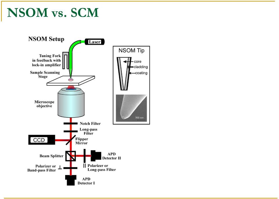
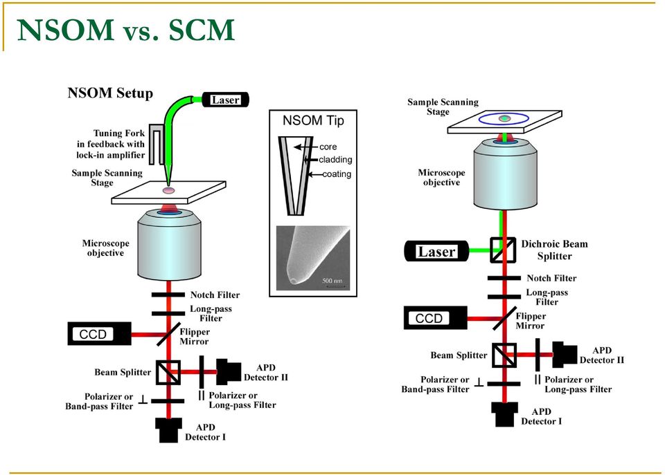
3 NSOM vs. SCM
4 NSOM vs. SCM
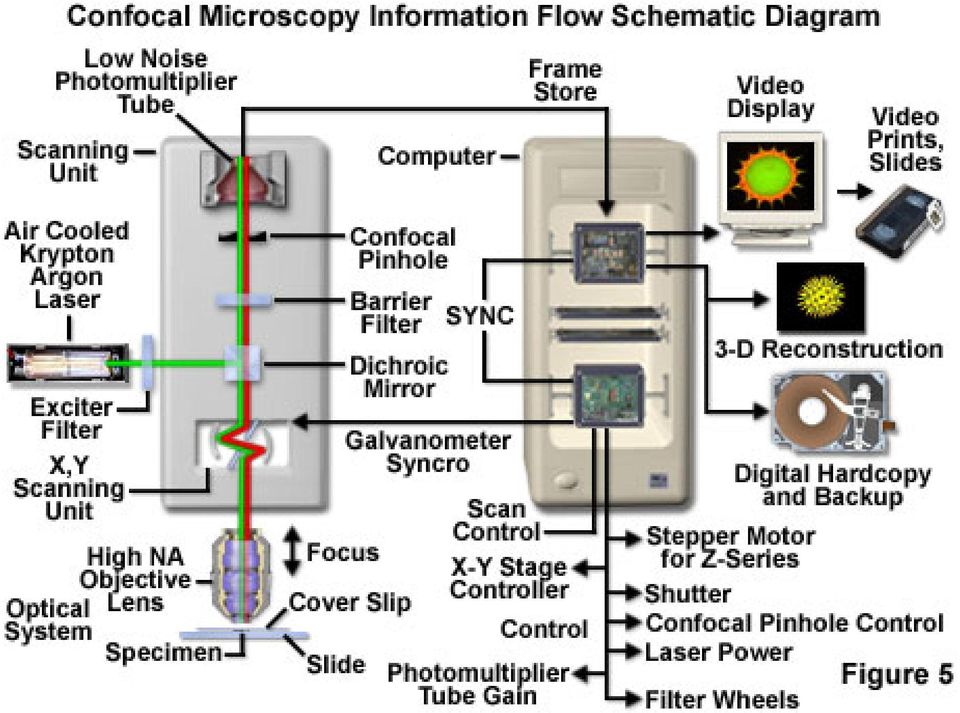
5 Optical paths of Scanning Confocal Microscope Co-focus

6 Confocal: removing scattering light (noise)

7 A good review on scanning confocal based single-molecule imaging Page 3597

8 THE 2008 WOLF PRIZE IN CHEMISTRY The Prize Committee for Chemistry has unanimously decided that the 2008 Wolf Prize be jointly awarded to William E. Moerner Stanford University Stanford, California, USA Allen J. Bard University of Texas Austin, Texas, USA for the ingenious creation of a new field of science, single molecule spectroscopy and electrochemistry, with impact at the nanoscopic regime, from the molecular and cellular domain to complex material systems.

9 Operation modes of SCM 1. Stage (sample) scanning --- tunable from a NSOM set up. Highly desirable for manufacturers of NSOM to compete with SCM optical manufacturers. Installation of a NSOM means an option of SCM. Major advantages: high resolution for scanning (0.1 nm). Slide. 2. Laser scanning --- sample not moving, scanning the laser beam with mirrors. Major advantages: large area scanning and multiple laser excitation for biological systems. Slide. 3. Nipkow disk --- maintaining both the stage and light source stationary, scanning the specimen with an array of light points transmitted through apertures in a spinning. Advantages: selective imaging, minimum damage to sample, no laser required. It is not as common as the first two. Slide.

10 Laser scanning and Nipkow disk imaging

11 Mechanism of laser scanning confocal small tilt of laser beam leads to out-of-focus

12 Laser scanning control system coincident moving of laser beam and optical accessories (e.g. pin holes)

13
14 Small illumination volume provides higher spatial resolution.

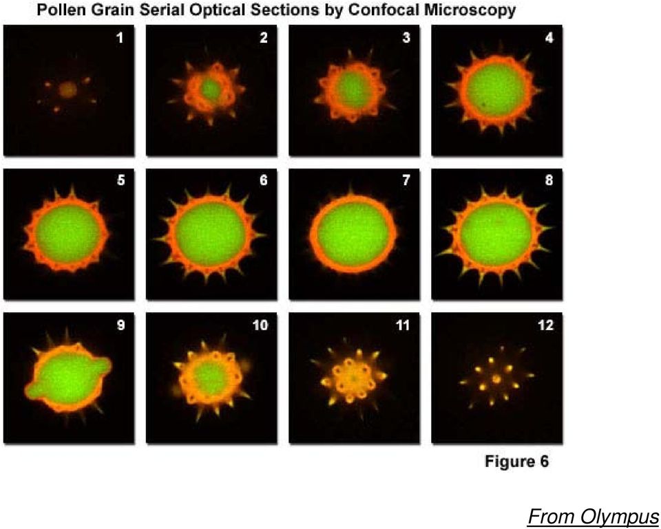
15 Fluorescently stained human medulla Fluorescently stained rabbit muscle fibers sunflower pollen grain From Olympus

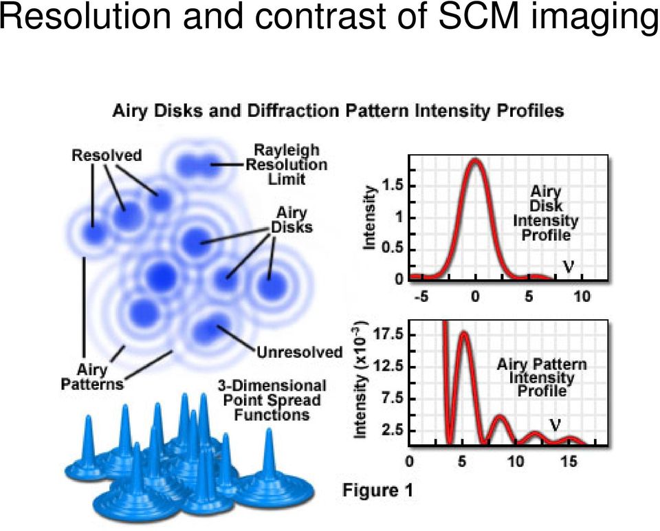
16 Resolution and contrast of SCM imaging
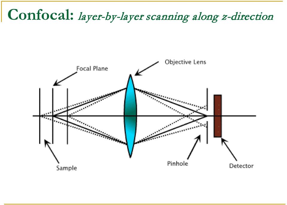
17 3-D imaging by Laser Scanning Confocal Microscopy Only the emission from the focal plane is detected through a pinhole. Synchronized scanning control of the laser beam enables the specimen to be optically sectioned along the z axis. Thus, 3-D imaging is allowable. See slide.

18 Confocal: layer-by-layer scanning along z-direction
19 Sunflower pollen grain Chinese hamster ovary (CHO) line mutated with GFP and HIV protein Mouse intestine section labeled with several fluorophores, created from stacking of 45 optical sections. From Olympus

20 From Olympus
21 Major optical components of SCM Major instrument component: scanner (stage), laser source (CW, pulsed), detector (PMT, APD, CCD (spectra, color image)). See a slide. Major optical components: 1. Objective --- focusing both excitation and emission. Large NA (numerical aperture) objectives (NA > 1.2) are normally used for efficient collection of emission. 2. Notch filter --- extremely narrow blocking of a specific wavelength (good for lasers). 3. Bandpass Filter --- A filter that transmits a defined region (or band) of wavelengths. 4. Shortpass (SP) Filter --- An optical interference or colored glass filter that attenuates longer wavelengths and transmits (passes) shorter wavelengths. 5. Longpass (LP) Filter --- An optical interference or colored glass filter that attenuates shorter wavelengths and transmits (passes) longer wavelengths. 6. Beamsplitter --- A common optical device used for separating an incident beam of light into two or more components that are subsequently projected in different directions. Beamsplitters are available in a variety of configurations to suit particular requirements. A). Prism beamsplitters, B) polarizing beamsplitters are composed of a crystalline birefringent material to produce linearly polarized light, C) dichromatic (often termed dichroic) mirrors act as beamsplitters to reflect excitation wavelengths back into the source while transmitting longer wavelength secondary fluorescence emission to the eyepieces or detector. 7. Neutral Density (ND) Filter - Extensively utilized for a variety of applications in optical microscopy, neutral density filters are neutral gray in color (resembling smoked glass) and are designed to reduce transmitted light intensity evenly across either a small number of wavelengths or the entire wavelength spectrum without altering the spectral profile of illumination. 8. Pinhole - placed near the light source and detector, enabling the microscope to produce thin optical sections of focal planes in the specimen.
22 A scheme of SCM Numerical Aperture (NA) of an objective and the effect on optical resolution and light collection
23 Two styles of SCM setup
24 Zeiss LSM 510
25 Olympus FV 1000
26 Many books and readings available And Google: scanning confocal + your topic
27 Major applications of SCM 1. Single-molecule imaging and spectroscopy (high throughput), particularly in solutions, where NSOM is hardly to be applied, and no spatial resolution required. 2. Single-molecule rotation and diffusion. 3. Protein folding and dynamic conformations. 4. In situ single-cell imaging of virus attack. 5. Nanocrystal optoelectronics. 6. Single-molecule Raman spectrometry.
28 SCM allows for detecting at different depths into sample Protection from oxygen and moisture is often required.
29 Lectures on SCM 1. Single-molecule imaging and spectroscopy, particularly in solutions, where NSOM is hardly to be applied, and no spatial resolution required. 2. Single-molecule rotation and diffusion. 3. Protein folding and dynamic conformations. 4. Cellular attack by virus. 5. Nanocrystal optoelectronics. 6. Single-molecule Raman spectrometry.
30 Advantages of SCM over NSOM High spatial resolution of NSOM --- critical for studying nanostructured materials, but not necessary for singlemolecule imaging, because the molecules can be dispersed such that they are far enough apart (> 300 nm) that diffraction limited optics can resolve individual ones. Historically, detection of the fluorescence from single molecules on a surface at room temperature was originally achieved with NSOM. Soon after this discovery, confocal microscopy had emerged as a more popular technique to detect single molecules. Although confocal microscopy is not capable of sub-wavelength resolution imaging, it has a number of advantages over NSOM. One representative person is Morner, slide. 1. Very high optical excitation powers are possible, whereas with NSOM, the upper limit of the excitation power is severely limited. The higher fluorescence intensities that accompany higher excitation powers raises the S/N and reduces the time integration needed for any given experiment. This is important for faster imaging rates and for looking at fluorescence signals which fluctuate on short (millisecond) time scales. 2. SCM is relatively easy to set up in comparison to NSOM, since no feedback system to be maintained. Setting up and optimizing shear force distance regulation is the most time consuming step in NSOM. 3. For NSOM, the position of the APD needs to be adjusted each time a new tip is used, and should also be optimized, or at least checked for optimization, each time a new region of the sample is imaged (i.e. when the course position of the sample is adjusted). This is because the tip, each time replaced or moved slightly, is not aligned confocal with the detector. In contrast to this, the focal spot of SCM objective does not change. Multiple samples can be imaged over days, and probably weeks, without any drifts substantial enough to require adjusting the position of the APD --- high throughput screening. 4. The excitation power of SCM is more easily measured. SCM is capable of power dependence studies, which simply cannot be done with NSOM because the high power regime is inaccessible. For NSOM, the differences in the throughput of each tip and differences in the efficiency of coupling junctions (typically, one which couples light from a free space laser beam to the single mode fiber, and another fiber-to-fiber splice to couple into the NSOM probe) prevent simply measuring the power of the input beam.
Basic principles and mechanisms of NSOM; Different scanning modes and systems of NSOM; General applications and advantages of NSOM.
 Lecture 16: Near-field Scanning Optical Microscopy (NSOM) Background of NSOM; Basic principles and mechanisms of NSOM; Basic components of a NSOM; Different scanning modes and systems of NSOM; General
5. Scanning Near-Field Optical Microscopy 5.1. Resolution of conventional optical microscopy
 5. Scanning Near-Field Optical Microscopy 5.1. Resolution of conventional optical microscopy Resolution of optical microscope is limited by diffraction. Light going through an aperture makes diffraction
Applications of confocal fluorescence microscopy in biological sciences
 Applications of confocal fluorescence microscopy in biological sciences B R Boruah Department of Physics IIT Guwahati Email: brboruah@iitg.ac.in Page 1 Contents Introduction Optical resolution Optical
Near-field scanning optical microscopy (SNOM)
") Adviser: dr. Maja Remškar Institut Jožef Stefan January 2010 1 2 3 4 5 6 Fluorescence Raman and surface enhanced Raman 7 Conventional optical microscopy-limited resolution Two broad classes of techniques
Adviser: dr. Maja Remškar Institut Jožef Stefan January 2010 1 2 3 4 5 6 Fluorescence Raman and surface enhanced Raman 7 Conventional optical microscopy-limited resolution Two broad classes of techniques
Confocal Microscopy and Atomic Force Microscopy (AFM) A very brief primer...
 A very brief primer...") Confocal Microscopy and Atomic Force Microscopy (AFM) of biofilms A very brief primer... Fundamentals of Confocal Microscopy Based on a conventional fluorescence microscope Fluorescent Microscope Confocal
Confocal Microscopy and Atomic Force Microscopy (AFM) of biofilms A very brief primer... Fundamentals of Confocal Microscopy Based on a conventional fluorescence microscope Fluorescent Microscope Confocal
Chapter 12 Filters for FISH Imaging
 Chapter 12 Filters for FISH Imaging Dan Osborn The application of in situ hybridization (ISH) has advanced from short lived, non-specific isotopic methods, to very specific, long lived, multiple color
Preface Light Microscopy X-ray Diffraction Methods
 Preface xi 1 Light Microscopy 1 1.1 Optical Principles 1 1.1.1 Image Formation 1 1.1.2 Resolution 3 1.1.3 Depth of Field 5 1.1.4 Aberrations 6 1.2 Instrumentation 8 1.2.1 Illumination System 9 1.2.2 Objective
Chapter 13 Confocal Laser Scanning Microscopy C. Robert Bagnell, Jr., Ph.D., 2012
 Chapter 13 Confocal Laser Scanning Microscopy C. Robert Bagnell, Jr., Ph.D., 2012 You are sitting at your microscope working at high magnification trying to sort out the three-dimensional compartmentalization
Zeiss 780 Training Notes
 Zeiss 780 Training Notes 780 Start Up Sequence Do you need the argon laser, 458,488,514nm lines? No Turn on the Systems PC Switch Turn on Main Power Switch Yes Turn on the laser main power switch and turn
Scanning Near Field Optical Microscopy: Principle, Instrumentation and Applications
 Scanning Near Field Optical Microscopy: Principle, Instrumentation and Applications Saulius Marcinkevičius Optics, ICT, KTH 1 Outline Optical near field. Principle of scanning near field optical microscope
CREOL, College of Optics & Photonics, University of Central Florida
 OSE6650 - Optical Properties of Nanostructured Materials Optical Properties of Nanostructured Materials Fall 2013 Class 3 slide 1 Challenge: excite and detect the near field Thus far: Nanostructured materials
DOE Solar Energy Technologies Program Peer Review. Denver, Colorado April 17-19, 2007
 DOE Solar Energy Technologies Program Peer Review Evaluation of Nanocrystalline Silicon Thin Film by Near-Field Scanning Optical Microscopy AAT-2-31605-05 Magnus Wagener and George Rozgonyi North Carolina
Acousto-optic modulator
 1 of 3 Acousto-optic modulator F An acousto-optic modulator (AOM), also called a Bragg cell, uses the acousto-optic effect to diffract and shift the frequency of light using sound waves (usually at radio-frequency).
Raman spectroscopy Lecture
 Raman spectroscopy Lecture Licentiate course in measurement science and technology Spring 2008 10.04.2008 Antti Kivioja Contents - Introduction - What is Raman spectroscopy? - The theory of Raman spectroscopy
It has long been a goal to achieve higher spatial resolution in optical imaging and
 Nano-optical Imaging using Scattering Scanning Near-field Optical Microscopy Fehmi Yasin, Advisor: Dr. Markus Raschke, Post-doc: Dr. Gregory Andreev, Graduate Student: Benjamin Pollard Department of Physics,
PHYSICAL METHODS, INSTRUMENTS AND MEASUREMENTS Vol. IV Femtosecond Measurements Combined With Near-Field Optical Microscopy - Artyom A.
 FEMTOSECOND MEASUREMENTS COMBINED WITH NEAR FIELD OPTICAL MICROSCOPY Artyom A. Astafiev, Semyonov Institute of Chemical Physics, Moscow, Russian Federation. Keywords: diffraction limit nearfield scanning
CONFOCAL LASER SCANNING MICROSCOPY TUTORIAL
 CONFOCAL LASER SCANNING MICROSCOPY TUTORIAL Robert Bagnell 2006 This tutorial covers the following CLSM topics: 1) What is the optical principal behind CLSM? 2) What is the spatial resolution in X, Y,
Scanning Near-Field Optical Microscopy for Measuring Materials Properties at the Nanoscale
 Scanning Near-Field Optical Microscopy for Measuring Materials Properties at the Nanoscale Outline Background Research Design Detection of Near-Field Signal Submonolayer Chemical Sensitivity Conclusions
Nano-Spectroscopy. Solutions AFM-Raman, TERS, NSOM Chemical imaging at the nanoscale
 Nano-Spectroscopy Solutions AFM-Raman, TERS, NSOM Chemical imaging at the nanoscale Since its introduction in the early 80 s, Scanning Probe Microscopy (SPM) has quickly made nanoscale imaging an affordable
Fiber Optics: Fiber Basics
 Photonics Technical Note # 21 Fiber Optics Fiber Optics: Fiber Basics Optical fibers are circular dielectric wave-guides that can transport optical energy and information. They have a central core surrounded
Specifying Plasma Deposited Hard Coated Optical Thin Film Filters. Alluxa Engineering Staff
 Specifying Plasma Deposited Hard Coated Optical Thin Film Filters. Alluxa Engineering Staff December 2012 Specifying Advanced Plasma Deposited Hard Coated Optical Bandpass and Dichroic Filters. Introduction
Lecture 4 Scanning Probe Microscopy (SPM)
") Lecture 4 Scanning Probe Microscopy (SPM) General components of SPM; Tip --- the probe; Cantilever --- the indicator of the tip; Tip-sample interaction --- the feedback system; Scanner --- piezoelectric
Lecture 4 Scanning Probe Microscopy (SPM) General components of SPM; Tip --- the probe; Cantilever --- the indicator of the tip; Tip-sample interaction --- the feedback system; Scanner --- piezoelectric
Raman Spectroscopy Basics
 Raman Spectroscopy Basics Introduction Raman spectroscopy is a spectroscopic technique based on inelastic scattering of monochromatic light, usually from a laser source. Inelastic scattering means that
WAVELENGTH OF LIGHT - DIFFRACTION GRATING
 PURPOSE In this experiment we will use the diffraction grating and the spectrometer to measure wavelengths in the mercury spectrum. THEORY A diffraction grating is essentially a series of parallel equidistant
Recording the Instrument Response Function of a Multiphoton FLIM System
 Recording the Instrument Response Function of a Multiphoton FLIM System Abstract. FLIM data analysis in presence of SHG signals or extremely fast decay components requires the correct instrument response
Molecular Spectroscopy
 Molecular Spectroscopy UV-Vis Spectroscopy Absorption Characteristics of Some Common Chromophores UV-Vis Spectroscopy Absorption Characteristics of Aromatic Compounds UV-Vis Spectroscopy Effect of extended
Module 13 : Measurements on Fiber Optic Systems
 Module 13 : Measurements on Fiber Optic Systems Lecture : Measurements on Fiber Optic Systems Objectives In this lecture you will learn the following Measurements on Fiber Optic Systems Attenuation (Loss)
Time out states and transitions
 Time out states and transitions Spectroscopy transitions between energy states of a molecule excited by absorption or emission of a photon hn = DE = E i - E f Energy levels due to interactions between
Principles of Microscopy and Confocal and Fluorescence Microscopy
 Principles of Microscopy and Confocal and Fluorescence Microscopy Content This course in Light Microscopy follows the series of successful courses in Light Microscopy, Confocal and Fluorescence Microscopy
Introduction to flow cytometry
 Introduction to flow cytometry Flow cytometry is a popular laser-based technology. Discover more with our introduction to flow cytometry. Flow cytometry is now a widely used method for analyzing the expression
Optical laser beam scanner lens relay system
 1. Introduction Optical laser beam scanner lens relay system Laser beam scanning is used most often by far in confocal microscopes. There are many ways by which a laser beam can be scanned across the back
Katharina Lückerath (AG Dr. Martin Zörnig) adapted from Dr. Jörg Hildmann BD Biosciences,Customer Service
 adapted from Dr. Jörg Hildmann BD Biosciences,Customer Service") Introduction into Flow Cytometry Katharina Lückerath (AG Dr. Martin Zörnig) adapted from Dr. Jörg Hildmann BD Biosciences,Customer Service How does a FACS look like? FACSCalibur FACScan What is Flow Cytometry?
Introduction into Flow Cytometry Katharina Lückerath (AG Dr. Martin Zörnig) adapted from Dr. Jörg Hildmann BD Biosciences,Customer Service How does a FACS look like? FACSCalibur FACScan What is Flow Cytometry?
Physics 441/2: Transmission Electron Microscope
 Physics 441/2: Transmission Electron Microscope Introduction In this experiment we will explore the use of transmission electron microscopy (TEM) to take us into the world of ultrasmall structures. This
Confocal Microscopy. Chapter 2
 Chapter 2 Confocal Microscopy This Chapter offers a brief introduction to confocal microscopy and to other experimental techniques employed in this thesis. Unraveling structure and dynamics by confocal
NEAR FIELD OPTICAL MICROSCOPY AND SPECTROSCOPY WITH STM AND AFM PROBES
 Vol. 93 (1997) A CTA PHYSICA POLONICA A No. 2 Proceedings of the 1st International Symposium on Scanning Probe Spectroscopy and Related Methods, Poznań 1997 NEAR FIELD OPTICAL MICROSCOPY AND SPECTROSCOPY
LBS-300 Beam Sampler for C-mount Cameras. YAG Focal Spot Analysis Adapter. User Notes
 LBS-300 Beam Sampler for C-mount Cameras P/N SP90183, SP90184, SP90185 and SP90186 YAG Focal Spot Analysis Adapter P/N SP90187, SP90188, SP90189, SP90190, SP90191 User Notes Ophir-Spiricon Inc. 60 West
Experiment 5. Lasers and laser mode structure
 Northeastern University, PHYS5318 Spring 2014, 1 1. Introduction Experiment 5. Lasers and laser mode structure The laser is a very important optical tool that has found widespread use in science and industry,
Confocal Fluorescence Microscopy
 Chapter 1 Confocal Fluorescence Microscopy 1.1 The principle Confocal fluorescence microscopy is a microscopic technique that provides true three-dimensional (3D) optical resolution. In microscopy, 3D
Introduction to Fourier Transform Infrared Spectrometry
 Introduction to Fourier Transform Infrared Spectrometry What is FT-IR? I N T R O D U C T I O N FT-IR stands for Fourier Transform InfraRed, the preferred method of infrared spectroscopy. In infrared spectroscopy,
Synthetic Sensing: Proximity / Distance Sensors
 Synthetic Sensing: Proximity / Distance Sensors MediaRobotics Lab, February 2010 Proximity detection is dependent on the object of interest. One size does not fit all For non-contact distance measurement,
Advanced Research Raman System Raman Spectroscopy Systems
 T600 Advanced Research Raman System Raman Spectroscopy Systems T600 Advanced Research Raman System T600 Triple stage Raman Spectrometer: The only solution for unprecedented stability and performance! Robust
Introduction to Flow Cytometry
 Introduction to Flow Cytometry presented by: Flow Cytometry y Core Facility Biomedical Instrumentation Center Uniformed Services University Topics Covered in this Lecture What is flow cytometry? Flow cytometer
Chapter 1 Parts C. Robert Bagnell, Jr., Ph.D., 2012
 Chapter 1 Parts C. Robert Bagnell, Jr., Ph.D., 2012 Figure 1.1 illustrates the parts of an upright compound microscope and indicates the terminology that I use in these notes. Figure 1.1. Parts of a Compound
Infrared Viewers. Manual
 Infrared Viewers Manual Contents Introduction 3 How it works 3 IR viewer in comparison with a CCD camera 4 Visualization of infrared laser beam in mid-air 4 Power Density 5 Spectral sensitivity 6 Operation
LabRAM HR. Research Raman Made Easy! Raman Spectroscopy Systems. Spectroscopy Suite. Powered by:
 LabRAM HR Research Raman Made Easy! Raman Spectroscopy Systems Powered by: Spectroscopy Suite Cutting-Edge Applications with the LabRAM HR Deeply involved in Raman spectroscopy for decades, HORIBA Scientific
LabRAM HR Evolution. Research Raman Made Easy!
 LabRAM HR Evolution Research Raman Made Easy! Cutting-Edge Applications with the LabRAM HR LabRAM HR Deeply involved in Raman spectroscopy for decades, HORIBA Scientific has been providing an extensive
Using light scattering method to find The surface tension of water
 Experiment (8) Using light scattering method to find The surface tension of water The aim of work: The goals of this experiment are to confirm the relationship between angular frequency and wave vector
The Basics of Scanning Electron Microscopy
 The Basics of Scanning Electron Microscopy The small scanning electron microscope is easy to use because almost every variable is pre-set: the acceleration voltage is always 15kV, it has only a single
AP Physics B Ch. 23 and Ch. 24 Geometric Optics and Wave Nature of Light
 AP Physics B Ch. 23 and Ch. 24 Geometric Optics and Wave Nature of Light Name: Period: Date: MULTIPLE CHOICE. Choose the one alternative that best completes the statement or answers the question. 1) Reflection,
Chapter 4. Microscopy, Staining, and Classification. Lecture prepared by Mindy Miller-Kittrell North Carolina State University
 Chapter 4 Microscopy, Staining, and Classification 2012 Pearson Education Inc. Lecture prepared by Mindy Miller-Kittrell North Carolina State University Microscopy and Staining 2012 Pearson Education Inc.
Microscopy. MICROSCOPY Light Electron Tunnelling Atomic Force RESOLVE: => INCREASE CONTRAST BIODIVERSITY I BIOL1051 MAJOR FUNCTIONS OF MICROSCOPES
 BIODIVERSITY I BIOL1051 Microscopy Professor Marc C. Lavoie marc.lavoie@cavehill.uwi.edu MAJOR FUNCTIONS OF MICROSCOPES MAGNIFY RESOLVE: => INCREASE CONTRAST Microscopy 1. Eyepieces 2. Diopter adjustment
Microscopy: Principles and Advances
 Microscopy: Principles and Advances Chandrashekhar V. Kulkarni University of Central Lancashire, Preston, United kingdom May, 2014 University of Ljubljana Academic Background 2005-2008: PhD-Chemical Biology
Laboratory #3 Guide: Optical and Electrical Properties of Transparent Conductors -- September 23, 2014
 Laboratory #3 Guide: Optical and Electrical Properties of Transparent Conductors -- September 23, 2014 Introduction Following our previous lab exercises, you now have the skills and understanding to control
Automatic and Objective Measurement of Residual Stress and Cord in Glass
 Automatic and Objective Measurement of Residual Stress and Cord in Glass GlassTrend - ICG TC15/21 Seminar SENSORS AND PROCESS CONTROL 13-14 October 2015, Eindhoven Henning Katte, ilis gmbh copyright ilis
Neuro imaging: looking with lasers in the brain
 Neuro imaging: looking with lasers in the brain Aim: To image life cells, label free, with cellular resolution in deep tissue Marloes Groot Vrije Universiteit Amsterdam Faculteit Exacte Wetenschappen Natuurkunde
Copyright 1999 2010 by Mark Brandt, Ph.D. 12
 Introduction to Absorbance Spectroscopy A single beam spectrophotometer is comprised of a light source, a monochromator, a sample holder, and a detector. An ideal instrument has a light source that emits
Holographically corrected microscope with a large working distance (as appears in Applied Optics, Vol. 37, No. 10, 1849-1853, 1 April 1998)
") Holographically corrected microscope with a large working distance (as appears in Applied Optics, Vol. 37, No. 10, 1849-1853, 1 April 1998) Geoff Andersen and R. J. Knize Laser and Optics Research Center
Holographically corrected microscope with a large working distance (as appears in Applied Optics, Vol. 37, No. 10, 1849-1853, 1 April 1998) Geoff Andersen and R. J. Knize Laser and Optics Research Center
Ion Beam Sputtering: Practical Applications to Electron Microscopy
 Ion Beam Sputtering: Practical Applications to Electron Microscopy Applications Laboratory Report Introduction Electron microscope specimens, both scanning (SEM) and transmission (TEM), often require a
Fundamentals of modern UV-visible spectroscopy. Presentation Materials
 Fundamentals of modern UV-visible spectroscopy Presentation Materials The Electromagnetic Spectrum E = hν ν = c / λ 1 Electronic Transitions in Formaldehyde 2 Electronic Transitions and Spectra of Atoms
Components for Infrared Spectroscopy. Dispersive IR Spectroscopy
 Components for Infrared Spectroscopy Mid-IR light: 00-000 cm - (5.5 m wavelength) Sources: Blackbody emitters Globar metal oxides Nernst Glower: Silicon Carbide Detectors: Not enough energy for photoelectric
ZEISS Microscopy Course Catalog
 ZEISS Microscopy Course Catalog ZEISS Training and Education Expand Your Possibilities Practical microscopy training has a long tradition at ZEISS. The first courses were held in Jena as early as 1907,
X-Rays and Magnetism From Fundamentals to Nanoscale Dynamics
 X-Rays and Magnetism From Fundamentals to Nanoscale Dynamics Joachim Stöhr Stanford Synchrotron Radiation Laboratory X-rays have come a long way 1895 1993 10 cm 10 µm 100 nm Collaborators: SSRL Stanford:
MEASURABLE PARAMETERS: Flow cytometers are capable of measuring a variety of cellular characteristics such as:
 INTRODUCTION Flow Cytometry involves the use of a beam of laser light projected through a liquid stream that contains cells, or other particles, which when struck by the focused light give out signals
Physics 10. Lecture 29A. "There are two ways of spreading light: to be the candle or the mirror that reflects it." --Edith Wharton
 Physics 10 Lecture 29A "There are two ways of spreading light: to be the candle or the mirror that reflects it." --Edith Wharton Converging Lenses What if we wanted to use refraction to converge parallel
Problem Set 6 UV-Vis Absorption Spectroscopy. 13-1. Express the following absorbances in terms of percent transmittance:
 Problem Set 6 UV-Vis Absorption Spectroscopy 13-1. Express the following absorbances in terms of percent transmittance: a 0.051 b 0.918 c 0.379 d 0.261 e 0.485 f 0.072 A = log P o /P = log1/t = - log T
h e l p s y o u C O N T R O L
 contamination analysis for compound semiconductors ANALYTICAL SERVICES B u r i e d d e f e c t s, E v a n s A n a l y t i c a l g r o u p h e l p s y o u C O N T R O L C O N T A M I N A T I O N Contamination
FTIR Instrumentation
 FTIR Instrumentation Adopted from the FTIR lab instruction by H.-N. Hsieh, New Jersey Institute of Technology: http://www-ec.njit.edu/~hsieh/ene669/ftir.html 1. IR Instrumentation Two types of instrumentation
Taking the Confusion out of Confocal Microscopy
 KEYWORDS: confocal microscopy, fluorescence imaging, three dimensional Special section on techniques: Taking the Confusion out of Confocal Microscopy Nana Rezai Pathology, University of British Columbia
Flow cytometry basics fluidics, optics, electronics...
 Title Flow cytometry basics fluidics, optics, electronics... RNDr. Jan Svoboda, Ph.D. Cytometry and Microscopy Core Facility IMB, CAS, v.v.i Vídeňská 1083 Fluorescence Fluorescence occurs when a valence
A Brief History of the Microscope and its Significance in the Advancement of Biology and Medicine
 Chapter 1 A Brief History of the Microscope and its Significance in the Advancement of Biology and Medicine This chapter provides a historical foundation of the field of microscopy and outlines the significant
3D Raman Imaging Nearfield-Raman TERS. Solutions for High-Resolution Confocal Raman Microscopy. www.witec.de
 3D Raman Imaging Nearfield-Raman TERS Solutions for High-Resolution Confocal Raman Microscopy www.witec.de 01 3D Confocal Raman Imaging Outstanding performance in speed, sensitivity, and resolution with
View of ΣIGMA TM (Ref. 1)
") Overview of the FESEM system 1. Electron optical column 2. Specimen chamber 3. EDS detector [Electron Dispersive Spectroscopy] 4. Monitors 5. BSD (Back scatter detector) 6. Personal Computer 7. ON/STANDBY/OFF
Overview of the FESEM system 1. Electron optical column 2. Specimen chamber 3. EDS detector [Electron Dispersive Spectroscopy] 4. Monitors 5. BSD (Back scatter detector) 6. Personal Computer 7. ON/STANDBY/OFF
Zeiss Axioimager M2 microscope for stereoscopic analysis.
 Zeiss Axioimager M2 microscope for stereoscopic analysis. This system is fully motorized and configured with bright field and multi-channel fluorescent. It works with Stereo Investigator, Neurolucida,
Webex-based remote instrument control guide
 Webex-based remote instrument control guide 1) Once a microscopic imaging experiment is scheduled, the host (i.e., Hunter Bioimaging Facility) will send an email containing a Webex meeting link to the
COMPENSATION MIT Flow Cytometry Core Facility
 COMPENSATION MIT Flow Cytometry Core Facility Why do we need compensation? 1) Because the long emission spectrum tail of dyes causes overlap like with the fluorophores FITC and PE. 2) For sensitivity reasons,
Interference. Physics 102 Workshop #3. General Instructions
 Interference Physics 102 Workshop #3 Name: Lab Partner(s): Instructor: Time of Workshop: General Instructions Workshop exercises are to be carried out in groups of three. One report per group is due by
A VERYbrief history of the confocal microscope 1950s
 Confocal Microscopy Confocal Microscopy Why do we use confocal microscopy? A brief history of the confocal microscope Advantages/disadvantages of a confocal microscope Types of confocal microscopes The
Lenses and Apertures of A TEM
 Instructor: Dr. C.Wang EMA 6518 Course Presentation Lenses and Apertures of A TEM Group Member: Anup Kr. Keshri Srikanth Korla Sushma Amruthaluri Venkata Pasumarthi Xudong Chen Outline Electron Optics
RAY TRACING UNIFIED FIELD TRACING
 RAY TRACING Start to investigate the performance of your optical system using 3D ray distributions, dot diagrams of ray positions and directions, and optical path length. GEOMETRIC FIELD TRACING Switch
Optical Communications
 Optical Communications Telecommunication Engineering School of Engineering University of Rome La Sapienza Rome, Italy 2005-2006 Lecture #2, May 2 2006 The Optical Communication System BLOCK DIAGRAM OF
The excitation in Raman spectroscopy is usually. Practical Group Theory and Raman Spectroscopy, Part II: Application of Polarization
 Electronically reprinted from March 214 Molecular Spectroscopy Workbench Practical Group Theory and Raman Spectroscopy, Part II: Application of Polarization In this second installment of a two-part series
Zecotek S Light Projection Network Marketing
 White Paper Zecotek Visible Fiber Laser Platform Enabling the future of laser technology Zecotek Photonics Inc. (TSX- V: ZMS; Frankfurt: W1I) www.zecotek.com is a Canadian photonics technology company
These particles have something in common
 These particles have something in common Blood cells Chromosomes Algae Protozoa Certain parameters of these particles can be measured with a flow cytometer Which parameters can be measured? the relative
Near-Field Scanning Optical Microscopy: a Brief Overview
 Near-Field Scanning Optical Microscopy: a Brief Overview Serge HUANT Laboratoire de Spectrométrie Physique (SPECTRO) Université Joseph Fourier Grenoble et CNRS Thanks to my former & present collaborators
Nano Optics: Overview of Research Activities. Sergey I. Bozhevolnyi SENSE, University of Southern Denmark, Odense, DENMARK
 Nano Optics: Overview of Research Activities SENSE, University of Southern Denmark, Odense, DENMARK Optical characterization techniques: Leakage Radiation Microscopy Scanning Near-Field Optical Microscopy
Reflectance Measurements of Materials Used in the Solar Industry. Selecting the Appropriate Accessories for UV/Vis/NIR Measurements.
 T e c h n i c a l N o t e Reflectance Measurements of Materials Used in the Solar Industry UV/Vis/NIR Author: Dr. Jeffrey L. Taylor PerkinElmer, Inc. 710 Bridgeport Avenue Shelton, CT 06484 USA Selecting
CSCI 4974 / 6974 Hardware Reverse Engineering. Lecture 8: Microscopy and Imaging
 CSCI 4974 / 6974 Hardware Reverse Engineering Lecture 8: Microscopy and Imaging Data Acquisition for RE Microscopy Imaging Registration and stitching Microscopy Optical Electron Scanning Transmission Scanning
Robert G. Hunsperger. Integrated Optics. Theory and Technology. Fourth Edition. With 195 Figures and 17 Tables. Springer
 Robert G. Hunsperger Integrated Optics Theory and Technology Fourth Edition With 195 Figures and 17 Tables Springer Contents 1. Introduction 1 1.1 Advantages of Integrated Optics 2 1.1.1 Comparison of
Observing a nanomachine at work: Single-molecule imaging or spectroscopy (SMI or SMS)
") Observing a nanomachine at work: Single-molecule imaging or spectroscopy (SMI or SMS) Principle: SMS allows one to observe the function and the motion of nano-objects in realtime in living systems. Usually,
Observing a nanomachine at work: Single-molecule imaging or spectroscopy (SMI or SMS) Principle: SMS allows one to observe the function and the motion of nano-objects in realtime in living systems. Usually,
Polarization Dependence in X-ray Spectroscopy and Scattering. S P Collins et al Diamond Light Source UK
 Polarization Dependence in X-ray Spectroscopy and Scattering S P Collins et al Diamond Light Source UK Overview of talk 1. Experimental techniques at Diamond: why we care about x-ray polarization 2. How
Characterizing Quantum Dots and Color Centers in Nanodiamonds as Single Emitters
 University of Rochester OPT253 Lab 3-4 Report Characterizing Quantum Dots and Color Centers in Nanodiamonds as Single Emitters Author: Nicholas Cothard Peter Heuer Professor: Dr. Svetlana Lukishova November
Diffraction of a Circular Aperture
 Diffraction of a Circular Aperture Diffraction can be understood by considering the wave nature of light. Huygen's principle, illustrated in the image below, states that each point on a propagating wavefront
Two-photon FCS Tutorial. Berland Lab Department of Physics Emory University
 Two-photon FCS Tutorial Berland Lab Department of Physics Emory University What is FCS? FCS : Fluorescence Correlation Spectroscopy FCS is a technique for acquiring dynamical information from spontaneous
Christine E. Hatch University of Nevada, Reno
 Christine E. Hatch University of Nevada, Reno Roadmap What is DTS? How Does it Work? What Can DTS Measure? Applications What is Distributed Temperature Sensing (DTS)? Temperature measurement using only
LASER SCANNING CONFOCAL MICROSCOPY
 LASER SCANNING CONFOCAL MICROSCOPY Nathan S. Claxton, Thomas J. Fellers, and Michael W. Davidson Department of Optical Microscopy and Digital Imaging, National High Magnetic Field Laboratory, The Florida
Leica TCS SP5 Confocal Laser Scanning Microscope User Guide 1. BASIC IMAGE ACQUISITION
 Leica TCS SP5 Confocal Laser Scanning Microscope User Guide 1. BASIC IMAGE ACQUISITION This manual is the FIRST section of a THREE part Leica TCS - SP5 User Guide edited by Donald Pottle Leica True Confocal
Measurement of Enhanced Specular Reflector (ESR) Films Using a LAMBDA 1050 UV/Vis/NIR Spectrometer and URA Accessory
 Films Using a LAMBDA 1050 UV/Vis/NIR Spectrometer and URA Accessory") FIELD APPLICATION REPORT UV/Vis/NIR Spectroscopy Author: Frank Padera Shelton, CT Contributor: Chris Lynch Shelton, CT Measurement of Enhanced Specular Reflector (ESR) Films Using a LAMBDA 1050 UV/Vis/NIR
FIELD APPLICATION REPORT UV/Vis/NIR Spectroscopy Author: Frank Padera Shelton, CT Contributor: Chris Lynch Shelton, CT Measurement of Enhanced Specular Reflector (ESR) Films Using a LAMBDA 1050 UV/Vis/NIR
UNIT I: INTRFERENCE & DIFFRACTION Div. B Div. D Div. F INTRFERENCE
 107002: EngineeringPhysics Teaching Scheme: Lectures: 4 Hrs/week Practicals-2 Hrs./week T.W.-25 marks Examination Scheme: Paper-50 marks (2 hrs) Online -50marks Prerequisite: Basics till 12 th Standard
Bio 321 Lightmicroscopy Electronmicrosopy Image Processing
 Bio 321 Lightmicroscopy Electronmicrosopy Image Processing Urs Ziegler Center for Microscopy and Image Analysis Light microscopy (Confocal Laser Scanning Microscopy) Light microscopy (Confocal Laser Scanning
Raman Spectroscopy. 1. Introduction. 2. More on Raman Scattering. " scattered. " incident
 February 15, 2006 Advanced Physics Laboratory Raman Spectroscopy 1. Introduction When light is scattered from a molecule or crystal, most photons are elastically scattered. The scattered photons have the