Diagnostic performance of MRI in differentiating metastatic from acute osteoporotic compression fractures of the spine
|
|
|
- Anna Sanders
- 7 years ago
- Views:
Transcription
1 Diagnostic performance of MRI in differentiating metastatic from acute osteoporotic compression fractures of the spine Poster No.: C-1399 Congress: ECR 2013 Type: Scientific Exhibit Authors: J. Martel, A. Bueno, M. Nogueras, M. Barxias, E. Pérez; Alcorcón/ ES Keywords: Musculoskeletal spine, Oncology, MR, MR-Diffusion/Perfusion, Diagnostic procedure, Comparative studies, Metastases, Osteoporosis DOI: /ecr2013/C-1399 Any information contained in this pdf file is automatically generated from digital material submitted to EPOS by third parties in the form of scientific presentations. References to any names, marks, products, or services of third parties or hypertext links to thirdparty sites or information are provided solely as a convenience to you and do not in any way constitute or imply ECR's endorsement, sponsorship or recommendation of the third party, information, product or service. ECR is not responsible for the content of these pages and does not make any representations regarding the content or accuracy of material in this file. As per copyright regulations, any unauthorised use of the material or parts thereof as well as commercial reproduction or multiple distribution by any traditional or electronically based reproduction/publication method ist strictly prohibited. You agree to defend, indemnify, and hold ECR harmless from and against any and all claims, damages, costs, and expenses, including attorneys' fees, arising from or related to your use of these pages. Please note: Links to movies, ppt slideshows and any other multimedia files are not available in the pdf version of presentations. Page 1 of 17

2 Purpose Differentiating benign from malignant vertebral marrow processes is a frequent and sometimes difficult problem, especially distinction between metastatic and acute osteoporotic compression fractures. Morphologic criteria may accurately predict benign from malignant fractures of the spine in up to 90% of cases, however it is not perfect and conventional MR sequences are nonspecific. Our purpose is to determine the contribution of the new sequences(diffusion, ADC values and in-phase/out-of-phase sequences) in differentiating between malignant and benign bone marrow lesions. Images for this section: Page 2 of 17

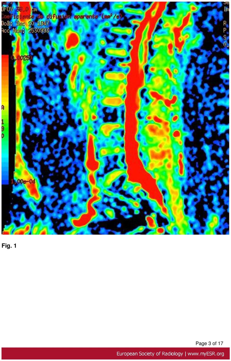
3 Fig. 1 Page 3 of 17
4 Methods and Materials Patient selection A total of 85 consecutive patients included 60 women and 25 men, ranging in age from 33 to 90 years old (mean 67) were prospectively studied. The patients presented vertebral collapse and/or altered signal intensity in one or more vertebral bodies on conventional MR sequences. A total of 213 vertebral bodies were analysed. From each patient, one normal vertebral body and one, two or three pathologic verterbal bodies were recorded. 26 patients had an history of a known primary tumor The different pathologies are showed in the table: Fig. 2: Table show diagnosis distribution Page 4 of 17

5 References: Diagnóstico por Imagen, Hospital Universitario Fundación Alcorcón Alcorcón/ES MR Images All MR studies were performed by using a 1.5 T unit (Signa Excite; GE Medycal Systems). The following pulse sequences were used for all patients: axial and sagittal T1-weighted spin-echo imaging; axial and sagittal T2-weighted fast spin-echo imaging; sagittal STIR imaging; sagittal in-phase and out-of-phase imaging and sagittal diffusion-weighted sequences (Echo planar imaging, b=400). Postprocessing and pathologic analysis All the images of each patient were interpreted in consensus by two radiologists. The signal intensity of the vertebral bodies were qualitatively evaluated on T1-weighted and T2-weighted images, STIR images and diffusion-weight images and were described as hypointense, isointense or hyperintense signal. The signal intensity values for in-phased an out-of-phase images were measured for each vertebral body or lesion. A cut-off of 0.8 was chosen with <0.8 defined as benign, defined as indeterminated an >1 defined as malignant. A computation of the bone marrow signal intensity ratio (SIR) measured on out-of-phase/ in-phase images was made. Apparent diffusion coefficent (ADC) values were also determined. Statistical analysis Sensibility, specificity and accuracy were estimated with 95% confidence interval. To evaluate discriminant capacity of ratio out-of-phase/in-phase and ADC values, area under ROC curve was estimated and the cut point with maximum sensibility and specificity were calculated. Results Page 5 of 17

6 Summary values of sensitivity, specificity and accuracy for changes in signal intensity and ratio (SIR) values, and ADC values for the diagnosis of metastasis are represented in the next table Fig. 3 References: Diagnóstico por Imagen, Hospital Universitario Fundación Alcorcón Alcorcón/ES The area under the curve shows the signal intentisity ratio (SIR) having better discriminant capacity than ADC values. Page 6 of 17
 having better discriminant capacity than ADC")
7 Fig. 4 References: Diagnóstico por Imagen, Hospital Universitario Fundación Alcorcón Alcorcón/ES A lesion in a vertebral body that shows hypointensity in T1-wi, hyperintensity on STIR and T2-wi, and a signal intensity ratio >0.8 has a sensitivity of 97.2%, specificity of 90% and accuracy of 91.2% respect the diagnosis of a malignat lesion. We summarize our results in the normal vertebra and different lesions: Normal vertebral bodies Page 7 of 17

8 All cases showed normal signal intensity in vertebral marrow on T1-weighted images, T2-weighted, diffusion-weighted or STIR images. Signal Intensity Ratio (SIR) were < ADC values were less than 0.6x10 mm /s Haemangioma Hemangiomas had high intensity on both T1- and T2-weighted images Fig. 5 on page 10. Three cases appeared as hypointense on T1-weighted images and hypo- or hyperintese on T2-weighted images (these cases were biopsied). Diffusion-weighted images were normal in all cases but three of them with a subtle high signal. SIR were > 0.8 in 50% of patients. Fig. 6 on page ADC values were > than 1x10 mm /s but < than 1.5x10 mm /s Fig. 7 on page 10 Espondilosis (endplate degeneration) All cases showed normal signal intensity on T1-weighted images, T2-weighted, diffusionweighted or STIR images, or showed typical findings of Modic lesions. SIR were < ADC values were less than 0.6x10 mm /s Osteoporotic fracture (chronic) Chronic osteoporotic fractures had low intensity on T1-weighted images. Signal on T2weighted images were variable. Page 8 of 17

9 STIR and diffusion-weighted images were normal. Fig. 8 on page 11 SIR were < ADC values were < than 1x10 mm /s Fig. 9 on page 11 Osteoporotic fracture (acute) Acute osteoporotic fractures had low intensity on T1-weighted images. Signal on T2weighted images were variable. STIR showed high signal and diffusion-weighted images showed subtle high signal. SIR were variable, almost 50% > 0.8 Fig. 10 on page ADC values were > than 1.5 x10 mm /s Fig. 11 on page 13 Metastasis Metastatic fractures had a low intensity on T1-weighted images. Signal on T2-weighted images were variable. STIR and diffusion-weighted images showed high signal. SIR > 0.8 in all cases ADC values were >than 1x10 mm /s but < 1.5x10 mm /s Fig. 12 on page 13 Miscellany Spondylodiscitis, Schmörl s nodes (Fig. 13 on page 14 Fig. 14 on page 15), calcified intraspongious disk herniation, and other entitys may simulate malignant lesion, but we have few cases to get statistical analysis. Page 9 of 17

10 Images for this section: Fig. 5: T2-(left) and T1-weighted images (right) show a hyperintense lesion in the vertebral body. Fig. 6: Out-of-phase (left) and in-phase MR images of the same patient. In this case, SIR was % of hemangiomas had SIR>0.8 Page 10 of 17
 and in-phase MR images of the same patient.")
11 Fig. 7: Difussion-weighted image (left) shows a very subtle hyperintensity in th vertebral body. ADC value (right) was 1.35 Fig. 8: Chronic fracture: T1-weighted, STIR and T2-weighted images. Lesion is hypointense in all sequences Page 11 of 17

12 Fig. 9: From left-to-right: out-of-phase and in-phase images. Lesion has a SIR of DW-EPI is normal and ADC values is 1.55 Fig. 10: Patient with breast cancer. T1-weighted image (left) shows a hypo intense lesion en L3 and another lesion in T1. In-phase and out-of-phase images (right). SIR is 0.52 Page 12 of 17
 shows a hypo intense lesion en L3 and another lesion")
13 Fig. 11: 63-year-old woman with acute osteoporotic fracture. DW-EPI shows hyperintense signal at b value of 400 s/mm2 Page 13 of 17

14 Fig. 12: 78-year-old man with lung cancer. T2-weighted image show an acute fracture in T12 and a hypointense lesion in L3. DW-EPI show hyperintense signal in both lesion. Biopsy was performed to confirm malignant fracture of T12 vertebral body. Page 14 of 17

15 Fig. 13: T2-, T1-weighted images; STIR and T1-weighted after gadolinium administration. A Schmörl s node is seen in the superior endplate. Fig. 14: Out-of-phase and in-phase images (left): SIR is 1.2 DW-EPI shows hyperintense lesion in the vertebral body. ADC is 1.5. Page 15 of 17
: SIR is 1.")
16 Conclusion MRI is crucial in the diagnosis and management of patients with vertebral lesions. The radiologist should know the semiology to make the differential diagnosis between osteoporotic fracture and metastasis, and the role of the new sequences. Distinction between metastatic and acute osteoporotic compression fractures could be made on the basis of these MR imaging findings in metastasis: Typically well marginated, hypointense on T1-weighted images and hyperintense on T2-weighted images A convex posterior border of the vertebral body Abnormal signal intensity of the pedicle or posterior element An epidural mass An encasing epidural mass A focal paraspinal mass Other spinal metastasis But acute osteoportic fractures (and some other lesions) may mimic the signal alterations observed in vertebra metastases. We have studied the utility of signal characteristics on T1-weighted images, STIR and diffusion-weighted images, out-of-phase/in-phase signal intensity ratio and apparent diffusion coefficent values to differentiate benign from malignant processes in the spine. MR imaging findings suggestive of malignat lesion were as follows: Hypointensity on T1-weighted images Hyperintensity on STIR Hyperintensity on diffsuion-weighted images Signal intensity ratio >0.8. Morphological aspects combined with all these findings are strongly suggestive of malignant etiology. ADC values play little role in distinguishing between malignat from benign lesions although increased values (>1.5) are more specific for acute osteoporotic fractures. Page 16 of 17

17 In our experience, the diagnosis of benign versus malignat lesion in a vertebral body is possible in more than 97% of patients. Biopsy is needed in certain selected cases. References Balliu E, Vilanova JC, Peláez I,et al. Diagnostic value of apparent diffusion coefficients to differentiate benign from malignant vertebral bone marrow lesions. European Journal of Radiology 2009; 69: Baur A, Stabler A, Brunning R,et al. Diffusion-weighted MR imaging of bone marrow: differentiation of benign versus pathologic compression fractures. Radiology 1998;207: Erly WK, Oh ES, Outwater EK. The Utility of In-Phase/Opposed-Phase Imaging in Differentiating Malignancy from Acute Benign Compression Fractures of the Spine. AJNR Am J Neuroradiol 2006; 27: Jung et al. Discriminationination of Metastatic from Acute Osteoporotic Compression Spinal Fractures with MR Imaging. RadioGraphics 2003; 23: Karchevsky Can diffusion-weighted imaging be used to differentiate benign from pathologic fractures? A meta-analysis. Skeletal Radiol 2008; 37: Khoo MMY et al. Diffsuion-weighted imaging (DWI) in musculoskeletal MRI: a critical review. Skeletal Radiol 2011; 40: Oztekin et al. SSH-EPI diffusion-weighted MR imaging of the spine with low b values: is it useful in differentiating malignant metastatic tumor infiltration from benign fracture edema?. Skeletal Radiol 2009; 38: Yuh WT, Zachar CK, Barloon TJ, et al. Vertebral compression fractures: distinction between benign and malignat causes with MR imaging. Radiology 1989;172: Zajick DC Jr, Morrison WB, Schweitzer ME,et al. Benign and malignant processes:normal Values and Differentiation with Chemical Shift MR Imaging in Vertebral marrow. Radiology 2005; 237: Personal Information Page 17 of 17

Differential diagnosis of vertebral compression fracture using in-phase/opposed-phase and Short TI inversion recovery imaging
 Differential diagnosis of vertebral compression fracture using in-phase/opposed-phase and Short TI inversion recovery imaging Poster No.: C-0795 Congress: ECR 2013 Type: Scientific Exhibit Authors: A.
CT findings in Differential Diagnosis between Tuberculous Pleurisy and Malignant Effusion
 CT findings in Differential Diagnosis between Tuberculous Pleurisy and Malignant Effusion Poster No.: E-0084 Congress: ESTI 2012 Type: Scientific Exhibit Authors: S. S. Shim, Y. Kim; Seoul/KR Keywords:
Diffuse infiltration in multiple myeloma treatment response assessment with "total-spine" contrast enhanced MR imaging
 Diffuse infiltration in multiple myeloma treatment response assessment with "total-spine" contrast enhanced MR imaging Poster No.: C-0276 Congress: ECR 2011 Type: Scientific Paper Authors: P. P. Arcuri,
Patterns of nodal spread in thoracic malignancies
 Patterns of nodal spread in thoracic malignancies Poster No.: C-0977 Congress: ECR 2010 Type: Educational Exhibit Topic: Chest Authors: R. dos Santos, M. Duarte, J. Alpendre, J. Castaño, Z. Seabra, Â.
Utilization management for successful process optimization in radiology
 Utilization management for successful process optimization in radiology Poster No.: C-0737 Congress: ECR 2011 Type: Scientific Paper Authors: H.-P. Busch 1, W. Frewer 1, A. van Est 2 ; 1 Trier/DE, 2 Eindhoven/NL
Pancreatic masses: What is there besides cancer
 Pancreatic masses: What is there besides cancer Poster No.: C-0201 Congress: ECR 2010 Type: Educational Exhibit Topic: Abdominal Viscera (Solid Organs) Authors: M. A. Portilha, C. Ruivo, I. Santiago, M.
OA related pain medication intake in subjects from the OAI incidence cohort - association with focal knee lesions and cartilage T2 measurements
 OA related pain medication intake in subjects from the OAI incidence cohort - association with focal knee lesions and cartilage T2 measurements Poster No.: C-0007 Congress: ECR 2012 Type: Scientific Exhibit
Comparison of radiation dose from X-ray, CT, and PET/ CT in paediatric patients with neuroblastoma using a dose monitoring program
 Comparison of radiation dose from X-ray, CT, and PET/ CT in paediatric patients with neuroblastoma using a dose monitoring program Poster No.: C-0591 Congress: ECR 2015 Type: Authors: Keywords: DOI: Scientific
Determination of bone age using MRI of hand/wrist: a pilot study
 Determination of bone age using MRI of hand/wrist: a pilot study Poster No.: C-0963 Congress: ECR 2011 Type: Scientific Exhibit Authors: E. Tomei, M. Marini, A. Stagnitti, A. sartori, L. bertana, N. Al
An Image Based Semiautomatic Online Cancer Staging Program for Common Female Pelvic Malignancies
 An Image Based Semiautomatic Online Cancer Staging Program for Common Female Pelvic Malignancies Poster No.: C-1050 Congress: ECR 2012 Type: Educational Exhibit Authors: R. Talanow; Lexington, KY/US Keywords:
Acute abdominal pain in the elderly patient: Impact of early MDCT examination on diagnosis and management
 Acute abdominal pain in the elderly patient: Impact of early MDCT examination on diagnosis and management Poster No.: C-1464 Congress: ECR 2010 Type: Topic: Scientific Exhibit GI Tract Authors: A. Pinto,
Needle crystal detector technology in mammography further dose reduction and clinical image quality with
 Needle crystal detector technology in mammography further dose reduction and clinical image quality with different beam qualities (W/Rh vs. Mo/Rh) Poster No.: C-1461 Congress: ECR 2013 Type: Scientific
Metastatic malignant melanoma revisited
 Metastatic malignant melanoma revisited Poster No.: C-2446 Congress: ECR 2010 Type: Educational Exhibit Topic: Musculoskeletal Authors: C. Paulino, B. Gonçalves, P. Marques, M. Gonçalo, F. CaseiroAlves;
Acute Osteoporotic and Neoplastic Vertebral Compression Fractures: Fluid Sign at MR Imaging 1
 Musculoskeletal Imaging Radiology Andrea Baur, MD Axel Stäbler, MD Susanne Arbogast, MD Hans Roland Duerr, MD Reiner Bartl, MD Maximilian Reiser, MD Index terms: Osteoporosis, 30.56 Spine, fractures, 30.411
Preoperative evaluation of future remnant liver function by the contrast enhance ratio in hepatocellular image.
 Preoperative evaluation of future remnant liver function by the contrast enhance ratio in hepatocellular image. Poster No.: C-0564 Congress: ECR 2011 Type: Scientific Exhibit Authors: S. Matsushima, Y.
MRI scanning of the claustrophobic patients
 MRI scanning of the claustrophobic patients Poster No.: C-0549 Congress: ECR 2014 Type: Educational Exhibit Authors: M. minov, A. Doreski, M. Popovska, G. Markoski, S. 1 2 3 2 2 1 2 2 3 Jovanoska, I. miladinovski
Development and implementation of a help desk system in a radiology department in a high complexity hospital: a South American experience
 Development and implementation of a help desk system in a radiology department in a high complexity hospital: a South American experience Poster No.: C-2409 Congress: ECR 2013 Type: Authors: Keywords:
Department of Diagnostic Radiology, Sohag Faculty of Medicine, Sohag University, Sohag, Egypt
 Journal of Orthopaedic Surgery 2011;19(2):145-50 Sensitivity, specificity and accuracy of magnetic resonance imaging for differentiating vertebral compression caused by malignancy, osteoporosis, and infections
Second look ultrasound examination for breast lesions: MRI and pathologic correlation
 Second look ultrasound examination for breast lesions: MRI and pathologic correlation Poster No.: C-0559 Congress: ECR 2015 Type: Scientific Exhibit Authors: E. Serrano Tamayo, E. López Soriano, M. Muñoz
MRI of Bone Marrow Radiologic-Pathologic Correlation
 MRI of Bone Marrow Radiologic-Pathologic Correlation Marilyn J. Siegel, M.D. Mallinckrodt Institute of Radiology Washington University School of Medicine St. Louis, MO and Visiting Scientist, AFIP, Washington,
Eye lens dose measurements in Interventional Cardiology
 Eye lens dose measurements in Interventional Cardiology Poster No.: 479 Congress: ESCR 2014 Type: Scientific Poster Authors: N. Kollaros 1, E. Carinou 2, C. Plemmenos 1, I. Stathopoulos 1, V. Keywords:
biij Diffusion weighted MR imaging in acute vertebral compression fractures: differentiation between malignant and benign causes
 Available online at http://www.biij.org/2006/2/e12 doi: 10.2349/biij.2.2.e12 biij Biomedical Imaging and Intervention Journal ORIGINAL ARTICLE Diffusion weighted MR imaging in acute vertebral compression
Attempts to differentiate benign and malignant. Magnetic Resonance Imaging Characteristics of Benign and Malignant Vertebral Fractures
 Original Article 808 Magnetic Resonance Imaging Characteristics of Benign and Malignant Vertebral Fractures Tsai-Sheng Fu, MD; Li-Hui Chen, MD; Jen-Chung Liao, MD;Po-Liang Lai, MD; Chi-Chien Niu, MD; Wen-Jer
Normal vascular variants of the upper extremity
 Normal vascular variants of the upper extremity Poster No.: C-1039 Congress: ECR 2014 Type: Educational Exhibit Authors: P. Paixao, A. P. Gomes, M. S. C. Sousa, I. Santiago, A. S. C. C. Germano; Amadora/PT
DICOM metadata-mining in PACS for computed radiography X-Ray exposure analysis: a mammography multisite study
 DICOM metadata-mining in PACS for computed radiography X-Ray exposure analysis: a mammography multisite study Poster No.: B-0276 Congress: ECR 2014 Type: Authors: Keywords: DOI: Scientific Paper M. R.
OVCF of the thoracic and lumbar spine can be a source of
 Published August 26, 2010 as 10.3174/ajnr.A2223 ORIGINAL RESEARCH A.O. Ortiz R. Bordia Injury to the Vertebral Endplate-Disk Complex Associated with Osteoporotic Vertebral Compression Fractures BACKGROUND
Radiologic Diagnosis of Spinal Metastases
 September 2002 Radiologic Diagnosis of Spinal Metastases Natalie J. M. Dailey, Harvard Medical Student Year III Our Patient s Presenting Story 70 year old male Presents to the hospital for laparascopic
Role of 3D volumetry CT in the correlation between postoperative gastric volume and weight loss in obese patients undergoing gastric sleeve surgery.
 Role of 3D volumetry CT in the correlation between postoperative gastric volume and weight loss in obese patients undergoing gastric sleeve surgery. Poster No.: C-2145 Congress: ECR 2014 Type: Scientific
In Practice Whole Body MR for Visualizing Metastatic Prostate Cancer
 In Practice Whole Body MR for Visualizing Metastatic Prostate Cancer Prostate cancer is the second most common cancer in men worldwide, accounting for 15% of all new cancer cases. 1 Great strides have
RESEARCH STUDY KCMJ 2014; 10(1): 11-17
: 11-17") RESEARCH STUDY KCMJ 4; 0(): -7 Nadia Hassan Ali ALSalihi (FICMS) ( diag. rad) Discrimination of Malignant from Acute Benign Compression Spinal Fractures with Magnetic Resonance imaging ARTICLE INFORMATION
RESEARCH STUDY KCMJ 4; 0(): -7 Nadia Hassan Ali ALSalihi (FICMS) ( diag. rad) Discrimination of Malignant from Acute Benign Compression Spinal Fractures with Magnetic Resonance imaging ARTICLE INFORMATION
Benign Versus Malignant Compression Fracture: A Diagnostic Accuracy of Magnetic Resonance Imaging
 Benign Versus Malignant Compression Fracture: A Diagnostic Accuracy of Magnetic Resonance Imaging Sopa Pongpornsup MD*, Phromphiang Wajanawichakorn MD*, Nasuda Danchaivijitr MD* * Department of Radiology,
Compression Fractures
 September 2006 Compression Fractures Eleanor Adams Harvard Medical School Year IV Overview Spine Anatomy Thoracolumbar Fractures Cases Compression Fractures, Ddx Radiologic Tests of Choice Treatment Options
Continuing Medical Education Article Imaging of Multiple Myeloma and Related Plasma Cell Dyscrasias JNM, July 2012, Volume 53, Number 7
 Continuing Medical Education Article Imaging of Multiple Myeloma and Related Plasma Cell Dyscrasias JNM, July 2012, Volume 53, Number 7 Authors Ronald C. Walker 1,2, Tracy L. Brown 3, Laurie B. Jones-Jackson
VERTEBRAL COMPRESSION FRACTURES: DIFFERENTIATION BETWEEN BENIGN AND MALIGNANT CAUSES
 VERTEBRAL COMPRESSION FRACTURES: DIFFERENTIATION BETWEEN BENIGN AND MALIGNANT CAUSES Kevin D. Harrington, M.D. Figure 1 Lateral radiograph of a sixty-six year old woman with known breast cancer and scintigraphically
Comparison between photoacoustic mammography images of breasts cancers and histological staining patterns of CD31 and carbonic anhydrase IX
 Comparison between photoacoustic mammography images of breasts cancers and histological staining patterns of CD31 and carbonic anhydrase IX Poster No.: C-1893 Congress: ECR 2013 Type: Scientific Exhibit
Angio-CT preoperative planning of inferior limb free flaps in plastic surgery
 Angio-CT preoperative planning of inferior limb free flaps in plastic surgery Poster No.: C-3046 Congress: ECR 2010 Type: Educational Exhibit Topic: Vascular Authors: L. Saba, M. Atzeni, D. Ribuffo, G.
Cervical Spine: Postmortem Assessment of Accident Injuries Comparison of Radiographic, MR Imaging, Anatomic, and Pathologic Findings
 Cervical Spine: Postmortem Assessment of Accident Injuries Comparison of Radiographic, MR Imaging, Anatomic, and Pathologic Findings 1 Radiology, November, 2001;221:340-346. Axel Stäbler, MD, Jurik Eck,
UMHS-PUHSC JOINT INSTITUTE
 Imaging Biomarkers for Staging and Assessing Response to Therapy in Multiple Myeloma Qian Dong, MD. Radiology University of Michigan Wei Guo, MD. Orthopedic Oncology Peking University Second Hospital Team
MR imaging of primary sclerosing cholangitis (PSC) using the hepatobiliary specific contrast agent Gd-EOB-DTPA
 using the hepatobiliary specific contrast agent Gd-EOB-DTPA") MR imaging of primary sclerosing cholangitis (PSC) using the hepatobiliary specific contrast agent Gd-EOB-DTPA Poster No.: C-0019 Congress: ECR 2010 Type: Educational Exhibit Topic: Abdominal Viscera (Solid
MR imaging of primary sclerosing cholangitis (PSC) using the hepatobiliary specific contrast agent Gd-EOB-DTPA Poster No.: C-0019 Congress: ECR 2010 Type: Educational Exhibit Topic: Abdominal Viscera (Solid
Abdominal CT Perfusion: Effects of Breath Control Technique
 Abdominal CT Perfusion: Effects of Breath Control Technique Poster No.: C-0068 Congress: ECR 2014 Type: Scientific Exhibit Authors: T. Yoshikawa, T. Kanda, Y. Ohno, Y. Fujisawa, N. Negi, M. 1 1 2 1 1 1
POST-OPERATIVE SPINE IMAGING M.Muto Chief Neuroradiology Dept Cardarelli Hospital Naples ITALY
 POST-OPERATIVE SPINE IMAGING M.Muto Chief Neuroradiology Dept Cardarelli Hospital Naples ITALY Postoperative spine imaging, either by surgery or by mini-invasive procedures, is a complex tool and depends
Case Report: Whole-body Oncologic Imaging with syngo TimCT
 Case Report: Whole-body Oncologic Imaging with syngo TimCT Eric Hatfield, M.D. 1 ; Agus Priatna, Ph.D. 2 ; John Kotyk, Ph.D. 1 ; Benjamin Tan, M.D. 1 ; Alto Stemmer 3 ; Stephan Kannengiesser, Ph.D. 3 ;
Subcutaneous desmoid tumors: characteristic "sun-burst" appearance on MRI and ultrasound
 Subcutaneous desmoid tumors: characteristic "sun-burst" appearance on MRI and ultrasound Poster No.: C-2241 Congress: ECR 2014 Type: Authors: Keywords: DOI: Scientific Exhibit R. Milos, T. Moritz, M. Bernathova,
Increase in Vertebral Body Height after Vertebroplasty
 AJNR Am J Neuroradiol 23:185 189, February 2003 Increase in Vertebral Body Height after Vertebroplasty Akio Hiwatashi, Toshio Moritani, Yuji Numaguchi, and Per-Lennart Westesson BACKGROUND AND PURPOSE:
Use of FDG-PET in differentiating benign from malignant compression fractures
 Skeletal Radiol (2008) 37:405 413 DOI 10.1007/s00256-008-0452-5 SCIENTIFIC ARTICLE Use of FDG-PET in differentiating benign from malignant compression fractures Miriam A. Bredella & Brendan Essary & Martin
Respiratory dynamic MRI for determining aortic invasion of thoracic neoplasms
 Respiratory dynamic MRI for determining aortic invasion of thoracic neoplasms Poster No.: C-0760 Congress: ECR 2013 Type: Educational Exhibit Authors: Y. J. Hong, J. Hur, H.-J. Lee, Y. J. Kim, B. W. Choi;
Approach to Lower Extremity Osteomyelitis. A radiologic tour of a patient encounter
 Approach to Lower Extremity Osteomyelitis A radiologic tour of a patient encounter David Guo,, HMS III Gillian Lieberman, MD BIDMC, October 2009 Our learning goals Review lower extremity anatomy Discuss
Diagnosis of Recurrent Prostate Tumor at Multiparametric Prostate MRI: Pearls and Pitfalls
 Diagnosis of Recurrent Prostate Tumor at Multiparametric Prostate MRI: Pearls and Pitfalls Mark Notley, MD; Jinxing Yu, MD; Ann S. Fulcher, MD; Mary A. Turner, MD; Don Nguyen, MD Virginia Commonwealth
MR Imaging of the Postoperative Lumbar Spine: Assessment with Gadopentetate Dimeglumine
 771 MR Imaging of the Postoperative Lumbar Spine: Assessment with Gadopentetate Dimeglumine Jeffrey S. Ross 1.2 Thomas J. Masaryk 1 2 Mauricio Schrader 1 Amilcare Gentili 1 Henry Bohlman 3 Michael T. Modic
MRI and Bone Scan Imaging in the Preoperative Evaluation of Painful Vertebral Fractures Treated with Vertebroplasty and Kyphoplasty
 MRI and Bone Scan Imaging in the Preoperative Evaluation of Painful Vertebral Fractures Treated with Vertebroplasty and Kyphoplasty SALVATORE MASALA 1, ORAZIO SCHILLACI 2, FRANCESCO MASSARI 1, ROBERTA
Extraspinal Malignancies Found Incidentally on Lumbar Spine MRI: Prevalence and Etiologies
 J Radiol Sci 2013; 38: 85-91 Extraspinal Malignancies Found Incidentally on Lumbar Spine MRI: Prevalence and Etiologies Chen-Ju Fu 1 Huan-Wu Chen 1,2 Chen-Te Wu 1 Lih-Huei Chen 3 Yon-Cheong Wong 1,2 Li-Jen
Characterization of small renal lesions: Problem solving with MRI Gary Israel, MD
 Characterization of small renal lesions: Problem solving with MRI Gary Israel, MD With the widespread use of cross-sectional imaging, many renal masses are incidentally found. These need to be accurately
bmr@ku.edu Category scientific abstract Relevant author disclosures - none
 SP IMPLICATIONS OF VERTEBRAL ENDPLATE DEFECTS AND OSSIFICATION PHENOMENA Rothschild Bruce, Jonhan Ho, Youssef Masharawi Northeast Ohio Medical University, Rootstown, OH, 7 USA Department of Dermatopathology,
Management of spinal cord compression
 Management of spinal cord compression (SUMMARY) Main points a) On diagnosis, all patients should receive dexamethasone 10mg IV one dose, then 4mg every 6h. then switched to oral dose and tapered as tolerated
MR images of prostate cancer site and normal prostate after radical radiotherapy
 MR images of prostate cancer site and normal prostate after radical radiotherapy Poster No.: C-1926 Congress: ECR 2015 Type: Scientific Exhibit Authors: J. Rembak-Szynkiewicz, K. Kansy, I. Gorczewska,
A Diagnostic Chest XRay: Multiple Myeloma
 Daniela Marinho Tridente, VI FCMSCSP October 2013 A Diagnostic Chest XRay: Multiple Myeloma Daniela Marinho Tridente, VI FCMSCSP Our Learning Agenda Introduction of our patient His imaging data and findings
Thoracic and Lumbar Spine Trauma
 Thoracic and Lumbar Spine Trauma Linda Gray, Robert Vandemark, and Matthew Hays Complete thoracolumbar trauma evaluation incorporates radiographs, computed tomography, and magnetic resonance imaging. Primarily
Wearing high heels and plantar fasciitis; MRI evaluation
 Wearing high heels and plantar fasciitis; MRI evaluation Poster No.: C-0809 Congress: ECR 2015 Type: Authors: Keywords: DOI: Scientific Exhibit S. A. Z. Khodair, R. L. Younes; Qwuesna/EG Extremities, Musculoskeletal
Quantitative Comparison of Conventional and Oblique MRI for Detection of Herniated Spinal Discs
 Quantitative Comparison of Conventional and Oblique MRI for Detection of Herniated Spinal Discs Doug Dean ENGN 2500: Medical Image Analysis Final Project Outline Introduction to the problem Based on paper:
Osteoporosis and Vertebral Compression (Spinal) Fractures Fact Sheet
 Fractures Fact Sheet") Osteoporosis and Vertebral Compression (Spinal) Fractures Fact Sheet About Osteoporosis Osteoporosis is estimated to affect 200 million women worldwide. 1 Worldwide, osteoporosis causes more than nine
Osteoporosis and Vertebral Compression (Spinal) Fractures Fact Sheet About Osteoporosis Osteoporosis is estimated to affect 200 million women worldwide. 1 Worldwide, osteoporosis causes more than nine
for breast cancer detection
 Microwave imaging for breast cancer detection Sara Salvador Summary Epidemiology of breast cancer The early diagnosis: State of the art Microwave imaging: The signal processing algorithm 2D/3D models and
Lumbar Spine MRI for Low Back Pain: Indications and Yield
 Best Practices Review Roudsari and Jarvik MRI of Lumbar Spine Best Practices Review Lumbar Spine MRI for Low Back Pain: Indications and Yield Bahman Roudsari 1,2 Jeffrey G. Jarvik1, 2, 3, 4 Roudsari B,
The Dedicated MRI Factbook. Esaote MRI: A world of difference
 The Dedicated MRI Factbook Esaote MRI: A world of difference Esaote Dedicated MRI Measures of Excellence Creativity in Healthcare is Esaote s motto which represents the philosophy that has been adopted
Spine DJD Nomenclature. Sonia K Ghei, MD
 Spine DJD Nomenclature Sonia K Ghei, MD Recommendations of the Combined Task Forces of the North American Spine Society, American Society of Spine Radiology, and American Society of Neuroradiology http://www.asnr.org/spine_nomenclature/
Metastatic Bone Disease and Multiple Myeloma 16000122-01
 Metastatic Bone Disease and Multiple Myeloma 16000122-01 1 Southwest Spine Institute Douglas S. Won, MD Spine Surgery Specialist Director of Southwest Spine Institute Clinical Asst. Professor, UT Southwestern
Metastatic Cervical Cancer s/p Radiation Therapy, Radical Hysterectomy and Attempted Modified Internal Hemipelvectomy
 Metastatic Cervical Cancer s/p Radiation Therapy, Radical Hysterectomy and Attempted Modified Internal Hemipelvectomy Sarah Hutto,, MSIV Marc Underhill, M.D. January 27, 2009 Past History 45 yo female
SITE IMAGING MANUAL ACRIN 6698
 SITE IMAGING MANUAL ACRIN 6698 Diffusion Weighted MR Imaging Biomarkers for Assessment of Breast Cancer Response to Neoadjuvant Treatment: A sub-study of the I-SPY 2 TRIAL Version: 1.0 Date: May 28, 2012
Accounts for approximately 46% of cervical spine injuries.
 Flexion Fracture of the Cervical Spine 2 Definition " Epidemiology Accounts for approximately 46% of cervical spine injuries. " Etiology, pathophysiology, pathogenesis Results from maximum flexion of the
Innovative RT SBRT. The variables with REQ in superscript are required.
 The variables with REQ in superscript are required. The variables with a are single-select variables; only one answer can be selected. The variables with a are multi-select variables; multiple answers
The File-Card-Browser View for Breast DCE-MRI Data
 The File-Card-Browser View for Breast DCE-MRI Data Sylvia Glaßer 1, Kathrin Scheil 1, Uta Preim 2, Bernhard Preim 1 1 Department of Simulation and Graphics, University of Magdeburg 2 Department of Radiology,
PET/CT-MRI First clinical experience
 20 th April 2013, Barcelona, Sp PET/CT-MRI First clinical experience Philippe Appenzeller, MD Staff Radiologist and Nuclear Medicine Physician Department Medical Imaging, University Hospital Zurich PET/CT-MR
Case Report. Central Neurocytoma. Fotis Souslian, MD; Dino Terzic, MD; Ramachandra Tummala, MD. Department of Neurosurgery, University of Minnesota
 1 Case Report Central Neurocytoma Fotis, MD; Dino Terzic, MD; Ramachandra Tummala, MD Department of Neurosurgery, University of Minnesota Case This is a previously healthy 20 year old female, with 3 months
Recommendations for cross-sectional imaging in cancer management, Second edition
 www.rcr.ac.uk Recommendations for cross-sectional imaging in cancer management, Second edition Breast cancer Faculty of Clinical Radiology www.rcr.ac.uk Contents Breast cancer 2 Clinical background 2 Who
Measure #405: Appropriate Follow-up Imaging for Incidental Abdominal Lesions National Quality Strategy Domain: Effective Clinical Care
 Measure #405: Appropriate Follow-up Imaging for Incidental Abdominal Lesions National Quality Strategy Domain: Effective Clinical Care 2016 PQRS OPTIONS FOR INDIVIDUAL MEASURES: CLAIMS, REGISTRY DESCRIPTION:
MRI of Benign Liver Lesions and Metastatic Disease Characterization with. Gadoxetate Disodium
 MRI of Benign Liver Lesions and Metastatic Disease Characterization with Gadoxetate Disodium Rocky C. Saenz, D.O. Department of Diagnostic Radiology, Botsford Hospital, Farmington Hills, MI Introduction
ARTICLES. Prevalence of Herniated Intervertebral Discs of the Cervical Spine in Asymptomatic Subjects Using MRI Scans: A Qualitative Systematic Review
 Please note that this electronic prepublication galley may contain typographical errors and may be missing artwork, such as charts, photographs, etc. Pagination in this version will differ from the published
Nomenclature and Standard Reporting Terminology of Intervertebral Disk Herniation
 167 Nomenclature and Standard Reporting Terminology of Intervertebral Disk Herniation Richard F. Costello, DO a, *, Douglas P. Beall, MD a,b MAGNETIC RESONANCE IMAGING CLINICS Magn Reson Imaging Clin N
MAGNETIC RESONANCE IMAGING (MRI) PATTERNS AMONG PATIENTS WITH LOW BACK PAIN ATTENDING MRI CENTRES IN ELDORET, KENYA. DR. JULIETTE A.
 PATTERNS AMONG PATIENTS WITH LOW BACK PAIN ATTENDING MRI CENTRES IN ELDORET, KENYA. DR. JULIETTE A.") MAGNETIC RESONANCE IMAGING (MRI) PATTERNS AMONG PATIENTS WITH LOW BACK PAIN ATTENDING MRI CENTRES IN ELDORET, KENYA. BY DR. JULIETTE A. OREGE STUDENT NO: SM/PGR/01/10 THESIS PRESENTED IN PARTIAL FULFILLMENT
MAGNETIC RESONANCE IMAGING (MRI) PATTERNS AMONG PATIENTS WITH LOW BACK PAIN ATTENDING MRI CENTRES IN ELDORET, KENYA. BY DR. JULIETTE A. OREGE STUDENT NO: SM/PGR/01/10 THESIS PRESENTED IN PARTIAL FULFILLMENT
Percutaneous vertebroplasty, initially conceived and developed
 ORIGINAL RESEARCH R.J. McDonald A.T. Trout L.A. Gray A. Dispenzieri K.R. Thielen D.F. Kallmes Vertebroplasty in Multiple Myeloma: Outcomes in a Large Patient Series BACKGROUND AND PURPOSE: Despite the
2011 Radiology Diagnosis Coding Update Questions and Answers
 2011 Radiology Diagnosis Coding Update Questions and Answers How can we subscribe to the Coding Clinic for ICD-9 guidelines and updates? The American Hospital Association publishes this quarterly newsletter.
CMS Limitations Guide Mammograms and Bone Density Radiology Services
 CMS Limitations Guide Mammograms and Bone Density Radiology Services Starting July 1, 2008, CMS has placed numerous medical necessity limits on tests and procedures. This reference guide provides you with
Indications for imaging in acute low back pain: workup of an unusual osteomyelitis. Amy Pasternack, HMS III Dr. Gillian Lieberman
 Indications for imaging in acute low back pain: workup of an unusual osteomyelitis Amy Pasternack, HMS III Dr. Gillian Lieberman Learning Objectives Anatomy and pathoanatomy of the lumbosacral spine Review
MALIGNANT SPINAL CORD COMPRESSION. Kate Hamilton Head of Medical Oncology Ballarat Health Services
 MALIGNANT SPINAL CORD COMPRESSION Kate Hamilton Head of Medical Oncology Ballarat Health Services OVERVIEW Background Epidemiology Pathophysiology Diagnosis Investigation Differential Diagnosis Management
CHARACTERSTIC RADIOGRAPHIC APPEARANCE
 OSTEOLYTIC LESIONS APPROACH AGE Metastatic Neuroblastoma in infant and young child. Metastases and multiple myeloma in middle aged and elderly. Ewing s sarcoma and simple bone cyst in children and teens.
Measure Title X RAY PRIOR TO MRI OR CAT SCAN IN THE EVAULATION OF LOWER BACK PAIN Disease State Back pain Indicator Classification Utilization
 Client HMSA: PQSR 2009 Measure Title X RAY PRIOR TO MRI OR CAT SCAN IN THE EVAULATION OF LOWER BACK PAIN Disease State Back pain Indicator Classification Utilization Strength of Recommendation Organizations
Medicare Part B. Mammograms - Updated Billing Guide for Screening and Diagnostic Tests
 Mammograms - Updated Billing Guide for Screening and Diagnostic Tests This article from Medicare B News Issue 223 dated October 21, 2005 is being updated and reprinted to ensure that the Noridian Administrative
New diagnostic criteria for myeloma
 New diagnostic criteria for myeloma Dr Guy Pratt Senior Lecturer/Honorary Consultant Haematologist University of Birmingham/Heart of England NHS Trust International Myeloma Working Group (IMWG) define
Bone Disease in Myeloma
 Bone Disease in Myeloma Washington, DC August 8, 2009 Brian G.M. Durie, M.D. Bone Disease in Myeloma Lytic Lesions Spike Bone Marrow Plasma Cells Collapse of Vertebrae Biology of Myeloma Vascular Cytokines
Kyphoplasty and Vertebroplasty
 Kyphoplasty and Vertebroplasty Policy Number: Original Effective Date: MM.06.007 01/11/2005 Line(s) of Business: Current Effective Date: HMO; PPO; QUEST 10/26/2012 Section: Surgery Place(s) of Service:
Sodium Fluoride PET/CT Bone Imaging: Theory and Practice
 Sodium Fluoride PET/CT Bone Imaging: Theory and Practice George Segall, M.D. Stanford University Why F-18 Fluoride? Faster Higher Resolution Anatomic Correlation Chemiadsorption Hydroxyapatite Ca10(PO4)6(OH)2
Aim of current study 05/09/2014. Spine J)
") Antibiotic treatment in patients with chronic low back pain and vertebral bone edema (Modic type I changes): a double-blind randomized clinical controlled trial of efficacy (Albert et al. 2013 Eur Spine
Antibiotic treatment in patients with chronic low back pain and vertebral bone edema (Modic type I changes): a double-blind randomized clinical controlled trial of efficacy (Albert et al. 2013 Eur Spine
Functional MRI (DCE-MRI) in the follow-up of prostate cancer after beam radiotherapy (EBRT)
 in the follow-up of prostate cancer after beam radiotherapy (EBRT)") Functional MRI (DCE-MRI) in the follow-up of prostate cancer after beam radiotherapy (EBRT) Poster No.: C-0748 Congress: ECR 2015 Type: Scientific Exhibit Authors: J. E. Méndez Escalante, D. Hernandez,
Functional MRI (DCE-MRI) in the follow-up of prostate cancer after beam radiotherapy (EBRT) Poster No.: C-0748 Congress: ECR 2015 Type: Scientific Exhibit Authors: J. E. Méndez Escalante, D. Hernandez,
Injuries of the Head and Spine sustained while Surf Board Riding
 Injuries of the Head and Spine sustained while Surf Board Riding Poster No.: R-0175 Congress: RANZCR-AOCR 2012 Type: Educational Exhibit Authors: S. Dimmick, D. Brazier, P. Wilson, S. Anderson Keywords:
The Lewin Group undertook the following steps to identify the guidelines relevant to the 11 targeted procedures:
 Guidelines The following is a list of proposed medical specialty guidelines that have been found for the 11 targeted procedures to be included in the Medicare Imaging Demonstration. The list includes only
Magnetic Resonance Imaging
 Magnetic Resonance Imaging North American Spine Society Public Education Series What Is Magnetic Resonance Imaging (MRI)? Magnetic resonance imaging (MRI) is a valuable diagnostic study that has been used
Evaluation and Treatment of Spine Fractures. Lara C. Portmann, MSN, ACNP-BC
 Evaluation and Treatment of Spine Fractures Lara C. Portmann, MSN, ACNP-BC Nurse Practitioner, Neurosurgery, Trauma Services, Intermountain Medical Center; Salt Lake City, Utah Objectives: Identify the
BRIEFING PAPER THE USE OF RED FLAGS TO IDENTIFY SERIOUS SPINAL PATHOLOGY THE CHRISTIE, GREATER MANCHESTER & CHESHIRE. Version:
 BRIEFING PAPER THE USE OF RED FLAGS TO IDENTIFY SERIOUS SPINAL PATHOLOGY THE CHRISTIE, GREATER MANCHESTER & CHESHIRE Procedure Reference: Document Owner: Jackie Turnpenney Lena Richards Version: Accountable