Diagnosis and Imaging of Nephrolithiasis in the Emergency Department
|
|
|
- Shavonne Barnett
- 8 years ago
- Views:
Transcription
1 November Diagnosis and Imaging of Nephrolithiasis in the Emergency Department Mark Bisanzo, Harvard Medical School Year- III MD

2 Agenda I. Epidemiology and risk factors II. Pathophysiology III. The Radiologist s Predicament IV. History of Nephrolithiasis Imaging Modalities V. Summary of Clinical Protocol 2

3 Epidemiology 12% of the US population will have a urinary tract stone at some point in their lifetime Peak incidence years old 2-3 % of population will experience acute renal colic 40% to 50% recur within 5 years 50% to 60% recur within 10 years 75% recur within 20 to 30 years. Adults: Incidence in men 3X that in women Children: Incidence males = females 3

4 Contributing Factors Heredity (RTA, cystinuria) Geography High incidence in: US, Britain, Scandinavia, northern India and Pakistan, Mediterranean countries, northern Australia, central Europe, and China. Low incidence in: Central and South America and most of Africa. Decreased H 2 O intake and urinary output in people predisposed to stone formation Diet which can increase urinary excretion of stone forming substances Sedentary life style Affluence 4

5 Mechanism of Stone Formation Related to the solubility product constant: If A (aq) + B (aq) AB (s) then [A][B] = K sp Calculation is more complicated in urine than in H 2 O, but similar principles apply If A x B > K sp then precipitate forms Once a crystal forms, it can act as a nidus for more precipitate formation as long as the solution continues to be supersaturated 5

6 Stone Composition Most consist of 95 % crystalline material and 5% non-crystalline matrix Matrix material consists of protein, cellular debris, and other organic materials. Most stones have only one or two crystalline components. 75% of urinary calculi contain calcium oxalate, calcium phosphate, or both. Struvite containing stones are seen in pts with chronic urea splitting organism infections 6

7 Stone Size and Obstruction Generations of medical students learn to quote the figure of 2 mm, as the size below which the stone is expected to pass spontaneously. This number is based on plain film and IVU assessment CT has demonstrated stones 1 mm or less that have resulted in obstruction 1 1 Radiologic Clinics North America Sep; 37(5):

8 Common Sites of Obstruction Three sites predominate where the ureter narrows in diameter: 1) The ureteropelvic junction (UPJ) 2) Iliac vessels bifurcation 3) The ureterovesical junction (UVJ) 8
 The ureterovesical")
9 Stone Site and Obstruction Some stones will spontaneously pass Varies depending on location A study by Morse and Resnick in which 60% of stones passed spontaneously showed: 22% for proximal ureteral stones, 46% for midureteral stones, 71% for distal ureteral stones. (These authors did not distinguish between distal ureteral stones and stones at the UVJ). 9

10 The Radiologists Predicament One cold evening, the night float radiologist is viewing films from ED Our Patient presented with Left flank/lower quadrant pain 10

11 The Radiologists Predicament The Differential for that is long. What do I need to look for? 11

12 DDX for LLQ/Flank Pain Diagnosis Ovarian Torsion Ovarian cyst Ectopic Pregnancy Diverticulitis Renal Stones Possible findings on KUB none none none Wall thickening Radiopaque stone in area of ureter 12

13 For Completeness, let s review DDX for RLQ/Flank Pain Diagnosis Appendicitis Ovarian Torsion Ectopic Pregnancy Ovarian cyst Right Sided Divertics Typhlitis Renal Stones Possible findings on KUB Appendicolith none none none wall thickening Cecal thickening Radiopaque stone in area of ureter 13

14 Menu of Tests Abdominal and pelvic ultrasound Abdominal and pelvic CT Abdominal and pelvic MRI IVU Barium GI studies 14

15 The Most Appropriate Test In some women, and all pregnant women and children US is usually ordered first In men a CT scan is often the first and definitive test Our patient is a 47 year old male, so a CT was obtained 15

16 Abdominal CT Scan 16
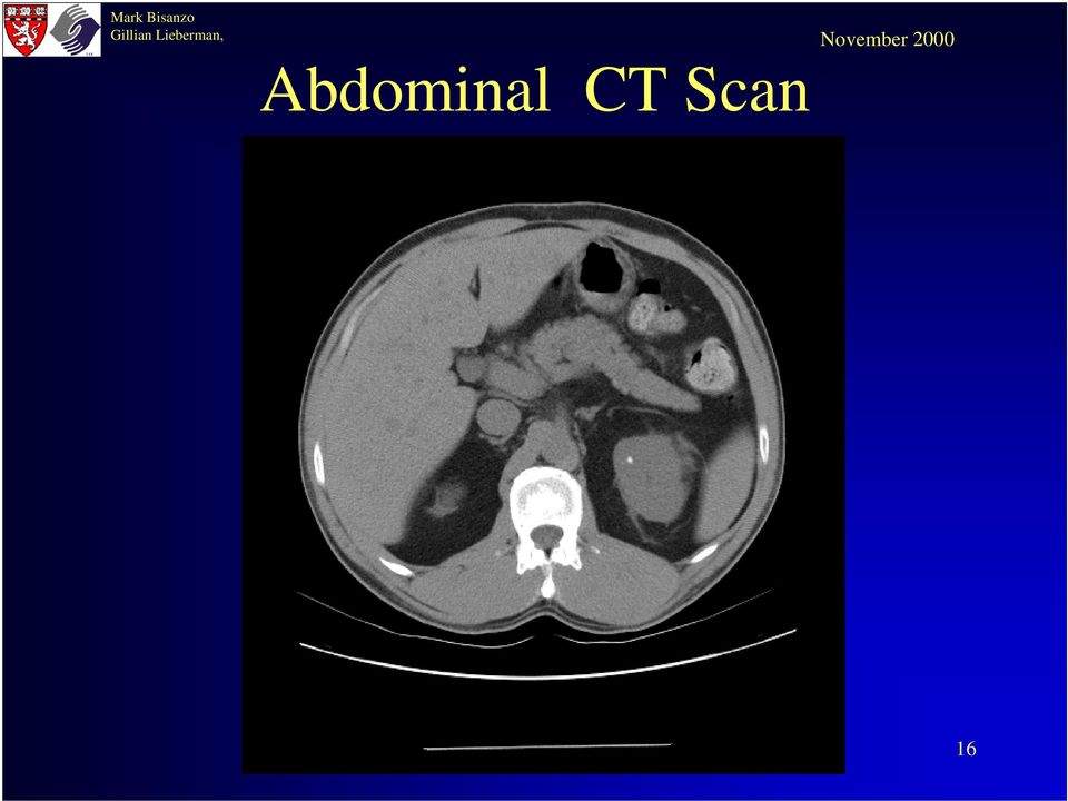
17 Abdominal CT Scan Without IV Contrast Liver IVC Pancreas Calcified Stone in Left Kidney (Not responsible for obstruction) spleen Right Kidney Left Kidney 17
 spleen Right Kidney Left")
18 Abdominal CT Scan Without IV Contrast Gallbladder Colon with stool Liver IVC Aorta Descending Colon Dilated collecting system Right Kidney Left Kidney Perinephric stranding No Stones Observed 18

19 Pelvic CT Scan Without IV Contrast Bladder Phleboliths Uretovesicular Junction (UVJ) No Stones Observed 19
 No Stones")
20 IV Contrast Enhanced Abdominal CT IVC Scan Gallbladder Aorta Liver Right Kidney Left Kidney No Stones Observed Stranding 20

21 Summary of CT Findings Delayed excretion and dilation of the renal pelvis and collecting system are c/w acute renal obstruction. Still no stone was visualized?! Almost all stones should be seen on unenhanced helical CT. 21
22 Plain Film following Abdominal CT 22
23 Plain Film following Abdominal CT Right Kidney Right ureter Left Kidney (enlarged, poorly functioning) Bladder 23
24 Findings Findings are consistant with left renal obstruction but again the site of obstruction was not seen. 24
25 Sonography Let s review the ultrasound appearance of renal obstruction 25
26 Sonography 26
27 Sonography Legend NL Right Kidney Fat **** 27
28 Sonography 28
29 Sonography Legend Liver Gallstones Fat *** Gallbladder Kidney 29
30 Sonography HYDRONEPHROTIC LEFT KIDNEY 30
31 Sonography Legend Dilated Collecting System (DC) Kidney ( ) Fat (***) 31
32 Sonography PELVIS 32
33 Sonography 33
34 The Appropriate Test An IVU and an Ultrasound were not indicated in this patient, as the CT scan coupled with the history made the diagnosis. So what was the history? 34
35 Back to the History The Radiologist, attending ER Doc and medical student met to review the imaging results. The Radiologist and ER Doc smiled knowingly. They suggested the medical student talk with the patient about his medical history, research nephrolithiasis on the web and then report why no stone was visualized. Here s what the medical student found. 35
36 Patient History PG is a 47 year old HIV+ male who has recently started taking Indinavir. His doctor asked him to drink at least 1.5 liters of fluid everyday, but he is too busy at work to comply He began experiencing left flank/llq pain approximately 12 hours ago which became intolerable. 36
37 History of Imaging Nephrolithiasis First image of renal calculus: April 1896 by John Macintyre Just a few months after Roentgen s discovery of x-rays Gained support quickly Orton in 1908 publishes on the diagnostic dilemma of the misdiagnosis of a phlebolith as renal calculus 37
38 Attempts to visualize the ureters Tuffier (1896) - 1 st opacification of the ureter on radiograph inserted a metal wire into a ureteral catheter radiopaque ureteral catheters were developed to diagnose ureteral stones. Other means of outlining the ureters on radiographs were soon attempted. Wittek (1903) - used air, but it did not gain popularity. Voelcker and von Lichtenberg (1905) - 1 st liquid contrast agent a colloidal suspension of silver (Collargol) instilled into the bladder same MDs opacified the entire collecting system 1 st technique of retrograde pyelogram Collargol deemed responsible for several cases of renal damage and even some deaths 38
39 Attempts to visualize the ureters Cameron (1918) - 1 st iodine-containing contrast material Solution of sodium and potassium iodide became the agent of choice for retrograde pyelography Retrograde pyelography, however, was still less than ideal given that cystoscopy and instrumentation were required. Weld (1919) reports a contralateral pyelogram in the contralateral kidney in patients undergoing unilateral retrograde pyelography. Assumed to be due to absorption of the contrast material into the circulation where it was filtered and excreted by both kidneys. Osborne (1923) - first use of IV sodium iodide to achieve a bilateral pyelogram Poor quality and consistency of images obtained in this manner. 39
40 Attempts to visualize the ureters Modern intravenous urography (IVU) Swick (1929) - iodinated pyridine compound (Selectan) Contrast agent of choice for the next 20 years. Iodinated benzoic Acid derivatives used in 1952 Much safer than pyridine-based agents Nonionic agents introduced s 40
41 The Plain Radiograph For almost a century after Macintyre s initial finding, the plain radiograph was felt to be the diagnostic imaging of choice for nephrolithiasis. This was supported by many reports throughout the years quoting excellent sensitivity values 41
42 The Plain Radiograph Many studies reported the sensitivity of the plain radiograph to be quite high: % of stones radiopaque (Twinem) % of stones radiopaque (Ravich) % of stones radiopaque (Herring) The 5th edition of Brenner and Rector s The Kidney quotes 85% are radiopaque 42
43 The Plain Radiograph Other studies reported less impressive numbers % radiopaque (Roth) % radiopaque (Mutgi) Problem with all of the studies: Used recovery of stones by the patient or a positive IVU to make a diagnosis of ureterolithiasis. This is not proof that the calcific density seen on the abdominal radiograph was the stone. 43
44 The Plain Radiograph The final answer on sensitivity: CT used as a gold standard to confirm the precise location of calcific densities seen on plain radiography (Levine et al) sensitivity of 59% for detecting ureteral calculi (95% CI for this value being 49% to 70%). Films viewed three times (twice blinded and once unblinded) 44
45 Stone composition and its appearance on plain radiograph Stone Composition Calcium phosphate Calcium oxalate Struvite Cystine Uric acid matrix stones Indinivar Appearance on Radiograph Most radiodense < Calcium phosphate < Calcium oxalate Mildly radiodense Radiolucent Radiolucent Radiolucent 45
46 Other Factors that Determine Visibility on Radiograph Peak kilovoltage used: low kilovoltage (peak) technique (60 to 70 kvp) is ideal, but this may not be possible especially with obese patients Presence of overlying bowel contents or bone. Size of the stone generally must be > 2 mm to be seen on plain radiographs 46
47 Visibility on Radiograph Bottom line: if a calcific density is seen along the anatomic course of the ureters on a plain radiograph, it cannot be definitively said to be in the ureter, because the ureter cannot be directly visualized. However, if the patient s clinical presentation suggests urolithiasis this calcification mandates a renal work up. 47
48 Advantages of IVU Can usually daignose ureteral obstruction Can image radiolucent stones Gives Rough estimate of renal function based on timing of opacification with contrast. 48
49 IVU Signs of obstruction include a delayed nephrogram delayed pyelogram dilatation of the collecting system, No literature that these findings correlate with true physiologic parameters, treatment outcome, or degree of residual renal impairment s/p obstruction. 49
50 Disadvantages of IVU IV iodinated contrast material - risk of adverse reactions GI: nausea, vomiting CP: bronchospasm, hypotension CNS: seizures Other: nephrotoxicity, uticaria, anaphylactoid reactions Indirect findings may be absent (acute partial obstruction). Multiple radiographs may be needed to determine level of obstruction leading to an increased dose of radiation. Cannot diagnoses nonrenal causes of flank pain. 50
51 IVU: Pyelogram Phase 51
52 IVU: Pyelogram Phase Right Calyx Left Calyx Bladder 52
53 US can detect Sonography dilatation of the collecting system changes in renal blood flow altered urine flow through the ureteral orifices in the bladder that may accompany obstruction. Stones may be visualized as an echogenic focus with or without acoustic shadowing. Stones within the ureter generally CANNOT be seen with US 53
54 Advantages of Sonography Patient is not exposed to radiation Stone visualization is not dependent on their composition Quick, inexpensive study 54
55 Disadvantages of Sonography Size of stones cannot be accurately measured. Diagnosis rests on indirect signs of obstruction, which may be unreliable. Delay of > 24 hours s/p onset of obstruction for collecting system dilatation and altered blood flow. Forniceal rupture with decompression of the pelvocalyceal system will yield ambiguous results. Therefore, high rate of false-negative US studies. Can be hard to image extra-ureteral causes of obstruction 55
56 CT as a modality to image ureteral calculi First report of successful use in 1994 and first published in Has since become the gold standard and is widely used in the ED to diagnose acute ureteral obstruction. Specialzed protocol using helical CT is referred to as a CT urogram (CTU). 56
57 Advantages of CTU May eliminate need for IV contrast Time: < 5 minutes Virtually all stones (including uric acid stones) can be readily visualized with CT Determines site and size of ureteral stones Secondary signs of obstruction allow diagnosis of recently passed stone Can diagnose other causes of acute flank pain 57
58 Accuracy of CT in determining stone size Neitlich et al. used spherical stone phantoms (diameters: 1 to 15 mm) & found that CT measurements had an error of 2% to 7% if the stone was 4 mm or greater and 6% to 12% if the diameter was less than 4 mm. * Size is crucial to determining management! * Presented at the annual meeting of the Society of Uroradiology, Santa Fe, New Mexico, June,
59 Stone Visible on CT In essence all stones are radiopaque (i.e. visualized) on CT scanning except. 59
60 Stones not visible on CT Indinavir (Crixovan) Ahah. That s the protease inhibitor that PG s on!. Associated with a 4% incidence of nephrolithiasis - calculi largely contain precipitated indinivar Can occur in patients who never had nephrolithiasis before indinivar therapy Asymptomatic crystalluria occurs as well (~20%) Treatment: hydration and drug withdrawal. Many restart drug. Pure Matrix stones (uncommon) 60
61 Secondary Signs Observed on CT 1) Ureteral dilatation - very important 2 o sign Ureter on obstructed side should have greater lumen diameter than unobstructed side at multiple levels! Studies report: sensitivity ~ 90%, specificity ~ 90%, Other causes of ureteral dilatation: acute or chronic diffuse or focal pyelonephritis Inflammatory processes adjacent to the ureter may can lead to decreased ureteral peristalsis both locally and diffusely, resulting in ureteral dilatation. Pts who have had prior obstruction, may remain dilated Extrinsic compression by an abdominal or pelvic mass 61
62 Secondary Signs Observed on CT 2) Soft tissue stranding of the perinephric fat Inflammatory change or fluid in the perinephric space May be most visible at the lower poles (dependent areas) Specificity ~ 90%, Sensitivity ~80% Relative amount of stranding may correlate with likelihood of passing the stone 3) Periureteral Stranding Less common than perinephric stranding 62
63 Secondary Signs Observed on CT 4) Collecting system dilatation NL prominence of the renal pelvis or an extrarenal pelvis should not be confused with dilatation Harder to assess than ureteral dilatation, but specificity and sensitivity are comparable 3) Unilateral renal enlargement 4) Decreased attenuation of the obstructed kidney 5) Rim sign: edema in ureteral wall at site of stone impaction (4-24 hours s/p impaction) 63
64 Alternate Diagnoses In a study by Dalrymple et al, in cases where CT was negative for stone disease, it provided evidence of the following diagnoses: GYN: ovarian masses that underwent torsion or hemorrhage GI: appendicitis, diverticulitis, choledocholithiasis, Crohn's disease, pancreatitis, ventral hernia, cholecystitis Cardiovascular : leaking abdominal aortic aneurysm, renal artery aneurysm Urinary tract: pyelonephritis and bladder outlet obstruction Masses: lymphoma with hemorrhage, retroperitoneal liposarcoma, a hemorrhagic liver hemangioma, vertebral metastases, and a subserosal uterine leiomyoma Other: ruptured spleen 64
65 Summary of Clinical Protocol Patient presents to the ED with Hx & Sx of Ureteral obstruction Order an unenhanced helical CT 1) Stone Identified Base Management on estimated size 2) No Stone seen; 2 o signs of obstruction are present Is patient taking Indinavir? yes R/O other causes of 2 o signs, consult PCP and taper indinavir No 3) No clear stone identified, but suspicious calcification present along ureter course Obtain overlapping reconstructions at and below level of calcification Proceed with work up of DDX 5) No stone identified and no 2 o signs of obstruction present Exclude stone disease and work up rest of DDX 4) Indeterminate result (usually 2 o to inadequate retroperitoneal fat) IV contrast and rescan 65
66 References Dalrymple NC, Verga M, Anderson KR, et al: The value of unenhanced helical CT in the management of patients with acute flank pain. J Urol 159: , 1997 Kopp J B, MD; Miller KD, MD; Mican JM, MD; et al: Crystalluria and urinary tract abnormalities associated with indinavir. Ann Internal Med. 127: , Levine JA, Neitlich JD, Verga M, et al: Identification of ureteral calculi on plain radiographs in patients with flank pain: Correlation with helical CT. Radiology 204:27-31, 1997 Morse R, Resnick M: Ureteral calculi: Natural history and treatment in an era of advanced technology. J Urol 145: , 1991 Smith RC; Levine J; Rosenfeld AT: Helical CT of urinary tract stones. Epidemiology, origin, pathophysiology, diagnosis, and management. Radiol Clin North Am 37(5): ,
67 Acknowledgments Special thanks to Dr. Martina Morrin for providing the index case; and Dr. Matthew Spencer for his expertise in interpreting CT and ultrasound. Thanks also to Beverlee Turner for her Power Point expertise and help. 67
Open the Flood Gates Urinary Obstruction and Kidney Stones. Dr. Jeffrey Rosenberg Dr. Emilio Lastarria Dr. Richard Kasulke
 Open the Flood Gates Urinary Obstruction and Kidney Stones Dr. Jeffrey Rosenberg Dr. Emilio Lastarria Dr. Richard Kasulke Nephrology vs. Urology Nephrologist a physician who has been trained in the diagnosis
Abdominal CT scan findings in Acute Appendicitis
 Abdominal CT scan findings in Acute Appendicitis Pathophysiology of acute appendicitis. Acute appendicitis occurs when the lumen is obstructed, leading to fluid accumulation, luminal distention, inflammation
NEPHROLITHIASIS Diagnosis & Treatment
 Learning Objectives NEPHROLITHIASIS Diagnosis & Treatment Jai Radhakrishnan, MD, MS Professor of Clinical Medicine Columbia University Management of the first episode of renal colic: Optimal Imaging Treatment
X-ray (Radiography) - Abdomen
 - Abdomen") Scan for mobile link. X-ray (Radiography) - Abdomen Abdominal x-ray uses a very small dose of ionizing radiation to produce pictures of the inside of the abdominal cavity. It is used to evaluate the stomach,
Scan for mobile link. X-ray (Radiography) - Abdomen Abdominal x-ray uses a very small dose of ionizing radiation to produce pictures of the inside of the abdominal cavity. It is used to evaluate the stomach,
Renal Cysts What should I do now?
 Renal Cysts What should I do now? Dr Edmund Chiong Asst. Professor & Consultant Department of Urology National University Hospital What are renal cysts? Fluid-filled structures in the kidney that are not
Radiographic Findings and Comments
 642 Abdomen (Cont.) Differential Diagnosis of Abdominal s E. Focal parenchymal calcification of the kidney Tuberculosis (Fig. 27.19) Adenocarcinoma (Fig. 27.20) Nephroblastoma (Wilms tumor) (Fig. 27.21)
Sonographic Diagnosis of Ureteral Tumors
 Sonographic Diagnosis of Ureteral Tumors Irith Hadas-Halpern, MD, micur Farkas, MD, Michael Patlas, MD, Ibrahim Zaghal, MD, Shoshana Sabag-Gottschalk, MD, Drora Fisher, MD We present our experience with
Primary Care Management of Male Lower Urinary Tract Symptoms. Matthew B.K. Shaw Consultant Urological Surgeon
 Primary Care Management of Male Lower Urinary Tract Symptoms Matthew B.K. Shaw Consultant Urological Surgeon NICE LUTS Guidelines Lower Urinary Tract Symptoms (LUTS) in men. NICE Clinical Guideline 97
53 X-rays and Diagnostic Radiology
 Learning Outcomes 53.1 Explain how x-rays are used for diagnostic and therapeutic purposes. 53-2 CHAPTER 53 X-rays and Diagnostic Radiology 53.2 Compare invasive and noninvasive diagnostic procedures.
US for Detecting Renal Calculi with Nonenhanced CT as a Reference Standard 1
 Genitourinary Imaging Keir A. B. Fowler, MD Julie A. Locken, MD Joshua H. Duchesne Michael R. Williamson, MD Index terms: Kidney, calculi, 811.811, 813.811 Kidney, CT, 81.12111, 81.12115 Kidney, US, 81.1298
thirteen, it was more marked and here one could see a certain degree of beading along the lines which radiated out from the calyx.
 CONGENITAL CYSTIC DILATATION OF THE RENAL COLLECTING TUBULES A NEW DISEASE ENTITY* VINCENT VERMOOTEN** In 1943, while I was in the U. S. Army, a young soldier was referred to me because of gross, painless,
THE KIDNEY. Bulb of penis Abdominal aorta Scrotum Adrenal gland Inferior vena cava Urethra Corona glandis. Kidney. Glans penis Testicular vein
 29 THE KIDNEY 9. Recurrent urinary tract infections Recurrent urinary tract infections The urinary tract consists of the urethra, the bladder, the ureters, the kidneys and in men the prostate gland. An
Positron Emission Tomography - For Patients
 Positron Emission Tomography - For Patients A physician s written order is required for any PET-CT tests. How should I prepare for my PET-CT? PET-CT is more complicated than most other tests you may be
06/10/09. 64 Slice BODY CT PROTOCOLS. Table of Contents
 06/10/09 64 Slice BODY CT PROTOCOLS Table of Contents General CT Comments 1 Protocol Routine Abdomen/Pelvis.. Routine Chest, Abdomen and Pelvis Retroperitoneal Hemorrhage Trauma. CT Cystography... Triple
surg urin Surgery: Urinary System 1
 Surgery: Urinary System 1 This section contains information to assist providers in billing for surgical procedures related to the urinary system. Extracorporeal Shock Wave Lithotripsy Medi-Cal covers Extracorporeal
To Whipple or Not to Whipple, that is the Question: Evaluating the Resectability of Pancreatic Adenocarcinoma
 August 2009 To Whipple or Not to Whipple, that is the Question: Evaluating the Resectability of Pancreatic Adenocarcinoma Christina Ramirez, Harvard Medical School Year III Gillian Lieberman, MD Agenda
Metastatic Prostate Cancer Causing Complete Obstruction of the IVC
 Department of Radiology Henry Ford Health System Detroit, Michigan Metastatic Prostate Cancer Causing Complete Obstruction of the IVC Jennifer Johnston MSIII, Wayne State Medical School Stage 4 Metastatic
X-Plain Kidney Stones Reference Summary
 X-Plain Kidney Stones Reference Summary Introduction Kidney stones are fairly common. Although they can be very painful, they are treatable, and in many cases preventable. This reference summary will help
BERGEN COMMUNITY COLLEGE DIAGNOSTIC MEDICAL SONOGRAPHY PROGRAM Division of Health Professions DMS 213 SYLLABUS
 BERGEN COMMUNITY COLLEGE DIAGNOSTIC MEDICAL SONOGRAPHY PROGRAM Division of Health Professions DMS 213 SYLLABUS Course Title: DMS 213 - Abdominal Sonography 2 2 lec. 3 lab. 3 credits (5 hours) Required
A Diagnostic Chest XRay: Multiple Myeloma
 Daniela Marinho Tridente, VI FCMSCSP October 2013 A Diagnostic Chest XRay: Multiple Myeloma Daniela Marinho Tridente, VI FCMSCSP Our Learning Agenda Introduction of our patient His imaging data and findings
Recurrent Kidney Stones
 Recurrent Kidney Stones Sean A. Pierre, MD; and Darren T. Beiko, MD, FRCSC As presented at the College of Canadian Family Physicians Annual Family Medicine Forum, Toronto, Ontario. Family physicians are
A912: Kidney, Renal cell carcinoma
 A912: Kidney, Renal cell carcinoma General facts of kidney cancer Renal cell carcinoma, a form of kidney cancer that involves cancerous changes in the cells of the renal tubule, is the most common type
Abdominal Pain in a Pregnant Patient
 January 2007 Abdominal Pain in a Pregnant Patient Megan Browning, Harvard Medical School Year III 1 HPI Ms.O is a 21yo pregnant female (23+6 weeks gestation) Woke with 5/10 crampy abdominal pain followed
Abdomen X-Ray (AXR) Collimation is ideally from diaphragms to lower border of the symphysis pubis and the lateral skin margins.
 Collimation is ideally from diaphragms to lower border of the symphysis pubis and the lateral skin margins.") Abdomen X-Ray (AXR) Collimation is ideally from diaphragms to lower border of the symphysis pubis and the lateral skin margins. LMP of child-bearing age female patients should be checked. 1. Acute abdomen
Abdomen X-Ray (AXR) Collimation is ideally from diaphragms to lower border of the symphysis pubis and the lateral skin margins. LMP of child-bearing age female patients should be checked. 1. Acute abdomen
Palm Beach Obstetrics & Gynecology, PA
 Palm Beach Obstetrics & Gynecology, PA 4671 S. Congress Avenue, Lake Worth, FL 33461 561.434.0111 4631 N. Congress Avenue, Suite 102, West Palm Beach, FL 33407 Urinary Tract Infection About one of every
Gallstone Ileus. Audrey C. Durrant,, M.D. SUNY Downstate Medical Center May 20, 2005
 Gallstone Ileus Audrey C. Durrant,, M.D. SUNY Downstate Medical Center May 20, 2005 Gallstone Ileus Diagnosis and Management Background Misnomer coined by Bartolin in 1654 Not a true ileus True mechanical
Table of Contents. User Instructions...Page 2. Group Administrator (Chief/Department Chair or Point of Contact (POC).Page 3
.Page 3") Table of Contents User Instructions......Page 2 Group Administrator (Chief/Department Chair or Point of Contact (POC).Page 3 Report Index and Sample Reports.Page 16 Example of Scoring..Page 19 1 RADPEER
Table of Contents User Instructions......Page 2 Group Administrator (Chief/Department Chair or Point of Contact (POC).Page 3 Report Index and Sample Reports.Page 16 Example of Scoring..Page 19 1 RADPEER
Published on: 07/04/2015 Page 1 of 5
 Bladder Stones A DNA test for Hyperunicosuria (HUU) to find the gene which is implicated in the development of URATE stones has been developed which work with Bulldogs and many other breeds including Black
Urinalysis and Body Fluids CRg. Automation: Introduction. Urine Automation. published by Bayer. Unit 3. Chemical Examination of Urine
 Urinalysis and Body Fluids CRg Unit 3 Chemical Examination of Urine Part 7, Automation, Function Tests, and Calculi Automation: Introduction Advantages Saves time Allows for standardization of procedures
Cardiovascular diseases. pathology
 Cardiovascular diseases pathology Atherosclerosis Vascular diseases A disease that results in arterial wall thickens as a result of build- up of fatty materials such cholesterol, resulting in acute and
Metastatic Cervical Cancer s/p Radiation Therapy, Radical Hysterectomy and Attempted Modified Internal Hemipelvectomy
 Metastatic Cervical Cancer s/p Radiation Therapy, Radical Hysterectomy and Attempted Modified Internal Hemipelvectomy Sarah Hutto,, MSIV Marc Underhill, M.D. January 27, 2009 Past History 45 yo female
The acute treatment of kidney stones (urolithiasis) addresses
 addresses") Renal Calculi: Emergency Department Diagnosis And Treatment Abstract The acute treatment of kidney stones (urolithiasis) addresses pain management and focuses on the effects of the morbidity associated
Renal Calculi: Emergency Department Diagnosis And Treatment Abstract The acute treatment of kidney stones (urolithiasis) addresses pain management and focuses on the effects of the morbidity associated
Urinary Tract Infections
 1 Infections in the urinary tract are relatively common. These infections are often referred to as bladder infections. They are also known as UTI s or urinary tract infections. When an infection is confined
URINARY (RENAL) STONE (NEPHROLITHOISIS) An Overview
 STONE (NEPHROLITHOISIS) An Overview") URINARY (RENAL) STONE (NEPHROLITHOISIS) An Overview UNIVERSITY OF PNG SCHOOL OF MEDICINE AND HEALTH SCIENCES DIVISION OF BASIC MEDICAL SCIENCES DISCIPLINE OF BIOCHEMISTRY AND MOLECULAR BIOLOGY PLB MBBS
URINARY (RENAL) STONE (NEPHROLITHOISIS) An Overview UNIVERSITY OF PNG SCHOOL OF MEDICINE AND HEALTH SCIENCES DIVISION OF BASIC MEDICAL SCIENCES DISCIPLINE OF BIOCHEMISTRY AND MOLECULAR BIOLOGY PLB MBBS
Amylase and Lipase Tests
 Amylase and Lipase Tests Also known as: Amy Formal name: Amylase Related tests: Lipase The Test The blood amylase test is ordered, often along with a lipase test, to help diagnose and monitor acute or
By Anne C. Travis, M.D., M.Sc. and John R. Saltzman, M.D., FACG Brigham and Women's Hospital Harvard Medical School Boston, MA
 SMALL BOWEL BLEEDING: CAUSES, DIAGNOSIS AND TREATMENT By Anne C. Travis, M.D., M.Sc. and John R. Saltzman, M.D., FACG Brigham and Women's Hospital Harvard Medical School Boston, MA 1. What is the small
Ovarian Torsion: Sonographic Evaluation
 J Clin Ultrasound 17:327-332, June 1989 Ovarian Torsion: Sonographic Evaluation Mark A. Helvie, MD,* and Terry M. Silver, MDI Abstract: The sonographic and clinical findings of 13 patients with surgically
Patient Information Booklet. Endovascular Stent Grafts: A Treatment for Abdominal Aortic Aneurysms
 Patient Information Booklet Endovascular Stent Grafts: A Treatment for Abdominal Aortic Aneurysms TABLE OF CONTENTS Introduction 1 Glossary 2 Abdominal Aorta 4 Abdominal Aortic Aneurysm 5 Causes 6 Symptoms
Pyelonephritis: Kidney Infection
 Pyelonephritis: Kidney Infection National Kidney and Urologic Diseases Information Clearinghouse U.S. Department of Health and Human Services NATIONAL INSTITUTES OF HEALTH What is pyelonephritis? Pyelonephritis
Women s Health Laparoscopy Information for patients
 Women s Health Laparoscopy Information for patients This leaflet is for women who have been advised to have a laparoscopy. It outlines the common reasons doctors recommend this operation, what will happen
An abdominal ultrasound produces a picture of the organs and other structures in the upper abdomen.
 Scan for mobile link. Ultrasound - Abdomen Ultrasound imaging of the abdomen uses sound waves to produce pictures of the structures within the upper abdomen. It is used to help diagnose pain or distention
Kidney Stones. This reference summary will help you understand kidney stones and how to treat and prevent them. Kidney
 Introduction A kidney stone is a solid piece of material that forms in the kidney from substances in the urine. Kidney stones are fairly common. Although kidney stones can be painful, they are treatable.
Acute abdominal conditions Key Points
 7 Acute abdominal conditions Key Points 7.1 ASSESSMENT AND DIAGNOSIS Referred abdominal pain Fore gut pain (stomach, duodenum, gall bladder) is referred to the upper abdomen Mid gut pain (small intestine,
PATIENT CONSENT TO PROCEDURE - ROUX-EN-Y GASTRIC BYPASS
 As a patient you must be adequately informed about your condition and the recommended surgical procedure. Please read this document carefully and ask about anything you do not understand. Please initial
Incontinence. What is incontinence?
 Incontinence What is incontinence? Broadly speaking, the medical term incontinence refers to any involuntary release of bodily fluids, but many people associate it strongly with the inability to control
Preventing Catheter Blockages: A Guide for Health Professionals
 Introduction Preventing Catheter Blockages: A Guide for Health Professionals and Long term catheterisation is common for someone with a spinal cord injury (SCI). However, it poses a concern as this method
The Lewin Group undertook the following steps to identify the guidelines relevant to the 11 targeted procedures:
 Guidelines The following is a list of proposed medical specialty guidelines that have been found for the 11 targeted procedures to be included in the Medicare Imaging Demonstration. The list includes only
6/3/2011. High Prevalence and Incidence. Low back pain is 5 th most common reason for all physician office visits in the U.S.
 High Prevalence and Incidence Prevalence 85% of Americans will experience low back pain at some time in their life. Incidence 5% annual Timothy C. Shen, M.D. Physical Medicine and Rehabilitation Sub-specialty
 P R E S E N T S Dr. Mufa T. Ghadiali is skilled in all aspects of General Surgery. His General Surgery Services include: General Surgery Advanced Laparoscopic Surgery Surgical Oncology Gastrointestinal
Metastatic Renal Cell Carcinoma: Staging and Prognosis of Three Separate Cases.
 Metastatic Renal Cell Carcinoma: Staging and Prognosis of Three Separate Cases. Abstract This paper describes the staging, imaging, treatment, and prognosis of renal cell carcinoma. Three case studies
What You Should Know About Cerebral Aneurysms
 What You Should Know About Cerebral Aneurysms From the Cerebrovascular Imaging and Interventions Committee of the American Heart Association Cardiovascular Radiology Council Randall T. Higashida, M.D.,
LIVER CANCER AND TUMOURS
 LIVER CANCER AND TUMOURS LIVER CANCER AND TUMOURS Healthy Liver Cirrhotic Liver Tumour What causes liver cancer? Many factors may play a role in the development of cancer. Because the liver filters blood
Nephrology (Renal Medicine)
") Nephrology (Renal Medicine) Dr Shabbir Moochhala Consultant Nephrologist, Royal Free Hospital, London Cystinuria Patient Day 1 st Feb 2014 Nephrologist Urologist The Kidney designed for the job The body
Nephrology (Renal Medicine) Dr Shabbir Moochhala Consultant Nephrologist, Royal Free Hospital, London Cystinuria Patient Day 1 st Feb 2014 Nephrologist Urologist The Kidney designed for the job The body
POAC CLINICAL GUIDELINE
 POAC CLINICAL GUIDELINE Acute Pylonephritis DIAGNOSIS COMPLICATED PYELONEPHRITIS EXCLUSION CRITERIA: Male Known or suspected renal impairment (egfr < 60) Abnormality of renal tract Known or suspected renal
Pediatric Upper GI Series New Patient
 Pediatric Upper GI Series New Patient Upper GI Series Thought to be malrotation, no evidence of midgut volvulus Needed to repeat UGI Series WHY? Repeat UGI Series Repeat UGI Series Repeat UGI Series No
A Patient s Guide to Minimally Invasive Abdominal Aortic Aneurysm Repair
 A Patient s Guide to Minimally Invasive Abdominal Aortic Aneurysm Repair Table of Contents The AFX Endovascular AAA System............................................ 1 What is an Abdominal Aortic Aneurysm
Four Important Facts about Kidney Cancer
 Volume Article.13-11 Publish Date: 24th June 2013 Author(s): MPUH - CRS Team Four Important Facts about Kidney Cancer ARTICLE hhh Muljibhai Patel Urological Hospital (MPUH) Centre For Robotic Surgery (CRS)
Smoking and misuse of certain pain medicines can affect the risk of developing renal cell cancer.
 Renal cell cancer Renal cell cancer is a disease in which malignant (cancer) cells form in tubules of the kidney. Renal cell cancer (also called kidney cancer or renal adenocarcinoma) is a disease in which
RENAL ANGIOMYOLIPOMA EMBOLIZATION
 RENAL ANGIOMYOLIPOMA EMBOLIZATION The information about renal angiomyolipomas on the next several pages includes questions commonly asked about the embolization procedure. Please take a few moments to
Guide to Abdominal or Gastroenterological Surgery Claims
 What are the steps towards abdominal surgery? Investigation and Diagnosis It is very important that all necessary tests are undertaken to investigate the patient s symptoms appropriately and an accurate
WHAT S WRONG WITH MY GALL BLADDER? GALL BLADDER POLYPS
 WHAT S WRONG WITH MY GALL BLADDER? GALL BLADDER POLYPS This is a patient information booklet providing specific practical information about gall bladder polyps in brief. Its aim is to provide the patient
KIDNEY FUNCTION RELATION TO SIZE OF THE TUMOR IN RENAL CELL CANCINOMA
 KIDNEY FUNCTION RELATION TO SIZE OF THE TUMOR IN RENAL CELL CANCINOMA O.E. Stakhvoskyi, E.O. Stakhovsky, Y.V. Vitruk, O.A. Voylenko, P.S. Vukalovich, V.A. Kotov, O.M. Gavriluk National Canсer Institute,
INTRAPERITONEAL HYPERTHERMIC CHEMOTHERAPY (IPHC) FOR PERITONEAL CARCINOMATOSIS AND MALIGNANT ASCITES. INFORMATION FOR PATIENTS AND FAMILY MEMBERS
 FOR PERITONEAL CARCINOMATOSIS AND MALIGNANT ASCITES. INFORMATION FOR PATIENTS AND FAMILY MEMBERS") INTRAPERITONEAL HYPERTHERMIC CHEMOTHERAPY (IPHC) FOR PERITONEAL CARCINOMATOSIS AND MALIGNANT ASCITES. INFORMATION FOR PATIENTS AND FAMILY MEMBERS Description of Treatment A major difficulty in treating
INTRAPERITONEAL HYPERTHERMIC CHEMOTHERAPY (IPHC) FOR PERITONEAL CARCINOMATOSIS AND MALIGNANT ASCITES. INFORMATION FOR PATIENTS AND FAMILY MEMBERS Description of Treatment A major difficulty in treating
Aehlert: Paramedic Practice Today PowerPoint Lecture Notes Chapter 50: Abdominal Trauma
 Aehlert: Paramedic Practice Today PowerPoint Lecture Notes Chapter 50: Abdominal Trauma Chapter 50 Abdominal Trauma 1 Describe the epidemiology, including morbidity, mortality rates, and prevention strategies,
Saint Francis Kidney Transplant Program Issue Date: 6/9/15
 Kidney Transplant Candidate Informed Consent Education Here are educational materials about Kidney Transplant. Please review and read these before your evaluation visit. The RN Transplant Coordinator will
Juvenile Dermatomyositis Joseph Junewick, MD FACR
 Juvenile Dermatomyositis Joseph Junewick, MD FACR 10/11/2015 History Child with several month history of weakness, arthralgias and palpable abnormalities at the knee Diagnosis Juvenile Dermatomyositis
Gallbladder Diseases and Problems
 Gallbladder Diseases and Problems Introduction Your gallbladder is a pear-shaped organ under your liver. It stores bile, a fluid made by your liver to digest fat. There are many diseases and problems that
Kidney Disease WHAT IS KIDNEY DISEASE? TESTS TO DETECT OR DIAGNOSE KIDNEY DISEASE TREATMENT STRATEGIES FOR KIDNEY DISEASE
 Kidney Disease WHAT IS KIDNEY DISEASE? Kidney disease is when your kidneys are damaged and not functioning as they should. When kidney disease is not going away it is called chronic kidney disease or CKD.
Gallbladder - gallstones and surgery
 Gallbladder - gallstones and surgery Summary Gallstones are small stones made from cholesterol, bile pigment and calcium salts, which form in a person s gall bladder. Medical treatment isn t necessary
Uterine Fibroid Symptoms, Diagnosis and Treatment
 Fibroids and IR Uterine Fibroid Symptoms, Diagnosis and Treatment Interventional radiologists use MRIs to determine if fibroids can be embolised, detect alternate causes for the symptoms and rule out misdiagnosis,
Patient Prep Information
 Stereotactic Breast Biopsy Patient Prep Information Imaging Services Cannon Memorial Hospital Watauga Medical Center Table Weight Limits for each facility Cannon Memorial Hospital Watauga Medical Center
Imaging of Thoracic Endovascular Stent-Grafts
 Imaging of Thoracic Endovascular Stent-Grafts Tariq Hameed, M.D. Department of Radiology and Imaging Sciences, Indiana University School of Medicine, Indianapolis, Indiana Disclosures: No relevant financial
Ovarian Cyst. Homoeopathy Clinic. Introduction. Types of Ovarian Cysts. Contents. Case Reports. 21 August 2002
 Case Reports 21 August 2002 Ovarian Cyst Homoeopathy Clinic Check Yourself If you have any of the following symptoms call your doctor. Sense of fullness or pressure or a dull ache in the abdomen Pain during
CHAPTER 20: URINARY SYSTEM
 OBJECTIVES: 1. Name the major function of the urinary system, and name and locate (on a diagram) the organs that compose the system. 2. Explain what the term renal refers to. 3. Define the term retroperitoneal.
X-ray (Radiography), Lower GI Tract
, Lower GI Tract") Scan for mobile link. X-ray (Radiography), Lower GI Tract What is Lower GI Tract X-ray Radiography (Barium Enema)? Lower gastrointestinal (GI) tract radiography, also called a lower GI or barium enema,
Scan for mobile link. X-ray (Radiography), Lower GI Tract What is Lower GI Tract X-ray Radiography (Barium Enema)? Lower gastrointestinal (GI) tract radiography, also called a lower GI or barium enema,
Gynecology Abnormal Pelvic Anatomy and Physiology: Cervix. Cervix. Nabothian cysts. cervical polyps. leiomyomas. Cervical stenosis
 Gynecology Abnormal Pelvic Anatomy and Physiology: (Effective February 2007) pediatric, reproductive, and perimenopausal/postmenopausal (24-28 %) Cervix Nabothian cysts result from chronic cervicitis most
Sample Learning Objectives for a Medical School Radiology Curriculum: Listed by Subjects
 Sample Learning Objectives for a Medical School Radiology Curriculum: Listed by Subjects This document lists sample learning objectives by subject matter The numerical ranking in parenthesis following
Introduction. Physiology of the Abdomen. Anatomy & Physiology. Abdominal Pain Introduction (2 of 2) Gastrointestional and Urologic Emergencies
 Gastrointestional and Urologic Emergencies") Gastrointestional and Urologic Emergencies Introduction Abdominal pain is a common complaint. Cause of abdominal pain is often difficult to determine. As an EMT: You do not need to determine exact cause.
Gastrointestional and Urologic Emergencies Introduction Abdominal pain is a common complaint. Cause of abdominal pain is often difficult to determine. As an EMT: You do not need to determine exact cause.
Case III. Disscussion. the UHP ultrasound protocol. Novel Ultrasound Approach to the Empiric Evaluation of the Undifferentiated Hypotensive Patient
 The UHP Ultrasound Protocol: A Novel Ultrasound Approach to the Empiric Evaluation of the Undifferentiated Hypotensive Patient JOHN S. ROSE, MD,* AARON E. BAIR, MD,* DIKU MANDAVIA, MD, AND DONNA J. KINSER,
Triple phosphates (Magnesium ammonium phosphates) (Struvite)
 (Struvite)") Amorphous phosphates Amorphous phosphates are the name given to a granular precipitate containing calcium and phosphate in an alkaline urine. Calcium phosphate crystals, regrouped under the term apatite,
Amorphous phosphates Amorphous phosphates are the name given to a granular precipitate containing calcium and phosphate in an alkaline urine. Calcium phosphate crystals, regrouped under the term apatite,
RBH GUIDELINES FOR ULTRASOUND REFERRAL
 RBH GUIDELINES FOR ULTRASOUND REFERRAL Introduction Ultrasound can be very useful as a first line investigation; it is typically non-invasive and does not involve ionising radiation. However, a significant
Identifying and treating long-term kidney problems (chronic kidney disease)
") Understanding NICE guidance Information for people who use NHS services Identifying and treating long-term kidney problems (chronic kidney disease) NICE clinical guidelines advise the NHS on caring for
Understanding NICE guidance Information for people who use NHS services Identifying and treating long-term kidney problems (chronic kidney disease) NICE clinical guidelines advise the NHS on caring for
Dual-energy CT refers to the acquisition
 bdominal dual-source dual-energy CT: Uses in clinical practice Marilyn J. Siegel, MD, Juan Carlos Ramirez-Giraldo, PhD, and nno Graser, MD Dual-energy CT refers to the acquisition of computed tomography
Total Abdominal Hysterectomy
 What is a total abdominal hysterectomy? Is the removal of the uterus and cervix through an abdominal incision (either an up and down or bikini cut). Removal of the ovaries and tubes depends on the patient.
SOD (Sphincter of Oddi Dysfunction)
") SOD (Sphincter of Oddi Dysfunction) SOD refers to the mechanical malfunctioning of the Sphincter of Oddi, which is the valve muscle that regulates the flow of bile and pancreatic juice into the duodenum.
SOD (Sphincter of Oddi Dysfunction) SOD refers to the mechanical malfunctioning of the Sphincter of Oddi, which is the valve muscle that regulates the flow of bile and pancreatic juice into the duodenum.
MANAGEMENT OF DIABETIC PATIENTS IN RADIOLGY. Edition 2
 MANAGEMENT OF DIABETIC PATIENTS IN RADIOLGY Edition 2 Author, Authority Clinical Director Date of Origin December 2007 Date of Review November 2009 Scope of Authority, (Trust, other) Trust Total pages
Surgical removal of fibroids through an abdominal incision-either up and down or bikini cut. The uterus and cervix are left in place.
 What is an abdominal myomectomy? Surgical removal of fibroids through an abdominal incision-either up and down or bikini cut. The uterus and cervix are left in place. When is this surgery used? Treatment
1. What is the prostate-specific antigen (PSA) test?
 test?") 1. What is the prostate-specific antigen (PSA) test? Prostate-specific antigen (PSA) is a protein produced by the cells of the prostate gland. The PSA test measures the level of PSA in the blood. The doctor
1. What is the prostate-specific antigen (PSA) test? Prostate-specific antigen (PSA) is a protein produced by the cells of the prostate gland. The PSA test measures the level of PSA in the blood. The doctor
The digestive system eliminated waste from the digestive tract. But we also need a way to eliminate waste from the rest of the body.
 Outline Urinary System Urinary System and Excretion Bio105 Lecture 20 Chapter 16 I. Function II. Organs of the urinary system A. Kidneys 1. Function 2. Structure III. Disorders of the urinary system 1
Consent for Treatment/Procedure Laparoscopic Sleeve Gastrectomy
 Patient's Name: Today's Date: / / The purpose of this document is to confirm, in the presence of witnesses, your informed request to have Surgery for obesity. You are asked to read the following document
Chapter 6 Gastrointestinal Impairment
 Chapter 6 Gastrointestinal This chapter consists of 2 parts: Part 6.1 Diseases of the digestive system Part 6.2 Abdominal wall hernias and obesity PART 6.1: DISEASES OF THE DIGESTIVE SYSTEM Diseases of
Colic is one of the most dreaded conditions horse owners and trainers encounter with
 Impaction Colic and Hydration Michele Frazer, DVM, DACVIM, DACVECC Impaction Colic Colic is one of the most dreaded conditions horse owners and trainers encounter with their horses. The term colic, however,
Kidney Stones removal Without surgery
 Patient Education Service Lithotripsy - The world s latest treatment for Kidney Stones removal Without surgery With COMPUTERISED high tech DIREX LITHOTRIPTER Experience counts PIONEERS in North INDIA -
Jessica Konvicka, MPAS, PA-C University Of Texas Southwestern Medical Center Dallas, TX
 Kidney Stones Jessica Konvicka, MPAS, PA-C University Of Texas Southwestern Medical Center Dallas, TX Objectives Recognize signs and symptoms of a kidney stone. Evaluate a patient suspected to have a kidney
Inferior Vena Cava filter and removal
 Inferior Vena Cava filter and removal What is Inferior Vena Cava Filter Placement and Removal? An inferior vena cava filter placement procedure involves an interventional radiologist (a specialist doctor)
Preparing for your laparoscopic pyeloplasty
 Preparing for your laparoscopic pyeloplasty Welcome We look forward to welcoming you to The Royal London Hospital. You have been referred to us for a laparoscopic pyeloplasty, which is an operation using
Registered Charity No: 326679 Caring for those with a rare, complex and lifelong disease www.behcets.org.uk
 Registered Charity No: 326679 Caring for those with a rare, complex and lifelong disease www.behcets.org.uk Behçet s Disease and the Kidneys How are the kidneys affected by Behçet s disease? Kidney disease
Understanding. Pancreatic Cancer
 Understanding Pancreatic Cancer Understanding Pancreatic Cancer The Pancreas The pancreas is an organ that is about 6 inches long. It s located deep in your belly between your stomach and backbone. Your
Laparoscopic Surgery of the Colon and Rectum (Large Intestine) A Simple Guide to Help Answer Your Questions
 A Simple Guide to Help Answer Your Questions") Laparoscopic Surgery of the Colon and Rectum (Large Intestine) A Simple Guide to Help Answer Your Questions What are the Colon and Rectum? The colon and rectum together make up the large intestine. After
Laparoscopic Surgery of the Colon and Rectum (Large Intestine) A Simple Guide to Help Answer Your Questions What are the Colon and Rectum? The colon and rectum together make up the large intestine. After
Long-term result of Memokath urethral sphincter stent in. spinal cord injury patients
 1 Long-term result of Memokath urethral sphincter stent in spinal cord injury patients Subramanian Vaidyanathan 1 E-mail: vaidyanathansiu@hotmail.com Bakul M Soni 1 E-mail: Bakul.Soni@mail.soh-tr.nwest.nhs.uk