Acute Effects of Cocaine on Human Brain Activity and Emotion
|
|
|
- Emily Allison
- 8 years ago
- Views:
Transcription
1 Neuron, Vol. 19, , September, 1997, Copyright 1997 by Cell Press Acute Effects of Cocaine on Human Brain Activity and Emotion Hans C. Breiter, 1,2,9 Randy L. Gollub, 2 contrast, regions that demonstrated early but sustained Robert M. Weisskoff, 1 David N. Kennedy, 1,3 signal maxima were more correlated with craving than Nikos Makris, 3 Joshua D. Berke, 5 Julie M. Goodman, 1,2 with rush ratings; such regions included the NAc/SCC, Howard L. Kantor, 1,6 David R. Gastfriend, 2,4 right parahippocampal gyrus, and some regions of lat- Jonn P. Riorden, 2,4 R. Thomas Mathew, 2,4,7 eral prefrontal cortex. Sustained negative signal change Bruce R. Rosen, 1 and Steven E. Hyman 2,8 was noted in the amygdala, which correlated with crav- 1 Nuclear Magnetic Resonance Center ing ratings. Our data demonstrate the ability of fmri Department of Radiology to map dynamic patterns of brain activation following 2 Department of Psychiatry cocaine infusion in cocaine-dependent subjects and 3 Center for Morphometric Analysis provide evidence of dynamically changing brain net- Department of Neurology works associated with cocaine-induced euphoria and Massachusetts General Hospital and Harvard cocaine-induced craving. Medical School Boston, Massachusetts Addictions Services Introduction Department of Psychiatry Cocaine is one of the most reinforcing drugs known, Massachusetts General Hospital and Harvard both in humans and in animals (Johanson and Fischman, Medical School 1989). With repetitive use, cocaine may produce a pro- Boston, Massachusetts found state of addiction in humans characterized by PrograminNeuroscience compulsive drug use and inability to control use despite Harvard University significant adverse consequences (Gawin, 1991; Ameri- Boston, Massachusetts can Psychiatric Association, 1994). Progress toward un- Department of Cardiology derstanding the neural substrates of addiction to co- Massachusetts General Hospital and Harvard caine has been substantial in recent years but has been Medical School focused on animal models that permit invasive studies. Boston, Massachusetts Noninvasive functional neuroimaging approaches, such as functional magnetic resonance imaging (fmri), now allow studies of neural circuit function to be extended Summary to the human. This has the advantage of being able to correlate subjective information about emotional and We investigated brain circuitry mediating cocaine- cognitive responses with observed patterns of brain acinduced euphoria and craving using functional MRI tivation. (fmri). During double-blind cocaine (0.6 mg/kg) and sa- Based on extensive investigations of rodent and priline infusions in cocaine-dependent subjects, the entire mate models, the mesoaccumbens dopamine pathway, brain was imaged for 5 min before and 13 min after extending from the ventral tegmentum of the midbrain infusion while subjects rated scales for rush, high, low, (VT) to the nucleus accumbens, appears to be the critical and craving. Cocaine induced focal signal increases in shared substrate of the reinforcing effects of cocaine nucleus accumbens/subcallosal cortex (NAc/SCC), cau- (Louilot et al., 1989; Williams, 1989; Apicella et al., 1991; date, putamen, basal forebrain, thalamus, insula, hippo- Schultz et al., 1992) and other addictive drugs (reviewed campus, parahippocampal gyrus, cingulate, lateral precumbens in Koob, 1996). Using nondrug stimuli, the nucleus ac- frontal and temporal cortices, parietal cortex, striate/ has also been shown to play a critical role in extrastriate cortices, ventral tegmentum, and pons and learning associated with reinforcement (Mirenowicz and produced signal decreases in amygdala, temporal pole, Schultz, 1996). Reinforcement in animals depends on and medial frontal cortex. Saline produced few positive the increase in synaptic dopamine levels in the mesoac- or negative activations, which were localized to lateral cumbens circuit produced by cocaine-like drugs via prefrontal cortex and temporo-occipital cortex.subjects blockade of the dopamine reuptake transporter (DAT) who underwent repeat studies showed good replication (DeWit and Wise, 1977; Ritz et al., 1987). In both aniof the regional fmri activation pattern following cocaine mals and humans, the acutely reinforcing effects of psy- and saline infusions, with activations on saline retest chostimulant drugs can produce a pattern of repeated that might reflect expectancy. Brain regions that exhibister cocaine to gain pleasure, to conform to peer behav- self-administration. Human users may initially self-adminited early and short duration signal maxima showed a higher correlation with rush ratings. These included the ior, or to relieve stress and other dysphoric feelings. An ventral tegmentum, pons, basal forebrain, caudate, cinmay produce increasing levels of dependence and, accelerated pattern of drug use in vulnerable individuals gulate, and most regions of lateral prefrontal cortex. In eventually, addiction (Hyman, 1996). 7 While the mesoaccumbens dopamine pathway has Present address: Tri-County Center for Substance Abuse Treatment, been most closely implicated in the acutely rewarding Wilmington, North Carolina, Present address: Office of the Director, National Institute of Mental actions of cocaine, other circuits have also been impli- Health, Bethesda, Maryland, cated in reward processes, including the basal fore- 9 To whom correspondence should be addressed. brain, which receives major afferents from the nucleus

2 Neuron 592 accumbens and itself receives dopaminergic input nucleus would exhibit blood oxygen level dependent (Heimer et al., 1997). Brain stimulation reward (BSR) (BOLD) signal changes (Ogawa et al., 1992) for cocaine experiments have directly implicated the basal forebrain and not for saline. We were also interested in studying in reinforcement (Rompre and Shizgal, 1986; Shizgal other regions associated with reward and reward-based et al., 1989; Arvanitogiannis et al, 1996). The nucleus memory (e.g., basal forebrain and amygdala) and paraaccumbens is also strongly linked to the amygdala (Ito limbic structures reported activated in animal studies et al., 1974; Yim and Mogenson, 1982; Russchen et of acute cocaine administration (Brown et al., 1992; Stein al., 1985; Amaral, et al., 1992), a linkage thought to be and Fuller, 1992, 1993; Graham and Porrino, 1995; Hamimportant for the formation of stimulus reward associa- mer et al., 1995; Lyons et al., 1996). We predicted that tions (Jones and Mishkin, 1972; Spiegler and Mishkin, fmri activation in the nucleus accumbens would be 1981; Gaffan and Harrison, 1987; Gaffan et al., 1988). correlated with behavioral reports of cocaine-induced Recently, PET scanning has demonstrated amygdala subjective rush and high in dependent subjects. The activation during cocaine craving in abstinent cocaine- results in this report will be focused on subcortical limbic abusing subjects relative to normal controls (Childress structures and paralimbic cortex, though all regions et al., 1996, Soc. Neurosci., abstract; Grant et al., 1996; found activated will be discussed in the text and in- Schweitzer et al., 1996, Soc. Neurosci., abstract). Thus, cluded as a supplemental table available on the Internet. according to current neurobiological models, the nucleus accumbens, amygdala, basal forebrain, and VT are central components of circuitry mediating brain pro- Results cesses underlying reward and memory of that reward. A number of human studies using cocaine infusions Clinical and Physiological Data (Fowler et al., 1989; London et al., 1990; Pearlson et al., Seventeen subjects were infused with cocaine while be- 1993; Volkow et al., 1997a) and withdrawing subjects ing scanned with fmri. Scans affected by uncorrectable (Volkow et al., 1990, 1991, 1992, 1993, 1997b) have impli- gross movement were rejected as uninterpretable. Of cated the striatum in human cocaine use, withdrawal, these 17 subjects, ten had interpretable fmri data for and craving. Given the spatial resolution of the tech- the cocaine infusions, and ten had interpretable data niques utilized, they may not have fully distinguished for the saline infusions after motion correction (seven the dorsal and ventral striatum, in particular the nucleus studies with usable matched infusions). accumbens. None of these studies reported specific Following the cocaine infusion (0.6 mg/kg over 30 s), sampling of other regions implicated with reward pro- there was an increase in heart rate (HR) within the first cesses, such as the VT, basal forebrain, or amygdala. minute, while the increase in mean blood pressure Only three of these studies approached the 1 2 min (MBP) was slower. Similarly, the drop in end-tidal carbon temporal resolution needed to resolve components of dioxide (ETCO 2 ) was also slower. Cocaine (N 17) cocaine-induced euphoria (Fowler et al., 1989; Pearlson caused the HR to increase rapidly from a preinfusion et al., 1993; Volkow et al., 1997a). value of 60 7 beats per minute (bpm) to bpm To investigate activity in reward circuitry in humans at 2 min postinfusion (p ), to bpm at during cocaine infusions and to associate this activity 5 min postinfusion (p ), to bpm at with subjective reports for both cocaine-induced eupho- 10 min postinfusion (p ). Normal sinus rhythm ria and postcocaine craving, we used fmri (Bandettini was observed in all subjects throughout the study (Golet al., 1992; Kwong et al., 1992; Ogawa et al., 1992) in lub et al., 1996). conjunction with physiological monitoring and online MBP rose slightly from torr before the infusion evaluation of computerized behavioral rating scales. to torr at 2 min postinfusion (p 0.11, NS), FMRI with a 1.5 T instrument has higher resolution than then up to torr at 5 min (p 0.002) before previous PET and SPECT studies of cocaine effects, starting to slowly decline. The ETCO 2 dropped slowly permitting investigation of regions with relatively small from a baseline of 39 4mmHgto36 4mmHgby volume, such as the nucleus accumbens and the amyg- 10 min (p 0.02). In all subjects scanned, these three dala. For these experiments, cocaine-dependent volunmeasures had returned to baseline by 2 hr, the interinfuteers underwent an unblinded cocaine infusion the night before the fmri experiment for clinical screening and sion interval (Gollub et al., 1996). Physiologic responses for training with behavioral assessments on scales of to the 0.6 mg/kg cocaine infusion are in close accord rush, high, low, and craving. During the subsequent doucaine abusers (Fischman and Schuster, 1982; Fischman with previously published studies in experienced co- ble-blind cocaine (0.6 mg/kg) and saline infusions, subet al., 1985; Foltin and Fischman, 1991). jects rated these four scales every 15 s during multiple fmri acquisitions (Figure 1). Pilot results from this study Plasma samples taken before the first infusion demon- have previously been presented (Breiter et al., 1996c; strated absence of residual cocaine at the time of the Gollub et al., 1996), and data regarding global versus first infusion in all of the subjects studied. Peak plasma regional cocaine effects is presented elsewhere (Gollub cocaine levels (C max ) following the cocaine infusion et al., submitted). ranged from 197 to 893 mcg/l with a mean of Based upon animal data (Koob and Bloom, 1988; (N 7 subjects with complete data). The time to Brown et al., 1992; Stein and Fuller, 1992, 1993), we set peak cocaine plasma concentration varied from 3 to out to study whether putative brain reward circuitry such 15 min for subjects in the initial series of experiments as the nucleus accumbens and VT along with other (mean SD: min) and the four subjects with in- known sites of cocaine binding such as the caudate terpretable retest experiments (mean SD: min).
 and paraaccumbens is also strongly linked to the amygdala (Ito limbic structures reported activated in animal studies et al.")
3 Acute Effects of Cocaine on Human Brain Activity 593 Figure 1. Experiment Design Over a 5 hr period, subjects participated in ten experimental scans. The experimental runs were grouped, five apiece, around each of the double-blind infusions. Details of each acquisition are presented in Experimental Procedures. Physiological recording along with behavioral ratings were initiated prior to the first FAIR scan and continued through the second FAIR scan of each infusion block. After the first infusion, the second doubleblind infusion could not be initiated until the 120 min blood sample had been collected. In between the sets of functional scans for each infusion, clinical scans were acquired for neuroradiolgical assessment. These scans included sagital T1 images, axial proton density and T2 images, and 3-D time-of-flight angiogram. Scores for the Profile of Mood States (POMS) inven- on any of the four measures. Ratings obtained for rush, tory, assessed before, between, and after the two infusions, high, low, and craving measures atthe 0.6 mg/kg blinded showed no change in five of the six POMS mea- cocaine dosage, given in the fmri scanner, were higher sures (i.e., tension, depression, vigor, fatigue, confusion) compared to those obtained at the unblinded 0.2 mg/ over the total scan time. Vigor increased in the second kg dosage administered in the Massachusetts General infusion for both cocaine and saline infusions. Spielberger Hospital (MGH) Mallinckrodt General Clinical Research scores assessed before, between, and after both Center (GCRC) (rush: ; high: ; low: infusions indicated no significant change in anxiety levels ; craving: ). For the four subjects across scans. These observations would be consis- with interpretable test retest cocaine data, behavioral tent with the interpretation that subjects did not experience measures were unchanged on average for the two con- increased discomfort or anxiety in the scanner ditions (retest results, rush: ; high: ; environment over the course of the experiment. low: ; craving: ). Behavioral Measures All ten subjects with interpretable cocaine fmri data Cocaine Infusion reported clear cocaine effects (see Figure 2). Both peak Foci of Signal Change rush (max score 3; mean SD ) and peak Cocaine caused regional signal increases (Kolmogorovhigh ( ) occurred, in the average data, 3 min Smirnov, p ) (see Tables 1 4 for multiple postinfusion. Peak low (primarily reports of dysphoria limbic and paralimbic regions, supplemental Table 6 and paranoia: ) occurred 11 min postinfusion, and while peak craving ( ) occurred 12 min postinfu- Figures 3a and 3b) in discrete foci in the nucleus accumbens/subcallosal sion. No subject reported effects from the saline infusion cortex (NAc/SCC), caudate nucleus, Figure 2. Graph of Average Behavioral Measures The rush, high, low, and craving ratings were averaged within category for the nine of ten subjects who had interpretable cocaine fmri data after motion correction and behavioral ratings time-locked to the scanner.

4 Neuron 594 Table 1. Characterization of Cocaine Effects on FMRI Signal in Subcortical Gray Structures Multiple Correlation % Signal Change Tal Coordinates Analysis P Value (Pre versus Proportion Individuals Anatomic Region R/L A/P S/I (KS Statistic) Post Drug) (p 0.001) Rush Craving NAc/SCC R /10 φ L /10 φ Caudate R /10 L /10 φ Putamen R /10 L NS ( ) 0.5 5/10 φ φ BF/GP R 22* 1* 6* NS φ 6/10 φ L /10 Thalamus athal R /10 φ φ L φ φ φ NS φ 7/10 φ φ pthal R /10 φ L 6* 31* 9* NS φ 8/10 φ LGN R NS ( ) 0.8 8/10 φ L /10 φ Table 2. Characterization of Cocaine Effects on FMRI Signal in Temporal Lobe Multiple Correlation % Signal Change Tal Coordinates Analysis P Value (Pre versus Proportion Individuals Anatomic Region R/L A/P S/I (KS Statistic) Post Drug) (p 0.001) Rush Craving Hippocampus ahip R 28* 18* 9* NS φ 9/10 φ L 28* 17* 16* NS φ 10/10 φ phip R /10 L NS ( ) /10 φ Insula ains R /10 φ L /10 φ pins R /10 φ L /10 Amygdala R NS ( ) 1.2 4/10 ( ), 5/10 ( ) φ L /10 ( ), 5/10 ( ) φ Table 3. Characterization of Cocaine Effects on FMRI Signal in Medial Paralimbic Cortices Multiple Correlation % Signal Change Tal Coordinates Analysis P Value (Pre versus Proportion Individuals Anatomic Region R/L A/P S/I (KS Statistic) Post Drug) (p 0.001) Rush Craving Cingulate G. acg (BA 24/32) R /10 φ (BA 32) R φ (BA 24) B /10 pcg (BA 31) R /10 φ (BA 31) L NS ( ) 0.5 5/10 Parahippocampal G. (BA 28) R /10 (BA 28) L /10 (BA 35) R 16* 40* 6* NS φ φ φ (BA 19) L 30* 50* 2* NS φ φ Table 4. Characterization of Cocaine Effects on FMRI Signal in Brainstem Multiple Correlation % Signal Change Tal Coordinates Analysis P Value (Pre versus Proportion Individuals Anatomic Region R/L A/P S/I (KS Statistic) Post Drug) (p 0.001) Rush Craving VT (SN) R /10 φ L /10 φ
 0.5 5/10 φ φ BF/GP R 22* 1* 6* NS φ 6/10 φ L 19 0 3 6 10 9 1.8 7/10 Thalamus athal R 3 18 13 6 10 8 0.")
5 Acute Effects of Cocaine on Human Brain Activity 595 putamen, basal forebrain, thalamus, insula, hippocam- Correlation Maps pus, parahippocampal region, cingulate, lateral frontal Multiple correlation analysis was used to determine cortices, lateral temporal cortex, parietal cortex, striate whether activations observed for the baseline versus and extrastriate cortices, along with regional decreases postinfusion comparison were associated with specific in signal in amygdala (see Tables 1 4 and Figure 3b), behavioral states. We calculated a correlation value (R) temporal pole, and medial frontal cortex (see supplemental for each behavioral measure to describe the strength of Table 7 similarity between the signal time course of each brain 19/3/591/T7). Negative activation in the temporal pole voxel to that behavioral measure. We used a multiple and medial frontal cortex lay in close proximity to re- correlation technique with the rush and the craving ratings, gions of susceptibility artifact. Positive signal change because these measures were the most temporally was also noted in the vicinity of the VT and the pons. distinct from each other (see Figure 2). Namely, rush Across most positive and negative activations with ratings had early and transient maxima, while craving cocaine, plots of signal intensity versus time showed ratings had a longer latency to reaching maximum following early signal maxima with rapid (starting within 1 min infusion. The resulting correlation data are shown of the signal maxima) decrease toward baseline. Some in Tables 1 4 and in Figure 5. activations, however, demonstrated early signal maxima Regions that correlated with rush had early and tran- that were sustained at a plateau level for time periods sient signal maxima. Strong correlations were noted for ranging from 5 min to the end of the scanning interval. the left basal forebrain and bilateral VT. In addition, many These differences in time course appeared to correlate other regions of brain activation on our KS maps due with different behavioral states. to cocaine were positively correlated with rush ratings, Large activations in a few individuals may result in including sections of the right cingulate gyrus, bilateral statistically significant activations in the averaged group insula, bilateral thalamus, bilateral caudate nucleus, bi- data. To determine the extent to which the averaged lateral pontine brainstem, and the majority of activations data reflects common activations, statistical maps were in the prefrontal cortex. analyzed for 16 subcortical regions (see Figure 4 for Regions that showed significant correlation with craving examples of anatomic definitions and Tables 1 4 for had early signal maxima (or minima for the negative results) in the ten subjects used for the average map. activations) followed by sustained signal change. The The data is presented as the ratio of the number of sustained signal change (see Figures 3a and 3b) in these subjects who showed activation in that structure at a regions produced the strong correlation with craving. less stringent p value threshold (p 0.001); this type of Thus, while regions hypothesized to be involved in brain analysis has previously been reported (Breiter et al., reward (NAc/SCC) and reinforcement-based memory 1996b). The individual data analysis strongly supports (amygdala) showed signal changes (positive and negathe average results in the NAc/SCC, thalamus, hippo- tive changes, respectively) early, at the time of subjective campus, insula, cingulate gyrus, and parahippocampal rush and high, both regions showed persistent sigcampus, gyrus with eight or nine of ten subjects contributing to nal changes that correlated significantly with subjective the group activation. Other regions including caudate, reports of craving and not rush. Another region that putamen, basal forebrain, and VT also reflected majority showed a positive correlation with craving was a region activation with six to eight subjects showing activation. of the right parahippocampal gyrus. One exception was noted: the amygdala demonstrated Finally, other regions demonstrated significant correlation response heterogeneity across individuals. This heterogeneity with both rush and craving ratings, identified in suggests caution in the interpretation of the neg- Tables 1 4 by a plus sign in the columns for both ratings. ative amygdala activation in the average map. Of these regions, those that overlapped with activations Tables 1 4 summarize activation due to cocaine infusion. Anatomic Region identifies the structure on the basis of subcortical location, lobe, gyrus (if medial paralimbic cortex), or placement in the brainstem. See Experimental Procedures for details regarding anatomic definitions and localization. BA indicates the probable Brodmann area, for cortical areas, of activation in the group average data as determined from the atlas of Talairach and Tournoux (1988). Activation laterality is denoted by R and L; when bilateral, a B is used. Tal Coordinates denotes the Talairach coordinates using the atlas of Talairach and Tournoux (1988) of the voxel with the maximum p value for the KS maps of pre- versus postinfusion time points and overlapping correlation regions (except where denoted by an asterisk, in which case the coordinates denote the location of the voxel with maximum p value for the correlational analysis when no significant activation was present in the KS maps of the group average data). Coordinates are expressed in mm from the anterior commissure: R/L, right ( )/left ( ); A/P, anterior ( )/posterior ( ); S/I, superior ( )/inferior ( ). P value indicates the maximum p value for each activated cluster of voxels on the unsmoothed Kolmogorov- Smirnov statistical map. Regions are listed as significant if p (see Experimental Procedures). When NS (nonsignificant) is followed by a p value in parentheses, this indicates a region of activation that did not meet our significance threshold, yet due to symmetric placement with respect to another activation in the opposite hemisphere was included. Percent signal change was determined for each activation by taking all voxels around the max vox with p 10 5 and comparing the first 38 fmri time points with the subsequent 98 time points. Proportion Individuals lists the number of subjects to the total number of subjects (N 10) who showed activation (p 10 3 for each voxel) in each anatomically defined region of interest; these regions of interest may include one or more activations from the group average statistical analysis. Correlation Analysis lists the results of a multiple correlational analysis of the fmri time data to the behavioral measures of rush and craving. A plus sign indicates a positive correlation, a minus sign indicates a negative correlation, and a φ indicates no correlation to the measure. To be tabulated, a correlation region had to have five voxels with R 0.70 for each voxel (see Experimental Procedures). The symbol indicates two nearby correlation regions of opposite sign in the same anatomic region. In Table 3, note that two plus signs are placed in the craving column for one activation; in this case, one correlation region was correlated to similar degree with both rush and craving measures, while the other correlation region was uniquely correlated to craving alone.
 temporal pole, and medial frontal cortex (see supplemental for each behavioral measure to describe the strength of Table 7 http://www.neuron.")
6 Neuron 596
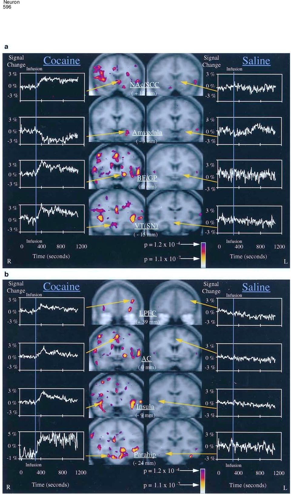
7 Acute Effects of Cocaine on Human Brain Activity 597 Figure 4. Selected Anatomic Definitions Used for Average and Individual Activation Localization Structural T1 images from one individual are shown in the same orientation as the Talairach coronal position. Regions of interest for localization of statistical map activations are indicated in white on top of these gray scale structural images. Coronal slice planes are given with respect to the anterior commissure. The abbreviations and definitions used in this image are defined in the Expermental Procedures section on anatomic localization of activations. It has to be pointed out that the nucleus accumbens (NAc in the figure) can be distinguished on this individual s image from the subcallosal cortex that is adjacent and medial to it. This distinction between nucleus accumbens and subcallosal cortex is not possible on averaged images and is not always possible on individual images. Therefore, we refer to a nucleus accumbens/ subcallosal cortex region of interest for both our averaged data and our individual data. conditions, subjects had been informed on several occasions that the identity of the first retest infusion did not imply the identity of the second retest infusion and that the MGH pharmacy maintained a double-blind ex- perimental design for subjects and researchers during all retest infusions. Regions of positive signal change that were similar between average maps of the test and retest cocaine infusions are listed in Table 5. Note that activations that overlapped did not necessarily have statistical maxima in the exact same anatomic region. However, their statistical maxima were within 1.5 cm of each other or the two activation clusters had overlapping voxels at a high statistical threshold. Twenty-six of seen in the comparison of preinfusion versus postinfusion time points include sections of the left parahippocampal gyrus, left cingulate gyrus, left insula, and right hippocampus. Test Retest Comparisons Seven subjects had retest infusions at times ranging from 3.5 to 4 months after the first experiment. Of these seven, four subjects had interpretable cocaine infusion data after motion correction for test retest comparison. These four subjects received their double-blind cocaine and saline infusions in the same order for the retest experiments as for the test experiments. To keep expectancies as similar as possible between test and retest Figure 3. Regional Brain Activation with Cocaine and Saline (a) Images of subcortical brain regions showing significant fmri signal changes after cocaine, but not after saline, infusions. On the left are Kolmogorov-Smirnov (KS) statistical maps at four coronal levels of pre- versus postinfusion time points for the average fmri data from ten subjects who received cocaine. These KS statistical maps are overlaid in pseudocolor on corresponding gray scale average structural maps. Activations with positive signal change include the NAc/SCC, BF/GP, and VT, while activations with negative signal change include the amygdala. The signal intensity versus time graph for the activations (for all voxels with p 10 6 within the named region) is placed next to each image. On the right are identical slice planes overlaid with the KS statistical map for the saline infusion; the saline signal intensity versus time graphs for the same anatomic regions active during cocaine are placed next to the saline images to demonstrate the absence of comparable change. (b) Images of other paralimbic and heteromodal cortex activations after cocaine and after saline infusions. Regions shown include the lateral prefrontal cortex (LPFC), anterior cingulate, insula, and parahippocampal gyrus. Image and graph layout follow the conventions described in (a).

8 Neuron 598 Figure 5. Multiple Correlation Images Correlation images associated with rush and with craving are displayed as coronal images, respectively, in the top row and the bottom row. Each correlation map is presented as a pseudocolor p value map superimposed on a gray scale structural image. Coronal images represent slices from 15 mm, 0 mm, 3 mm, 18 mm, and 24 mm with respect to the anterior commissure. Regions highlighted in this figure were more strongly correlated with one behavioral measure than another and include the NAc/SCC, BF/GP, Amygdala, VT, and parahippocampal gyrus. thirty-two postcocaine activations in the test sample by previous test activations, but 22 of these directly were matched by similar activations in the retest experiments, matched activations seen with the total cohort of ten including subcortical regions originally hypothe- subjects. Other factors that might contribute to the het- sized to be activated, namely the NAc/SCC (see Figure erogeneity include the current level of cocaine usage or 6), basal forebrain, and caudate. For regions such as altered anxiety or positive expectancy related to the the NAc/SCC, the percent signal change for voxels prior experience with our experimental procedures. meeting the threshold of p 10 5 in the test condition (left 3.8%, right 2.4%) was marginally higher than the percent signal change for the retest condition (left Saline Infusion 2.3%, right 2.1%), though more voxels met the p Foci of Signal Change 10 5 threshold on retest. In the ten subjects with interpretable data, saline infusions Other areas of activation that matched between test produced no positive signal change in limbic or and retest conditions included parahippocampal re- paralimbic regions. One focus of negative signal change gions, thalamic, insular, and cingulate regions. For the was noted in the left temporal pole, which approximated average map of four subjects in the test condition, there a similar activation for the cocaine infusion. For areas were fewer activations overall (N 32) than for the outside of limbic and paralimbic regions, positive signal average map with ten subjects (N 92 activations with changes were noted in the inferior frontal gyrus, inferior/ positive signal change). This raises the possibility that middle temporal gyri, and extrastriate region (see sup- the smaller cohort had insufficient power to identify plemental Table 8 smaller magnitude signal changes, thus the test cohort s full/19/3/591/t8) and negative signal changes in lateral activation profile may be a subset of the larger group s frontal cortex, superior temporal gyri, and extrastriate activation profile. In support of this possibility is the cortex (see supplemental Table 9 observation that of the 56 regions observed with the org/cgi/content/full/19/3/591/t9). All five positive activations cocaine retest experiment, 28 regions were not matched with saline matched the location of activations

9 Acute Effects of Cocaine on Human Brain Activity 599 Table 5. Test Retest Cocaine Infusions: Regions of Similarity for Foci of Positive Signal Change Test Retest Tal Coordinate Tal Coordinate Anatomy Anatomy Proximity (Region/BA) R/L A/P S/I Vox # (Region/BA) R/L A/P S/I Vox # ( 1.5 cm) Subcortical Gray Structures Caudate/NAc Caudate Caudate/NAc GO a BF/GP BF/GP Thalamus/pThal Caudate Cingulate a Temporal Lobe Lateral and Intrasylvian Surfaces GTm a GTm a a Insula Insula Insula GTm a Insula Insula Insula Insula a Medial Paralimbic Cortices Cingulate a Cingulate a Cingulate a23/ Cingulate a Cingulate a Parahip a GF a GF a20/ a Parahip a28/ Thalamus/pThal Parahip a35/ Table 5 shows which activations were similar between test and retest conditions for the cocaine infusions. Specific anatomic regions are described using the nomenclature discussed in Experimental Procedures with the exception of the following terms: GTm (gyrus temporalis medius), GF (gyrus fusiformis), GO (gyrus orbitales). BA indicates the probable Brodman area, for cortical areas, of activation. Under Tal Coordinate are the Talairach coordinates (Talairach and Tournoux, 1988) of the voxel with the maximum p value as determined from the KS maps (Breiter et al., 1996b). Coordinates are expressed in mm from the anterior commissure: R/L, right ( )/left ( ); A/P, anterior ( )/posterior ( ); S/I, superior ( )/inferior ( ). The number of voxels around the max vox that meet the p value threshold of p 10 6 are listed under Vox #. Proximity lists whether the voxels with the maximum p values for each activation are within 1.5 cm of each other; thus a plus sign is placed in the last column if they are 1.5 cm apart, or a minum sign is placed if they are more than 1.5 cm apart. a Indicates there is no overlap, but the max vox of the two activations are within 1.5 cm of each other. S/I 3) and the right insula (Talairach coordinates: R/L 40, A/P 15, S/I 0). The saline retest NAc/SCC (Figure 7) and insula activations closely approximated the same activations seen for the initial cocaine infusion in the total cohort (see Tables 1 4) and the cocaine NAc/SCC activation that correlated more with maximum ratings of craving than with rush (Tables 1 4). Moreover, these NAc/SCC and insula activations were similar to the same activations in the cocaine condition that showed good cocaine test retest reproducibility (Table 5). Compared to the bilateral NAc/SCC activations seen with cocaine test retest infusions, the bilateral NAc/SCC activations seen with saline retest infusion demonstrated a lower percent signal change (left 0.4%, right 1.5%) for all voxels meeting the threshold of p On the basis of location of activation maxima, 11 of the 16 new activa- tions seen with saline retest infusion in the NAc/SCC, the frontal cortex, and the temporal cortex were seen with either the cocaine test or retest infusions. Discussion Following an infusion of cocaine under double-blind conditions, cocaine-dependent subjects demonstrated significant increases in HR and MBP and decreases in ETCO 2. Cocaine plasma concentration reached maxi- mum at 7 min after infusion. Subjects reported early seen in the cocaine maps; only one negative saline signal change, in the superior temporal gyrus, matched the location of an activation with negative signal change following cocaine infusion. Test Retest Comparisons As with the cocaine test retest comparisons, four of seven subjects had interpretable saline infusion data for test retest comparison after motion correction. For the saline test retest comparison with four individuals, no limbic or paralimbic regions were activated. For regions outside of limbic and paralimbic regions, six of the test activations were also similar to those seen with retest. Of these six activations, four of the six approximated activations seen with the average saline map of ten individuals, suggesting that the subgroup of four represent a good approximation of the group of ten. The saline retest data evidenced multiple new activations not seen during the first saline test. The majority of these (10/16) were in the striate, extrastriate, and ventral temporal cortex implicated in ventral stream information processing for vision (Tootell et al., 1995). Eleven of the sixteen activations were similar to activations seen with the initial cocaine infusion for the total cohort and the retest cocaine infusions in the subgroup of four individuals (Table 5). Most striking was the appearance of activations in the bilateral NAc/SCC (Talairach coordinates: R/L 9, A/P 18, S/I 3; R/L 9, A/P 15,
 Subcortical Gray Structures Caudate/NAc 25 27 18 8 Caudate 18 12 21 77 Caudate/NAc 9 15 3 81 GO a11 3 15 6 160 BF/GP 21 0 6 10 BF/GP 15 3 0 23 Thalamus/pThal 6 27 12 62 Caudate 18 12 21 77")
10 Neuron 600 Figure 6. Test Retest Cocaine Experiments The Kolmogorov-Smirnov (KS) statistical maps (unsmoothed) for the average fmri data in Talairach space from four subjects who had test retest cocaine infusions are displayed in pseudocolor on top of gray scale structural images from these same subjects. Coronal slices are identified by their relationship in mm to the anterior commissure (AC). be associated with rush and with craving ratings (Figure 8). Brain activation correlated with rush ratings was noted in a subset of regions associated in animal experiments with brain reward such as the VT, left basal fore- brain, midbrain and pontine brainstem, bilateral caudate nucleus, and right cingulate gyrus. All of these regions are directly connected with the VT or the nucleus accumbens. Other brain activations in regions not previously implicated in animal models of drug self-administration or BSR, which showed a similar pattern of early transient signal maxima, included regions of prefrontal, parietal, temporal, and occipital cortex. Brain activation corre- lated with craving measures was noted in the NAc/SCC and right parahippocampus. A negative correlation with craving was also noted in the amygdala (a region with negative fmri signal change on the average maps). maxima 3 min after infusion for behavioral ratings of rush and high and later maxima for behavioral ratings of craving and low. Thus, maximal subjective euphoria was reported during the distribution phase of cocaine plasma kinetics, before maximal intravascular cocaine levels had been attained. Brain regions that showed focal increases in BOLD signal at the time of onset of subjective measures of euphoria included putative brain reward circuitry (NAc/ SCC, basal forebrain, and VT), caudate, putamen, thalamus, medial temporal and paralimbic regions (hippocampus, parahippocampal gyrus, cingulate cortex, and insula), brainstem (pons), and neocortical regions such as the lateral prefrontal cortex, lateral temporal cortex, parietal cortex, and occipital cortex. Decreases in fmri signal were also noted in the amygdala, temporal pole, and medial frontal cortex, although the latter regions were in close proximity to areas of susceptibility artifact (see below). In comparison to cocaine, saline produced few regions of fmri signal increase, limited to lateral prefrontal and temporo-occipital cortex. Small regions of signal decrease were also noted in the lateral prefrontal cortex and temporal cortex. All of the four positive temporal lobe activations seen during the saline infusion, along with the negative temporal pole activation, were also seen in the cocaine condition. Multiple correlational analysis of averaged behavioral ratings with averaged cocaine fmri data indicated differences in the temporal pattern of activation, which can Limitations In this study, subjects exhibited head movement following both cocaine and placebo infusions despite use of a bite bar. Complete movement correction was possible for some subjects with maximal displacements up to 3 mm, although in some subjects, complex movements of similar magnitude produced unacceptable residual motion artifacts. As with our previous experience with a psychiatric population (Breiter et al., 1996b), a significant proportion of studies (6/17 cocaine infusions and 4/15 saline infusions) had to be discarded due to uncorrectable movement.
.")
11 Acute Effects of Cocaine on Human Brain Activity 601 Figure 7. Test Retest Saline Experiments The Kolmogorov-Smirnov (KS) statistical maps (unsmoothed) are displayed as described in Figure 6, but for the four subjects who had test retest saline infusions. Note the NAc/SCC activaton in the cocaine test (Figure 6) but not the saline test condition (this figure). By the retest, NAc/SCC activation could also be observed in response to the saline condition. Despite this generalization of response, NAc/SCC activation still represented a larger percent signal change for both the test and retest cocaine conditions. potential for magnetic susceptibility artifact, primarily seen on echo-planar images as signal dropout. Given unpredictable effects on T2*-weighted signal change from regions with high susceptibility, especially with concurrent motion, we checked and confirmed that activations seen with the cocaine and saline infusions did not overlap regions of susceptibility artifact on the functional images. For this reason, regions such as the me- dial frontal cortex and temporal pole, which showed large negative signal changes that were proximal to ar- eas of susceptibility artifact, cannot be considered reliable activations. Two issues regarding experimental design need to be mentioned. This study incorporated double-blind condi- tions and subject instructions designed to equalize cocaine expectancy for each infusion, in both test and retest conditions. Despite these precautions, since subjects knew they would only receive a single infusion per scan and could feel the infusion volume as it was administered, the blind only lasted a few minutes past the infusion, after which all subjects knew whether they had received cocaine or saline. This was clearly re- flected, for both test and retest conditions, in the absolutely uniform zero ratings for rush and high following saline. Secondly, in the design of the overall study, multiple attempts were made to distinguish BOLD fmri signal Motion correction must also be considered in the context of its contribution to altered spatial resolution. Our voxel size during imaging was mm, which would appear adequate to resolve some subnuclei of larger subcortical structures. But, the combination of (1) motion correction, (2) transformation into Talairach space, and (3) averaging alter our effective spatial resolution to approximately 1 cm 3. Activations from averaged data in our study, thus, cannot be attributed with certainty to specific subnuclei of larger gray matter structures. Indeed, any anatomic localization with averaged data sets and superimposition of different structural and functional data acquisitions must be considered in probabilistic terms. This is the case even for individual statistical maps superimposed on structural images, albeit to a lesser degree, since images are superimposed from different acquisitions with the potential for movement between them, as well as different spatial warping and signal-to-noise characteristics. These issues are apparent with the test retest data, where some activations that overlap do not necessarily have statistical maxima in the identical anatomic spot. This could be due to limitations imposed by our effective spatial resolution or noise of physiological origin in our underlying fmri measurements. Some regions, such as the NAc/SCC, basal forebrain, hypothalamus, and amygdala, are near areas with a high

12 Neuron 602 Figure 8. Summary Schematic of Limbic and Paralimbic Brain Regions that Correlate with Euphoria (in Red) Versus Those Regions that Correlate with Craving (in Green) Above these summary schematics is a schematic of the brain regions (in yellow) we predicted to be active after the infusion of cocaine. Two other brainstem monoaminergic regions, potentially encompassed in a pontine activation seen in our baseline versus postinfusion comparison, are also illustrated in blue. This pontine activation did correlate with behavioral ratings for rush. or output from the NAc/SCC, which would be associated with a regional increase in neuronal cell body activity. The linkage of fmri signal to underlying neuronal activity remains an area of continuing research, which is needed to connect more directly the results of this neuroimaging study and others to the body of basic substance abuse research using animals. Regions with Short Duration Signal Change and Relation to Euphoria Two of the behavioral ratings used in this experiment, rush and high, described separable features of the sub- jective experience of euphoria or pleasure. Such subjective measures can only be obtained with human subjects. With animal experiments, behavioral scientists have been limited to investigation of the effects of re- warding stimuli on observable behavior (White et al., 1987) based on repeated approach behaviors or re- sponse repetitions. It can be hypothesized that acutely rewarding behavioral stimuli (i.e., cocaine) administered to a conscious (human) subject produce not only behavioral effects (reinforcement) and lead to encoding of emotional memories but also produce subjective pleasure that can be reported. Behavioral research with animal models has shown that increased dopamine transmission in the nucleus accumbens is associated with behavioral responses to rewards. However, the exact relationship of mesoac- cumbens dopamine function to the action of a reward changes due to region specific activation from changes due to systemic physiological or direct vascular effects of cocaine. Each infusion scan (see Figure 1) was bracketed by control experiments to determine whether regionally specific primary visual cortex activation was altered by the cocaine or placebo infusion, and how much change there was in global cerebral blood flow (Gollub et al., submitted). The results of these control experiments clearly support our interpretation of focal regional activation following cocaine infusion. Focal primary visual cortical activation was quantitatively unchanged following cocaine or saline. Moreover, although the flow-sensitive scan revealed an approximate 14% decrease in flow-related signal in cortical gray matter, no such change was measured during BOLD scanning. Even with these reassurances, though, a strong caveat should be added to the results of local gray matter changes related to cocaine; namely, that they were observed in the presence of significant cardiovascular and respiratory effects from cocaine. Finally, a general issue regarding the linkage of fmri signal to underlying neuronal activity must be mentioned. At this time, the relationship of BOLD signal to pre- and postsynaptic mechanisms of neurotransmission is unknown. Thus, it is conceivable that increased fmri signal in the NAc/SCC, for example, could be due to increased activity of inhibitory (GABAergic) nerve terminals, which produce a decrease in neuronal cell body activity in the NAc/SCC rather than excitatory input to

13 Acute Effects of Cocaine on Human Brain Activity 603 as an incentive or as a reinforcement has been an area of outputs of both the nucleus accumbens and the amygdala controversy (Richardson and Gratton, 1996). The implicit (Heimer et al., 1997). assumption in relating dopamine transmission in the In our study, extensive brainstem activation distinct nucleus accumbens to cocaine use is that dopamine from the VT was also observed, including activation in transmission is a central correlate of the reinforcing ac- the vicinity of regions for other monoaminergic systems tions of rewards (Wise et al., 1978; Wise, 1982; Koob, such as the serotonergic system (primarily the dorsal 1992). Thus, in humans, we postulated that an acute raphe) and the noradrenergic system (the locus coeruleus). change in fmri activation in the nucleus accumbens This other brainstem activation also demonstrated area (NAc/SCC) would be correlated with behavioral early signal maxima with rapid return to basal levels that measures of euphoria. The pattern of fmri signal change correlated with rush ratings. FMRI BOLD scans in the could not be predicted, since dopamine produces com- brainstem are confounded by cardiac-induced motion. plex modulatory effects on postsynaptic neurons and Thus, replication of our brainstem observations using a because the relationship of the BOLD signal to pre- recently developed technique of cardiac gating with a and postsynaptic mechanisms of neurotransmission is subsequent T1 correction algorithm (Guimares et al., unknown. What we found was that fmri activation in the 1996) will be important. VT (the source of dopamine for the nucleus accumbens), basal forebrain, pontine brainstem, caudate, insula, cingulate gyrus, and prefrontal cortex were correlated with Regions of Sustained Signal Change and Possible behavioral measures of euphoria. Relation to Cocaine-Induced Craving Computational models of the output of VT neurons In this study, no brain region showed statistically signifisuggest that they code for a deviation between the expechange cant signal intensity changes that directly paralleled the rienced reward and the previous predictions for reward. in behavioral ratings for cocaine-induced crav- VT neurons would accordingly report ongoing prediction ing or dysphoria (i.e., with slow onset of signal change errors for reward and deliver a signal to forebrain targets and peak effects after approximately 10 min). To a first to alter ongoing processing of reward predictions and approximation, the ramp function of the craving ratings the direction of reward-maximizing actions (Schultz et is the same as the average motion displacement de- al., 1997). Thus, in the naive state, VT neuronal firing is tected by the AIR algorithm. Since our baseline drift increased in nonhuman primates early in the acquisition correction removed signal changes that would be corre- of lever pressing behavior (Schultz, 1986; Nishino et al., lated with this motion, the absence of brain regions 1987; Romo and Schultz, 1990; Schultz and Romo, 1990; with time course changes specific for cocaine-induced Ljungberg et al., 1992; Schultz et al., 1993). However, craving is a matter to be interpreted with caution. after several lever press trials in the same experiments, Some brain regions did however show sustained acti- simulating a more chronic state of drug use, VT neurons vation that led to a higher degree of correlation with demonstrate electrophysiological decreases in response. the craving ratings. Our observation, for instance, of In our fmri study, subjects were chronic users but were sustained signal change in the NAc/SCC is the explananaive to cocaine use in the fmri environment. They tion for its stronger association with craving than with showed a pattern of early but transient signal change rush ratings. In general, the differences at high thresh- in VT, which is analogous to the response of primates olds between the rush and craving correlation maps naive to cocaine. Perhaps the novelty of cocaine adminearly reflect a distinction between behavioral ratings with istration in the fmri setting may have contributed to peaks and shorter duration (i.e., rush) and ratings the observed VT activation. This formulation regarding with prolonged time courses (i.e., craving; see signal learning effects could be confirmed by serial retest studsubtraction time courses in Figures 3a and 3b). It is significant that ies with larger cohorts than our current study. of fmri time courses with early maxima and Regions to which VT input might be important in mediwould short duration from those with prolonged time courses ating the subjective concomitants of reward and that produce a time course closely resembling that of demonstrated activation with early signal maxima and the craving ratings. This suggests a possible model for short duration and thus correlation with rush ratings craving in humans. Craving may not be mediated by include the cingulate gyrus and the basal forebrain. The one or two distinct brain regions; rather, postcocaine cingulate gyrus has been associated both with euphoric craving may reflect a change over time in the pattern of experiences in humans induced by procaine (Ketter et brain activation from cocaine. Many brain regions are al., 1996) but also, in direct contrast, with the emotional active at the time that subjects report euphoria. Over intensity of aversive events (Talbot et al., 1991; Sikes time, though, only a few brain regions remain activated; and Vogt, 1992; Coghill et al., 1994; Casey et al., 1996; this change in the pattern of brain regions activated Craig et al., 1996). The basal forebrain has also been may be causally related to the subjective experience of implicated in affective function, in that it has been di- craving. rectly implicated in the results from BSR experiments. The observation of sustained activation in the NAc/ Since BSR was first observed by Olds and Milner (1954), SCC, which occurred over the time interval that subjects evidence has accumulated that regions such as VT, latlinks experienced cocaine-induced rush and then craving, eral hypothalamus (Murray and Shizgal, 1991, 1996), and the NAc/SCC with both reinforcement and with basal forebrain (Rompre and Shizgal, 1986; Shizgal et incentive functions. This contrasts with the simple view al., 1989; Arvanitogiannis et al., 1996) contain the neuron that dopamine transmission in the nucleus accumbens somata that generate this effect. It is important to note area (NAc/SCC) is the central correlate of reward and that the basal forebrain constitutes one of the primary therefore subjective euphoria. Although some studies

14 Neuron 604 the current double-blind infusion experiment, order effects in each of the cue-conditioned craving experi- ments (Childress et al., 1996, Soc. Neurosci., abstract; Grant et al., 1996; Schweitzer et al., 1996, Soc. Neurosci., abstract), and differences in neuroanatomical resolution between our fmri work and the PET studies of other investigators (Childress et al., 1996, Soc. Neurosci., abstract; Grant et al., 1996; Schweitzer et al., 1996, Soc. Neurosci., abstract), further work will be needed to assert that differences exist between craving during acute cocaine withdrawal and craving elicited by cues. Signal Changes During Saline and Possible Relation to Expectancy and Craving The lateral prefrontal and temporo-occipital activations observed with saline infusion were similar to activations after cocaine infusion and might represent a common effect from expectation or a chance similarity given the number of regions activated during the cocaine condition. It is unlikely they represent chance, since they were mostly replicated in saline retest experiments. The saline activations might be considered in the context of the results from a recent PET study of cue-induced cocaine craving in cocaine addicts (Grant et al., 1996), if one considers the saline infusion as a potential cue. In the study of Grant and colleagues (1996), increased glucose metabolism was reported in the lateral prefrontal and temporo-occipital cortices. It is interesting to note the similarity of regional activation between studies, even though our subjects rated craving at zero throughout the saline infusion. The issue of cocaine expectancy also arises with re- gard to new activations observed with the saline retest experiments. Orbital cortex activation was noted bilater- ally on saline retest; this region has been implicated in the suppression of expectancies in animals (Morgan et al., 1993; Morgan and LeDoux, 1995). Bilateral NAc/SCC activation was also observed with saline retest. Given theorized involvement of the nucleus accumbens with the processing of predictions of reward (Schultz et al., 1997) and observations of altered conditioned responses in animals after only one cocaine dose (Weiss et al., 1989), it is possible the NAc/SCC activation on saline retest may represent one-trial learning. Further work will be needed to evaluate the time course of change in regions such as the NAc/SCC to determine if these rep- resent learning effects (Schultz et al., 1997). Conclusions During double-blind infusion experiments in cocainedependent subjects, we observed dramatic effects from cocaine in physiology, behavioral report, and fmri brain activation that were not found following saline. A significant feature of this study was the continuous sampling of brain blood oxygenation changes to intravenous co- caine over 18 min, which was exploited for multiple correlational analysis with the behavioral data. Several brain regions showed short duration of activation that was well correlated with the reinforcement-related rating of rush, while other regions showed sustained activa- tion, demonstrating some of the features associated have reported an association between feeding or consummatory behavior and elevated dopamine levels in the nucleus accumbens (Heffner et al., 1980; Hernandez and Hoebel, 1988; Radhakishun et al., 1988; Yoshida et al., 1992), other studies suggest that the increases in nucleus accumbens dopamine transmission do not result from consummatory behavior (Blackburn et al., 1986, 1989, 1992; Chance et al., 1987; Weatherford et al., 1991; McCullough and Salamone, 1992; Elbaz et al., 1993, Soc. Neurosci., abstract; McCullough et al., 1993; Phillips et al., 1993; Salamone et al., 1994) and that mesoaccumbens dopamine neurons respond to incentive rather than to reinforcing components of rewards (Kiyatkin and Gratton, 1994; Richardson and Gratton, 1996). Our fmri data, showing a correlation between cocaine-induced craving and sustained activation in the NAc/SCC, supports a complex role for the NAc/SCC in the human, with a potential role in incentive as well as reinforcement. Other brain regions with sustained signal change after early signal maxima included several lateral prefrontal regions and one section of the parahippocampal gyrus (see Figure 3b), though other parahippocampal activa- tions did not display this behavior (see Tables 1 4). The parahippocampal gyrus has efferents to the nucleus ac- cumbens and amygdala, is a primary input source for the hippocampus, and has been implicated not only in explicit memory (Squire and Knowlton, 1995) but also in the association of context to emotionally relevant stimuli during fear conditioning (LeDoux, 1993). The common sustained activation of the NAc/SCC and para- hippocampal gyrus, along with relatively discrete sections of lateral prefrontal cortex, points to a distributed network of brain regions involved with the cocaineinduced craving. Sustained negative signal change in the left amygdala was also observed. This left amygdala signal change needs to be discussed with the caveat that inspection of individual maps showed some heterogeneity of acti- vation, in that three subjects evidenced positive signal change and only five subjects displayed negative signal change. Such heterogeneity in amygdala activation re- sembles electrophysiologic findings in rodents, in that acute intravenous cocaine produces mixed suppression and excitation in amygdalar neurons (Cunningham, 1995). In contrast, microiontophoretic application of cocaine in the amygdala uniformly produces suppression of spontaneous neuronal discharges (Cunningham, 1995), indicating that functional connectivity is important for mediating amygdala response to cocaine. Our left amygdala data appear to contrast with other reports of positive correlation between amygdala activa- tion and cue-elicited craving (Childress et al., 1996, Soc. Neurosci., abstract; Grant et al., 1996; Schweitzer et al., 1996, Soc. Neurosci., abstract). The amygdala has been implicated in the orientation to and remembering of affectively salient stimuli for social interaction (Leonard et al., 1985; Rolls, 1992; Breiter et al., 1996d). Interpretation of our current negative amygdala activation in the con- text of this other work can only be speculative. It is possible that cocaine-induced craving represents a distinct process from that of cue-conditioned craving (Everitt, 1997). Given the potential for expectancy effects in

15 Acute Effects of Cocaine on Human Brain Activity 605 with the incentive-related measure of craving. In particudeficiency or cardiac disease. All subjects tested negative for human immuno- lar, the VT and basal forebrain correlated more strongly virus (HIV). Women were not pregnant by HCG testing and were scanned at the midfollicular phase of their menstrual cycle. with rush measures, while the NAc/SCC and amygdala All subjects fulfilled criteria for cocaine dependence, with or without correlated more strongly with craving measures, even comorbid alcohol or marihuana abuse, by Mini-Structured Clinical though these latter two regions had early signal maxima Interview for DSM-IV (SCID) (American Psychiatric Association, as seen with the rush measures. Early but sustained 1994). Our subjects were selected to be heavy, long-term cocaine activation in the NAc/SCC implies that it is activated users (mean years; days of cocaine use in 30 days prior during both rush and craving experiences, which confor to experiment days). Current monetary expenditure cocaine was $ over the week prior to the experi- trasts with the general view of circuitry mediating reinment. No subjects wereseeking orreceiving treatment for substance forcement (Wise et al., 1978; Wise, 1982; Koob, 1992) abuse at the time of the study. To be accepted into the imaging and suggests the NAc/SCC is also involved with incen- protocol, during screening, subjects had to have one positive urinalysis tive functions. to confirm recent cocaine use, but had to be abstinent from In contrast to cocaine effects, saline produced activamately cocaine and alcohol for at least 18 hr before the infusion. Approxitions in prefrontal cortex and lateral temporo-occipital 18 hr before each imaging session, subjects underwent a screening IV test dose of 0.2 mg/kg in the MGH Mallinckrodt GCRC cortex, most of which were also found active with counder the supervision of a cardiologist and psychiatrist to ascertain caine, and resembled findings from other investigators cardiac and neurological tolerance of the experimental procedures. during cue-induced craving in abstinent cocaine-depen- They were subsequently monitored in the GCRC until the time of dent subjects (Childress et al., 1996, Soc. Neurosci., scanning. All subjects gave informed consent to participate in these abstract; Grant et al., 1996; Schweitzer et al., 1996, Soc. procedures following the rules of the Subcommittee on Human Stud- Neurosci., abstract). The observation of NAc/SCC acticol ies at MGH. Subjects were reimbursed for participation in this protovation on the basis of days in the hospital and could earn a bonus for on saline retest infusion raises the possibility that completion of all scans. Reimbursement was with noncash vouchers generalization of expectancy across the two infusion (e.g., nontransferrable food coupons). conditions may occur within one trial; this hypothesis needs confirmation. Experimental Design Our cocaine results provide evidence in the human Subjects were admitted to the MGH GCRC for the screening procefor a functional integration of circuits involved with reinin dures; those meeting all criteria were boarded overnight on the unit preparation for imaging the following day. The following morning, forcement and circuits involved with drug craving (see the subject had bilateral intravenous catheters placed (right forearm Figure 8). The known anatomic interconnections befor cocaine or saline infusion, left forearm for serial venous blood tween limbic and paralimbic regions with short duration sampling for quantitative cocaine levels). Scanning was performed versus sustained alteration in BOLD signal converge on between 11 AM and 3 PM, during which the subject was in the a core set of brain regions: the NAc/SCC, basal fore- scanner for two periods of time, each lasting from 45 to 90 min. brain, amygdala, and VT. From animal research, there is During each scanning period, one infusion was given, either cocaine (0.6 mg/kg, maximum dose 40 mg) or saline (both in a volume of 10 evidence that the VT is necessary for reward prediction ml given over 30 s IV) in a randomized, double-blind order. Five (summarized in Schultz et al., 1997), the amygdala for different scans were performed during each period. The infusion orienting to and remembering affectively significant itself was made 5 min into an 18-minute-long BOLD scan. The BOLD stimuli (Everitt et al., 1991; LeDoux, 1992; Hatfield et al., infusion scan was bracketed by flow-sensitive alternating inversion 1996) and attentional modulation of perceptional functhese recovery (FAIR) and visual stimulation BOLD scans (the data from tion (Leonard et al., 1985; Rolls, 1992), the nucleus acchanges scans were used to delineate the global versus regional signal from cocaine and are reported in a separate manuscript cumbens for determination and modulation of motoric [Gollub et al., submitted]). The time interval between functional responses toward perceptual stimuli and internal hoscans within a period was kept to a minimum. The entire sequence meostatic needs (Le Moal et al., 1977; Kelley and Stinus, of five functionalscans was completed within45 60 min. The subject 1985; Fibiger and Phillips, 1986; White, 1986; Blackburn was removed from the scanner for a min rest and then was et al., 1989), and the basal forebrain for attention to returned to magnet and the sequence was repeated for the second internal state and attribution of primary reward (Shizgal infusion. A minimum of 2 hr had to pass between each double-blind et al., 1989; Arvanitogiannis et al., 1996). Future research infusion. will be important for determining whether or not these Subject Instructions regions function in this manner in humans and how these For the preexperiment test infusion with 0.2 mg/kg cocaine on the functions produce incentive and reward. night before scanning, subjects were informed they would receive a small dose of intravenous cocaine in the presence of a cardiologist and a psychiatrist to screen for medical side effects from intravenous Experimental Procedures cocaine and to train them in making behavioral ratings of their experience. Subjects For experiments performed in the magnet, subjects were informed Of the 17 subjects who completed the experimental protocol, 13 they would receive two infusions to which both they and the experiwere men and four were women (mean age years; menters were blind. Infusions could either be saline or 0.6 mg/kg education years; weight kg; Addiction of cocaine in saline; the experience of one infusion did not imply Severity Index [McLellan et al., 1980] Composite Score [0 to 1.00] what would be the identity of the other. Subjects were further asked on the drug dimension and on the alcohol dimension to continue behavioral ratings throughout the FAIR and BOLD infu ; Hamilton Anxiety Scale [0 to 54] ; Hamilton sion scans ( 40 min in total) and to remain as motionless as possible Depression Scale [0 to 52] ). All subjects were rightcould to minimize fmri movement artifacts. All subjects understood they handed. Except for cocaine addiction, they were medically and neurologically terminate the experiment at any time without explanation. normal by physical exam, review of systems, blood work including electrolytes, liver function tests, cell blood count, and Plasma/Urine Monitoring toxicology. No subject had a history of head trauma with loss of Sequential 4 ml venous blood samples were collected immediately consciousness or had any family history of sudden cardiac death before and at 1, 3, 5, 10, 15, 30, 60, 90, and 120 min following each

16 Neuron 606 infusion. The 120 min sample for the first infusion was also the the planning andimplementation of physicalactivity. Thus,by definition, preinfusion sample for the second infusion. only craving was defined as a motivational state. In general, rush experiencesinvolved physical sensations of elevated heart rate Physiological Monitoring and sweating, along with internal feelings variously characterized Physiologic monitoring was conducted using an InVivo OmniTrak as speeding sensations and sensations of being out of control patient monitoring system (Orlando, FL) modified to permit In contrast, the high experience was generally associated with feelon-line computer acquisition of physiologic measurements. Each ings of self-confidence, well-being, and sociability. The low experisubject was fitted with chest leads to record the electrocardiogram ence encompassed all negative subjective feelings potentially asso- (ECG) and to measure heart rate (HR), a nasal cannula to measure ciated with cocaine use, such as anxiety, paranoia, dysphoria, or respiratory rate and ETCO 2, and a blood pressure cuff to measure anhedonia; the majority of subjects in this study discussed the low noninvasively systemic MBP. The temporal resolution of the system in terms of dysphoric effect distinct from a diminishment in the high for sampling blood pressure was once every 2 min. The InVivo system experience. sampled and displayed updated values for each of the other parameters once per second except for the ECG trace, which was Imaging digitized at a rate of 100 Hz. Scanning was performed with a quadrature head coil and a 1.5 T The measured physiologic parameters wereported to a Macintosh MR scanner (General Electric) modified for echo-planar imaging Power PC 7100 running a custom National Instruments LabView (Advanced NMR). Imaging involved the following protocol. First, a data acquisition program. This program allowed the simultaneous sagittal localizer scan (conventional T1-weighted spoiled gradient acquisition of (1) the digitized analog ECG trace signal acquired refocused gradient echo [SPGR] sequence; through-plane resoluusing a National Instruments MIO16L board, (2) the GE scanner J8 tion 2.8 mm; 60 slices) was performed to orient, for subsequent trigger pulse that indicated when the gradient coils of the magnet scans, 15 contiguous axial slices covering the whole brain. This were firing, and (3) serial port read of ASCII characters reporting scan was also used as the structural scan for Talairach transformaphysiologic measures from the InVivo system. tion. Next, an automated shimming technique was used to optimize Precautions taken to ensure safe conduct of the study included B 0 homogeneity (Reese et al., 1995). This was followed by an SPGR use of ACLS trained personnel, frequent running of mock codes T1-weighted flow-compensated scan (resolution 1.6 mm 1.6 with clocked performance of tasks and strict definition of individual mm 8 mm), which was primarily obtained to aid Talairach transfortasks, and presence of a cardiologist at the time of all infusions mation during data analysis (see Breiter et al., 1996b). The fourth whose sole responsibility was to monitor subject safety. Before and scan was a T1-weighted echo-planar inversion recovery sequence after completion of both infusions, subjects underwent a 12-lead (TI 1200 ms, in-plane resolution 1.57 mm) for high resolution ECG to determine the absence of any interval change from the structural images to be used in preliminary statistical maps but experiments and to clear them for discharge home. Because of not with Talairach transformed or averaged maps. Finally, BOLD magnetohydrodynamic effects on the ECG tracing, a baseline imaging was performed using an asymmetric spin echo T2*- rhythm strip was obtained prior to each drug infusion and all subseweighted sequence (TR 8000, TE 50, 180 refocusing pulse quent tracings were compared to that one. offset by 25 ms; FOV cm; in-plane resolution mm; through-plane resolution 8 mm; 15 contiguous axial slices Behavioral Monitoring covering the whole brain) to measure activation (local changes in For both infusions, analog scales for behavioral response were problood flow and oxygenation) (Bandettini et al., 1992; Kwong et al., jected via the LabView program and a back projection television 1992; Ogawa et al., 1992). Images were acquired interleaved for 136 system (Sharp Liquid Crystal, RU2000) outside the Faraday shield time points for each infusion. of the scanner. These projected stimuli were then focused via a biconvex lens (Buhl Optical) inside the Faraday shield onto a rear projection screen that was viewed through an overhead mirror in Data Analysis the magnet bore. For both infusions, subjects viewed images prior Plasma/Urine Levels to actual experimentation so that images could be focused and Cocaine quantitative assays were performed by the MGH Clinical centered in each subject s visual field. Chemistry Laboratory using a liquid chromatography with photodi- During FAIR and BOLD infusion scans, behavioral measures of ode array detection method they developed (Puopolo et al., 1992), rush, high, low, and craving were obtained in a continuous sequence with minor modifications (flow rate increased from 2.0 to 2.6 ml/min each minute. Thus, over each 15 s epoch, one rating scale would and LCPCN column length increased from 150 to 250 mm). Intraassay be projected for the subject s response. Given four scales, it took imprecision at 100, 20, and 10 mg/l for cocaine is 5.1%, 5.7%, 1 min to cycle through the complete set of scales. Timing of scan and 6.6%, respectively. initiation, infusion onset and offset, and scan completion were linked Physiological Data with ongoing behavioral reports to allow subsequent correlational The data analysis and graphing program IGOR (WaveMetrics, Inc.) analysis between behavioral ratings and fmri acquisitions. Behavioral was used to analyze the data. Data were first analyzed by a two- responses were acquired with a four-button button press that way ANOVA with drug treatment (saline, cocaine) and time of mea- had been adapted to the magnet environment by construction with surement as factors. When significant F values were obtained for nonmagnetic components and filtering of its output at the Faraday one of the physiologic measures, individual time points were compared shield. by posthoc t tests to determine if (and at what times) the To obtain meaningful behavioral ratings during scanning, subjects change from baseline was significant. The Bonferroni correction for were trained beforehand. The day before scanning, subjects were multiple comparisons was used; the criteria for significance at the interviewed in depth by one of two board-certified psychiatrists to 0.05 level was p describe fully their experience of cocaine intake. These descriptions Behavioral Data were then categorized by the psychiatrist and subject into four The integer output for each behavioral rating was segregated by components: the rush, high, low, and craving that were to be rated category of rush, high, low, and craving. For the group data in Figure on an integer scale of 0 (none) to 3 (maximum). The individualized 2, the 18 measures for each behavioral category obtained during conventions for description of subjective responses were then the 18 min BOLD infusion scan were averaged for the nine subjects tested, during the unblinded preinfusion with 0.2 mg/kg cocaine, with both interpretable behavioral data andfmri data. This averaged on a portable computer with a program simulating that used in the data was then utilized in the correlational analysis of the cocaine MRI. fmri data. Of the four behavioral measures, only craving was defined operationally BOLD Data for Initial fmri Experiments in terms of the action the individual wanted to engage in (to and for Test/Retest Experiments get more cocaine). The other three behavioral measures, rush, high, MOTION CORRECTION. To reduce head motion, each subject was posi- and low, were defined in terms of subjective feelings that were not tioned using a bite bar, and echo-planar data was motion corrected necessarily associated with a behavioral output or associated with using an algorithm (Jiang et al., 1995) adapted from Woods et al.

17 Acute Effects of Cocaine on Human Brain Activity 607 (1992) and described elsewhere (Breiter et al., 1996b). Motion cor- NEUROANATOMICAL ANALYSIS. We used a combined approach to anarection of the BOLD saline infusion data revealed an average maxi- tomic localization of functional data. The group average data (GAD) mal displacement of mm, resulting in a mean correction were mapped using an approach focused on Talairach coordinates. per time point of mm. For the cocaine infusion data, there In addition, the individual data (ID) were mapped using a region was an average maximal displacement of mm, resulting of interest-based approach, focused on the limbic and paralimbic in a mean correction per time point of mm. After motion areas. correction, time series data were inspected to assure that no data Anatomic Localizationof GAD. Statistical maps of group averaged set evidenced residual motion in the form of cortical rim or ventricu- data were superimposed over high resolution conventional T1- lar artifacts 1 voxel. There was no statistically significant difference weighted images that had been transformed into the Talairach doin maximal displacement between paired groups of saline versus main and averaged. Primary anatomic localization of activation foci cocaine infusions (p 0.4). was performed by inspection of these coronally resliced T1- TALAIRACH TRANSFORMATION. Each individual s set of infusion-re- weighted scans and via the Talairach coordinates of the maximum lated functional images, along with the associated conventional voxel from each activation cluster (see section on determination of structural scans, were transformed into Talairach space (Talairach activation clusters). Subcortical localization of activations followed and Tournoux, 1988; Breiter et al., 1996b, 1996d) and resliced in the the region of interest conventions described below and demoncoronal orientation over 57 slices with isotropic voxel dimensions strated in Figure 4. All activations were checked against the func- (x, y, z mm). Because of possible movement between acquitional image data to ascertain that they did not overlap areas of sitions of structural and functional scans, functional data were fursusceptibility artifact. Such overlap was determined by whether or ther fit to the structural scan by translation of exterior contours. For not a voxel s signal intensity during the baseline was less than the the cocaine and saline infusions, two subjects evidenced movement average voxel in its slice by 50% of the difference between the between structural and functional scans of 2 voxels in magnitude, average voxel signal intensity in the slice and the average voxel and, therefore, were discarded from further analysis. signal intensity outside of the slice. NORMALIZATION, AVERAGING, AND CONCATENATION. For cocaine and sa- Anatomic Localization of ID. To assess the degree to which subline infusions, Talairach-transformed functional data were intensity cortical activations seen in the group represent common activations scaled (i.e., normalized relative to a standard preinfusion epoch) so across the population, as opposed to the effect of strong activations that all mean baseline raw magnetic resonance signals were equal. in a few subjects, each individual s Talairach-transformed T1 high Talairach-transformed structuraland functional data were then averresolution scan was inspected andregions of interest (ROIs) defined. aged by run across the ten subjects with interpretable cocaine infu- Visual inspection of the superimposed KS statistical maps, threshsion data andthe ten subjects with interpretable saline infusion data; olded at a liberal statistical threshold (KS, p 0.001), was then similar averaging of Talairach-transformed structural and functional performed to determine if activation was present in each of the data was performed for the four subjects used in the test/retest anatomic structures discussed below. These results were tabulated analysis (Breiter et al., 1995, Proc. Soc. Magn. Reson./Euro. Soc. as a ratio of individuals showing lateralized activation in that struc- Magn. Reson. Med. Biol., abstract; Breiter et al., 1995, Proc. Soc. ture to the total number of subjects evaluated (N 10). As our Neurosci., abstract; Brieter et al., 1996b, 1996d). predictions involved only subcortical structures, our individual anal- VOXEL-BY-VOXEL STATISTICAL MAPPING. Unsmoothed Kolmogorovysis also focused on noncortical regions, with the exception of me- Smirnov (KS) statistical images were constructed (Breiter et al., dial paralimbic and intrasylvian cortices. 1996b, 1996d) from these averaged data sets comparing baseline The methods used for definition of the subcortical ROIs followed (N 38 time points) and postinfusion (N 98 time points) epochs. the conventions of the MGH Center for Morphometric Analysis. Drift correction (i.e., removal of a first-order linear function) was These ROIs were defined by use of specific anatomic landmarks incorporated in the statistical calculation but not for the signal intenidentified by direct visualization of each individual Talairach-transsity time courses shown. Subsequently, clusters of activation were formed T1 anatomic scan. These coronal scans had voxel dimendetermined on data that were smoothed by a 0.7 pixel gaussian sions of x, y, z mm, a matrix of , and were filter ( Hamming filter in the spatial domain). To guide the determiviewed on the computer monitor with a size of 38 mm 31 mm nation of activation clusters, smoothed data sets were subjected to a cluster-growing algorithm (Jiang et al., 1996, Neuroimage, abincluded the anterior commissure (AC), posterior commissure (PC), (see Figure 4). Key landmarks necessary for anatomic localization stract; Bush et al., 1996, Neuroimage, abstract) and activation cluslateral geniculate nucleus (LGN), mammillary body (MB), substantia ters listed that met a corrected p value threshold. The cluster-grownigra (SN), anterior and posterior extents of amygdala, anterior and ing algorithm was set to select activations with maximum p values below p 10 5 and to separate activations with pixels of p 10 4 posterior extents of hippocampus, posterior extent of pulvinar, col- between them. All activation clusters were then evaluated on the lateral sulcus, and splenium of corpus callosum. unsmoothed data to ascertain that they met cluster constraints, did Sixteen ROIs were defined to encompass the following structures: not overlap areas of susceptibility, had time courses consistent with the caudate nucleus (Cau), the nucleus accumbens and subcallosal the experimental paradigm, and could be anatomically localized cortex (NAc/SCC), the putamen (Put), the pallidum (GP), the amyg- (see below for details). The correction for multiple comparisons of dala (Amy), the anterior and posterior insula (ains and pins), the this data, in order to maintain an overall 0.05, was the Bonferroni anterior and posterior hippocampus (ahip and phip), the parahippo- correction for all gray matter voxels sampled in the brain, or p campal gyrus (Parahip), the precommissural and postcommissural (Breiter et al., 1996d). To be tabulated, activations had cingulate gyrus (acgand pcg), the basal forebrain (BF), theprecomto meet cluster constraints on the unsmoothed KS statistical maps missural and postcommissural thalamus (athal and pthal), the latas follows: for subcortical gray matter, three contiguous voxels with eral geniculate nucleus (LGN), and the ventral tegmentum (VT: in- one voxel at p and two voxels at p 10 5 ; and for cluding SN and surrounding region). Definitions for each of these cortical activations, five contiguous voxels with one voxel at p ROIs were as follows: Cau extents reached from the anterior tip of and four voxels at p The effect of such cluster its head to the part of its body corresponding at the coronal level constraints on statistical significance have been discussed prethe of the LGN. NAc/SCC was identified at the inferior junction between viously by our group (Breiter et al., 1996b). head of Cau and the Put. It was delimited superiorly by a line The time course of signal change was evaluated for each putative connecting the inferior corner of the lateral ventricle and the inferiactivation identified on statistical maps of averaged data by the ormost point of the internal capsule abutting NAc/SCC and laterally cluster-growing algorithm. These signal intensity versus time curves by a vertical line passing from the latter point. Put, GP, VT, LGN, were assessed to ascertain that activation did not precede infusion and Amy were directly visualized, and the posterior extent of Amy onset. All activations had to meet this constraint, along with anatomposterior was at the identical coronal plane as the anterior tip of ahip. The ical constraints that the Talairach coordinate of their maximum voxel extent of the ahip was the coronal plane in front of the (i.e., the voxel with the lowest p value) was in the brain as assessed PC; the PC plane was the anterior border of phip. The posterior by the Talairach atlas (Talairach and Tournoux, 1988) and that the border of the phip was identified by direct visualization. Parahip activation, when thresholded at p 10 5, did not extend outside the was limited superiorly by the hippocampus or the amygdala and brain when superimposed over the Talairach-transformed structural inferiorly by the collateral sulcus. By convention, we did not consider images. Parahip activation behind the posterior end of the hippocampus.
 were mapped using a region was an average maximal displacement of 1.1 0.")
18 Neuron 608 The insula was directly identified on the coronal plane throughout We thank Terry Campbell and Mary Foley for technical assistance, its anteroposterior extent; its anterior portion (ains) continued to Drs. Peter Fox, Nora Volkow, Anna-Rose Childress, Ron Hammer, the coronal plane before the AC while its posterior extent (pins) and Steve Grant for critical review of the pilot data for this study, included the coronal plane with the AC. The precommissural cingulate Drs. Joseph Coyle and Barry Kosofsky for critical comments on the (acg) extended from the paracingulate sulcus anteriorly to the manuscript, the clinical staff of the MGH GCRC for their clinical and coronal plane anterior to the posterior commissure. Its superior technical assistance, Dr. Verne Caviness for consultation regarding border was determined by the paracingulate sulcus through the anatomic localization of activation foci, Dr. Alex Guimares for technical coronal slice containing the AC and, behind this plane, the cingulate assistance initiating these experiments, along with Monica sulcus. Its inferior border was defined by the paracingulate sulcus Strauss, Elizabeth Huffman, Etienne Benson and William Kennedy (curving portion) anteriorly and the callosal sulcus posteriorly. The for assistance with data analysis and manuscript preparation. postcommissural cingulate (pcg) extended from the coronal plane of the PC anteriorly to the subparietal sulcus posteriorly. Its superior Received August 22, 1997; revised September 8, border was defined by the cingulate sulcus and the subparietal sulcus, whereas its inferior border was the anterior portion of the References calcarine sulcus (Damasio, 1995; Caviness et al., 1996). BsFor region extended anteroposteriorly from the NAc level to the SN coronal Amaral, D.G., Price, J.L., Pitkanen, A., and Carmichael, S.T. (1992). section and medially to the hypothalamus (which extended antero- Anatomical organization of the primate amygdala complex. In The posteriorly from AC to include posteriorly the MB, having a vertical Amygdala, J.P. Aggleton, ed. (New York: John Wiley-Liss), pp line at the level of the optic tract or the lateralmost extent of the American Psychiatric Association. (1994). DSM-IV: Diagnostic and optic chiasm of the internal capsule as its lateral border and the Statistical Manual of Mental Disorders, 3rd Ed. Rev. (Washington, interhemispheric midline as its medial border). The thalamus was DC: American Psychiatric Association). divided anteroposteriorly in two sectors. The athal extended from Apicella, P., Ljungberg, T., Scarnati, E., and Schultz, W. (1991). Rethe anterior tip of the thalamus to the coronal plane anterior to the sponses to reward in monkey dorsal and ventral striatum. Exp. Brain posterior commissure, and pthal extended posterior to the PC, Res. 85, including the PC coronal section. The thalamic ROIs were defined inferiorly by the hypothalamic fissure. Arvanitogiannis, A., Waraczynski, M., and Shizgal, P. (1996). Effects Correlational Analysis of BOLD Data of excitotoxic lesions of the basal forebrain on MFB self-stimulation. from the FMRI Experiments Physiol. Behav. 59, A multiple correlational analysis was performed between group- Bandettini, P.A., Wong, E.C., Hinks, R.S., Tikofsky, R.S., and Hyde, averaged behavioral data (N 9), and group-averaged fmri data J.S. (1992). Time course EPI of human brain function during task (N 10). In one subject, the behavioral data was not time locked activation. Magn. Reson. Med. 25, to the scanner due to computer malfunction, thus these data were Blackburn, J., Phillips, A., Jakubovic, A., and Fibiger, H. (1986). not used in the group average of behavioral data. The multiple Increased dopamine metabolism in the nucleus accumbens and correlation technique involved cross-correlation of the group averstriatum following consumption of a nutritive meal but not a palatage behavioral ratings for rush and craving with the group average able non-nutritive saccharine solution. Pharmacol. Biochem. Behav. fmri data to generate correlation coefficient (R value) maps and 25, transformation of the R value maps via the Fischer transform into Blackburn, J., Phillips, A., Jakubovic, A., and Fibiger, H. (1989). p value maps. To be tabulated, an activation had to have five contig- Dopamine and preparatory behavior: II. a neurochemical analysis. uous voxels with R 0.70 for each voxel. For 136 time points, an Behav. Neurosci. 103, R 0.70 corresponds to a p Because we averaged ten subjects, this R 0.70 corresponds to an R 0.22 in the individual. Blackburn, J., Pfaus, J., and Phillips, A. (1992). Dopamine functions The resultant maps illustrated thebrain regions whosesignal change in appetitive and defensive behaviors. Prog. Neurobiol. 3, resembled the time course of rating change for each category of Breiter, H.C., Berke, J.D., Kennedy, W.A., Rosen, B.R., and Hyman, subjective rating. S.E. (1996a). Activation of striatum and amygdala during reward Comparison of BOLD Data from Test Experiments conditioning: an fmri study. NeuroImage 3, S220. with Retest Experiments Breiter, H.C., Rauch, S.L., Kwong, K.K., Baker, J.R., Weisskoff, R.M., For the subgroups of subjects that had interpretable repeat cocaine Kennedy, D.N., Kendrick, A.D., Davis, T.L., Jiang, A., Cohen, M.S., infusion scan data (N 4) and those with interpretable repeat saline et al. (1996b). Functional magnetic resonance imaging of symptom infusion scan data (N 4), data analysis involving Talairach transforprovocation in obsessive-compulsive disorder. Arch. Gen. Psychiamation, signal normalization, averaging, and statistical mapping foltry 53, lowed the procedures described above. Also, as above, activations were determined using the same cluster-growing algorithm; activa- Breiter, H.C., Gollub, R., Weisskoff, R., Kennedy, W., Kantor, H., tions were interrogated regarding proximity to susceptibility, relaal. (1996c). Activation of human brain reward circuitry by cocaine Gastfriend, D., Berke, J., Riorden, J., Matthew, T., Makris, N., et tionship to experimental paradigm, and anatomy in similar manner. Activations were tabulated with the number of unsmoothed voxels observed using fmri. Proc. Soc. Neurosci. 3, in each activation cluster that met the criteria of p Similarity Breiter, H.C., Etcoff, N.L., Whalen, P.J., Kennedy, W.A., Rauch, S.L., of activation between test and retest conditions was determined by Buckner, R.L., Strauss, M.M., Hyman, S.E., and Rose, B.R. (1996d). the proximity of the maximum voxels (i.e., with reference to p value Response and habituation of the human amygdala during visual threshold) for each activation and by overlap between the set of processing of facial expression. Neuron 17, voxels in each cluster that met the threshold of p 10 6 (note: the Brown, E.E., Robertson, G.S., and Fibiger, H.C. (1992). Evidence for Bonferroni threshold for multiple comparisons is p ). To conditional neuronal activation following exposure to a cocainebe considered similar activations, they had to have their maximum paired environment: role of forebrain limbic structures. J. Neurosci. voxels within 1.5 cm of each other or have at least one overlapping 12, voxel at the strict p 10 6 threshold. Casey, K.L., Minoshima, S., Morrow, T.J., and Koeppe, R.A. (1996). Acknowledgments Comparison of human cerebral activation pattern during cutaneous warmth, heatpain, and deep cold pain. J.Neurophysiol. 76, This work was supported by grants to H. C. B., R. L. G., and Caviness, V.S., Meyer, J., Makris, N., and Kennedy, D.N. (1996). MRI- B. R. R. from the National Institute of Drug Abuse (grants #00265, based topographic parcellation of human neocortex: an anatomi- #00275, and #09467) and to B. R. R. from the Heart, Lung, and Blood cally specified method with estimate of reliability. J. Cog. Neurosci. Institute of the NIH (grant #39810), Bethesda, Maryland. This work 8, was also supported by a Young Investigator Award to H. C. B. from Chance, W.T., Foli-Nelson, T., Nelson, J.L., and Fischer, J.E. (1987). the National Alliance for Research on Schizophrenia and Depression Neurotransmitter alterations associated with feeding and satiety. (NARSAD). Brain Res. 416,

19 Acute Effects of Cocaine on Human Brain Activity 609 Coghill, R.C., Talbot, J.D., Evans, A.C., Meyer, E., Gjedde, A., Bush- Heimer, L., Harlan, R.E., Alheid, G.F., Garcia, M.M., and DeOlmos, nell, M.C., and Duncan, G.H. (1994). Distributed processing of pain J. (1997). Substantia innominata: a notion which impedes clinical- and vibration by the human brain. J. Neurosci. 14, anatomical correlations in neuropsychiatric disorders. Neuroscience Craig, A.D., Reiman, E.M., Evans, A., and Bushnell, M.C. (1996). 76, Functional imaging of an illusion of pain. Nature 384, Hernandez, L., and Hoebel, B. (1988). Food reward and cocaine Cunningham, K.H. (1995). Modulation of serotonin function by acute increase extracellular dopamine in the nucleus accumbens as meaand chronic cocaine: neurophysiological analyses. In The Neurobiology sured by microdialysis. Life Sci. 42, of Cocaine, R.P. Hammer, ed. (Boca Raton, FL: CRC Press), Hyman, S.E. (1996). Addiction to cocaine and amphetamine. Neuron pp , Damasio, H. (1995). Human Brain Anatomy in Computerized Images. Ito, N., Ishida, H., Miyakawa, F., Naito, H. (1974). Microelectrode (Oxford: Oxford University Press). study of projections from the amygdaloid complex to the nucleus DeWit, H., and Wise, R.A. (1977). Blockade of cocaine reinforcement accumbens in the cat. Brain Res. 67, in rats with the dopamine receptor blocker pimozide, but not with Jiang, A., Kennedy, D.N., Baker, J.R., Weisskoff, R.M., Tootell, the noradrenergic blockers phentolamine or phenoxybenzamine. R.B.H., Woods, R.P., Benson, R.R., Kwong, K.K., Brady, T.J., Rosen, Can. J. Psychol. 31, B.R., and Belliveau, J.W. (1995). Motion detection and correction in Everitt, B.J. (1997). Craving cocaine cues: cognitive neuroscience functional MR imaging. Hum. Brain Mapp. 3, meets drug addiction research. Trends Cogn. Sci. 1, 1 2. Johanson, C.E., and Fischman, M.W. (1989). The pharmacology of Everitt, B.J., Morris, K.A., O Brien, A., and Robbins, T.W. (1991). The cocaine related to its abuse. Pharmacol. Rev. 41, basolateral amygdala-ventral striatal system and conditioned place Jones, B., and Mishkin, M. (1972). Limbic lesion and the problem preference: further evidence of limbic-striatal interactions underlying of stimulus-reinforcement association. Expl. Neurol. 36, reward-related processes. J. Neurosci. 42, Kelley, A., and Stinus, L. (1985). Disappearanceof hoardingbehavior Fibiger, H., and Phillips, A. (1986). Reward, motivation, cognition: after 6-hydroxydopamine lesions of the mesolimbic dopamine neupsychobiology of neotelencephalic dopamine systems. In Hand- rons and its reinstatement with L-dopa. Behav. Neurosci. 99, book of Physiology: The Nervous System, Vol. 4, V.B. Mountcastle, F.E. Bloom, and S.R. Geiger, eds. (Bethesda, MD: American Physio- Ketter, T., Andreason, P., George, M., Lee, C., Gill, D., Parekh, P., logical Society), pp Willis, M., Herscovitch, P., and Post, R. (1996). Anterior paralimbic Fischman, M.W., and Schuster, C.R. (1982). Cocaine self-administration mediation of procaine-induced emotional and psychosensory expe- in humans. Fed. Proceed. 41, riences. Arch. Gen. Psychiatry 53, Fischman, M.W., Schuster, C.R., Javaid, J., Hatano, Y., and Davis, Kiyatkin, E., and Gratton, A. (1994). Electrochemical monitoring of J. (1985). Acute tolerance development to the cardiovascular and extracellular dopamine in nucleus accumbens of rats lever-pressing subjective effects of cocaine. J. Pharm. Exper. Ther. 235, for food. Brain Res. 652, Foltin, R.W., and Fischman, M.W. (1991). Smoked and intravenous Koob, G.F. (1992). Neurobiological mechanisms of cocaine and opiate cocaine in humans: acute tolerance, cardiovascular and subjective dependence. In Addictive States, C.P. O Brien and J.H. Jaffe, effects. J. Pharm. Exper. Ther. 257, eds. (New York: Raven Press), pp Fowler, J.S., Volkow, N.D., Wolf, A.P., et al. (1989). Mapping cocaine Koob, G.F. (1996). Drug addiction: the yin and yang of hedonic binding sites in human and baboon brain in vivo. Synapse 4, homeostasis. Neuron 16, Koob, G.F., and Bloom, F.E. (1988). Cellular and molecular mechanisms Gaffan, D., and Harrison, S. (1987). Amygdalectomy and disconnection of drug dependence. Science 242, in visual learning for auditory secondary reinforcement by Kwong, K.K., Belliveau, J.W., Chesler, D.A., Goldberg, I.E., Weiskoff, monkeys. J. Neurosci. 7, R.M., Poncelet, B.P., Kennedy, D.N., Hoppel, B.E., Cohen, M.S., Gaffan, D., and Harrison, S. (1988). Disconnection of the amygdala Turner, R., et al. (1992). Dynamic magnetic resonance imaging of from visual association cortex impairs visual reward-association human brain activity during primary sensory stimulation. Proc. Natl. learning in monkeys. J. Neurosci. 8, Acad. Sci. USA 89, Gawin, F.H. (1991). Cocaine addiction: psychology and neurophysi- LeDoux, J.E. (1992). Emotion and the amygdala. In The Amygdala: ology. Science 251, Neurobiological Aspects of Emotion, Memory, and Mental Dysfunc- Gollub, R., Breiter, H., Weisskoff, R., Kennedy, W., Kantor, H., Gastfriend, tion, J.P. Aggleton, ed. (New York: John Wiley-Liss), pp D., Berke, J., Riorden, J., Matthew, T., Makris, N., et al. (1996). LeDoux, J.E. (1993). Emotional memory: in search of systems and Global responses to cocaine during fmri do not obscure regional synapses. Ann. N Y Acad. Sci. 702, activation. Proc. Soc. Neurosci. 3, Le Moal, M., Stinus, L., Simon, H., Tassin, J., Thierry, A., Blanc, G., Graham, J.H., and Porrino, L.J. (1995). Neuroanatomical basis of Glowinski, J., and Cardo, B. (1977). Behavioral effects of a lesion in cocaine self-administration. In The Neurobiology of Cocaine, R.P. the ventral mesencephalic tegmentum: evidence for involvement of Hammer, ed. (New York: CRC Press, Inc.), pp A10 dopaminergic neurons. In Advances in Biochemical Psycho- Grant, S., London, E., Newlin, D., Villemagne, V., Liu, X., Contoreggi, pharmacology, Vol. 16, E. Costa and G.L. Gessa, eds. (New York: C., Phillips, R., and Margolin, A. (1996). Activation of memory circuits Raven Press), pp during cue-elicited cocaine craving. Proc. Natl. Acad. Sci. USA 93, Leonard, C.M., Rolls, E.T., Wilson, F.A.W., and Baylis, G.C. (1985) Neurons in the amygdala of the monkey with responses selective Guimares, A.R., Melcher, J.R., Talavage, T.M., Baker, J.R., Rosen, for faces. Behav. Brain Res. 15, B.R., and Weisskoff, R.M. (1996). Detection of inferior colliculus Ljungberg, T., Apicella, P., and Schultz, W. (1992). Responses of activity during auditory stimulation using cardiac gated functional monkey dopamine neurons during learning of behavioral reactions. MRI with T1 correction. NeuroImage 3, S9. J. Neurophysiol. 67, Hammer, R.P., Young, B.B., and Thomas, W.L. (1995). Regional metabolic London, E.D., Cascella, N.G., Wong, D.F., Phillips, R.L., Dannals, manifestations of cocaine sensitization, tolerance, and with- R.F., Links, J.M., Herning, R., Grayson, R., Jaffe, J.H., and Wagner, drawal. In The Neurobiology of Cocaine, R.P. Hammer, ed. (New H.N. (1990). Cocaine-induced reduction of glucose utilization in hu- York: CRC Press, Inc.), pp man brain. Arch. Gen. Psychiatry 47, Hatfield, T., Han, J.-S., Conley, M., Gallagher, M., and Holland, P. Louilot, A., Taghzouti, K., Simon, H., and Le Moal, M. (1989). Limbic (1996). Neurotoxic lesions of basolateral, but not central, amygdala system, basal ganglia and dopaminergic neurons. Brain Behav. Evol. interfere with pavlovian second-order conditioning and reinforcer 33, devaluation effects. J. Neurosci. 16, Lyons, D., Friedman, D.P., Nader, M.A., and Porrino, L.J. (1996). Heffner, T., Hartman, J., and Seidan, L. (1980). Feeding increases Cocaine alters cerebral metabolism within the ventral striatum and dopamine metabolism in the rat brain. Science 208, limbic cortex of monkeys. J. Neurosci. 16,
. Food reward and cocaine Cunningham, K.H. (1995).")
20 Neuron 610 McCullough, L., and Salamone, J. (1992). Involvement of nucleus Rompre, P.-P., and Shizgal, P. (1986). Electrophysiological characteristics accumbens dopamine in the motor activity induced by periodic food of neurons in forebrain regions implicated in self-stimulation presentation: a microdialysis and behavioral study. Brain Res. 592, of the medial forebrain bundle in the rat. Brain Res. 364, Russchen, F.T., Bakst, I., Amaral, D.G., and Price, J.L. (1985). The McCullough, L., Cousins, M., and Salamone, J. (1993). The role amygdalostiatal projections in the monkey. An anterograde tracing of nucleus accumbens dopamine in responding on a continuous study. Brain Res. 329, reinforcement operant schedule: a neurochemical and behavioral Salamone, J., Cousins, M., McCullough, L., Carriero, D., and Berstudy. Pharmacol. Biochem. Behav. 46, kowitz, R. (1994). Nucleus accumbens dopamine release increases McLellan, A.T., Luborsky, L., and Woody, G.E. (1980). An improved during instrumental lever pressing for food but not free food condiagnostic evaluation instrument for substance abuse patients: the sumption. Pharmacol. Biochem. Behav. 49, addiction severity index. J. Nerv. Ment. Disord. 168, Schultz, W. (1986). Responses of midbrain dopamine neurons to Mirenowicz, J., and Schultz, W. (1996). Preferential activation of behavioral trigger stimuli in the monkey. J. Neurophysiol. 56, 1439 midbrain dopamine neurons by appetitive rather than aversive stim uli. Nature 379, Schultz, W., and Romo, R. (1990). Dopamine neurons of the monkey Morgan, M.A., and LeDoux, J.E. (1995). Differential contribution of midbrain: contingencies of responses to stimuli eliciting immediate dorsal and ventral medial prefrontal cortex to the acquisition and behavioral reactions. J. Neurophysiol. 63, extinction of conditioned fear inrats. Behav. Neurosci. 109, Schultz, W., Apicella, P., Scarnati, E., and Ljungberg, T. (1992). Neu- Morgan, M.A., Romanski, L.M.., and LeDoux, J.E. (1993). Extinction ronal activity in monkey ventral striatum related to the expectation of emotional learning: contribution of medial prefrontal cortex. Neu- of reward. J. Neurosci. 12, rosci. Lett. 163, Schultz, W., Apicella, P., and Ljungberg, T. (1993). Responses of Murray, B., and Shizgal, P. (1991). Anterolateral lesions of the medial monkey dopamine neurons to reward and conditioned stimuli during forebrain bundle increases the frequency threshold for self-stimula- successive steps of learning a delayed response task. J. Neurosci. tion of the lateral hypothalamus and ventral tegmental area in the 13, rat. Psychobiology 19, Schultz, W., Dayan, P., and Montague, P.R. (1997). A neural sub- Murray, B., and Shizgal, P. (1996). Attenuation of medial forebrain strate of prediction and reward. Science 275, bundle reward by anterior lateral hypothalamic lesions. Behav. Brain Shizgal, P., Schindler, D., and Rompre, P.-P. (1989). Forebrain neu- Res. 75, rons driven by rewarding stimulation of the medial forebrain bundle Nishino, H., Ono, T., Muramoto, K., Fukuda, M., and Sasaki, K. (1987). in the rat: comparison of psychophysical and electrophysiological Neuronal activity in the ventral tegmental area during motivated bar estimates of refractory periods. Brain Res. 499, press feeding in the monkey. Brain Res. 413, Sikes, R.W., and Vogt, B.A. (1992). Nociceptive neurons in area 24 Ogawa, S., Tank, D.W., Menon, R., Ellermann, J.M., Kim, S.G., Mer- of rabbit cingulate cortex. J. Neurophysiol. 68, kle, H., and Ugurbil, K. (1992). Intrinsic signal changes accompa- Spiegler, B.J., and Mishkin, M. (1981). Evidence for the sequential nying sensory stimulation: functional brain mapping using MRI. participation of inferior temporal cortex and amygdala in the acquisi- Proc. Natl. Acad. Sci. USA 89, tion of stimulus-reward associations. Behav. Brain Res. 3, Olds, J., and Milner, P.M. (1954). Positive reinforcement produced Squire, L.R., and Knowlton, B.J. (1995). In The Cognitive Neurosciby electrical stimulation of septal area and other regions of rat brain. ences, M.S. Gazzaniga, ed. (Cambridge, MA: MIT Press), pp. J. Comp. Physiol. Psychol. 47, Pearlson, G.D., Jeffery, P.J., Harris, G.J., Ross, C.A., Fischman, Stein, E.A., and Fuller, S.A. (1992). Selective effects of cocaine on M.W., and Camargo, E.E. (1993). Correlation of acute cocaineregional cerebral blood flow in the rat. J. Pharmacol. Exp. Ther. 262, induced changes in local cerebral blood flow with subjective effects Am. J. Psych. 150, Stein, E.A., and Fuller, S.A. (1993). Cocaine s time action profile on Phillips, A., Atkinson, L., Blackburn, J., and Blaha, C. (1993). Inregional cerebral blood flow in the rat. Brain Res. 626, creased extracellular dopamine in the nucleus accumbens of the rat elicited by a conditional stimulus for food: an electrochemical Talairach, J., and Tournoux, P. (1988). C0-planar Stereotaxic Atlas study. Can. J. Physiol. Pharmacol. 71, of the Human Brain (New York: Thieme Medical Publishers). Puopolo, P.R., Chamberlin, P., and Flood, J.G. (1992). Detection Talbot, J.D., Marrett, S., Evans, A.C., Meyer, E., Bushell, M.C., and and confirmation of cocaine and cocaethylene in serum emergency Duncan, G.H. (1991). Multiple representations of pain in human cere- toxicology specimens. Clin. Chem. 38, bral cortex. Science 251, Radhakishun, F., van Ree, J., and Westerink, B. (1988). Scheduled Tootell, R.B.H., Reppas, J.B., Kwong, K.K., Malach, R., Born, R.T., eating increases dopamine release in the nucleus accumbens of Brady, T.J., Rosen, B.R., and Belliveau, J.W. (1995). Functional anal- food-deprived rats as assessed with on-line brain dialysis. Neurosci. ysis of human MT and related visual cortical areas using magnetic Lett. 85, resonance imaging. J. Neurosci. 15, Reese, T.G., Davis, T.L., and Weisskoff, R.M. (1995). Automated Volkow, N.D., Fowler, J.S., Wolf, A.P., Schlyer, D., Chyng-Yann, S., shimming at 1.5T using echo planar image frequency maps. J. Magn. Alpert, R., Dewey, S., Logan, J., Bendriem, B., Christman, D., et al. Reson. Imaging 5, (1990). Effects of chronic cocaine abuse on postsynaptic dopamine Richardson, N., and Gratton, A. (1996). Behavior-relevant changes receptors. Am. J. Psychiatry 147, in nucleus accumbens dopamine transmission elicited by food reinglucose Volkow, N.D., Fowler, J.S., and Wolf, A.P. (1991). Changes in brain forcement: an electrochemical study in rat. J. Neurosci. 16, 8160 metabolism in cocaine dependence and withdrawal. Am J. Psychiatry 148, Ritz, M.C., Lamb, R.J., Goldberg, S.R., and Kuhar, M.J. (1987). Cocaine Volkow, N.D., Hitzemann, R., and Wang, G.-J. (1992). Long-term receptors on dopamine transporters are related to self-admin- frontal brain metabolic changes in cocaine abusers. Synapse 11, istration of cocaine. Science 237, Rolls, E.T. (1992). Neurophysiology and function of the primate Volkow, N.D., Fowler, J.S., and Wang, G.-J. (1993). Decreased dopamine amygdala. In The Amygdala: Neurobiological Aspects of Emotion, D2 receptor availability is associated with reduced frontal meamygdala. Memory, and Mental Dysfunction, J.P. Aggleton, ed. (New York: tabolism in cocaine abusers. Synapse 14, John Wiley-Liss), pp Volkow, N.D., Wang, G.-J., Fischman, M.W., Foltin, R.W., Fowler, Romo, R., and Schultz, W. (1990). Dopamine neurons in the monkey J.S., Abumrad, N.N., Vitkun, S., Logan, J., Gatley, S.J., Pappas, N., midbrain: contingencies of responses to acute touch during self- et al. (1997a). Relationship between subjective effects of cocaine initiated arm movements. J. Neurophysiol. 63, and dopamine transporter occupancy. Nature 386,
. The role amygdalostiatal projections in the monkey. An anterograde tracing of nucleus accumbens dopamine in responding on a continuous study. Brain Res.")
NEURO M203 & BIOMED M263 WINTER 2014
 NEURO M203 & BIOMED M263 WINTER 2014 MRI Lab 1: Structural and Functional Anatomy During today s lab, you will work with and view the structural and functional imaging data collected from the scanning
Slide 1: Introduction Introduce the purpose of your presentation. Indicate that you will explain how the brain basically works and how and where
 Slide 1: Introduction Introduce the purpose of your presentation. Indicate that you will explain how the brain basically works and how and where drugs such as heroin and cocaine work in the brain. Tell
Obtaining Knowledge. Lecture 7 Methods of Scientific Observation and Analysis in Behavioral Psychology and Neuropsychology.
 Lecture 7 Methods of Scientific Observation and Analysis in Behavioral Psychology and Neuropsychology 1.Obtaining Knowledge 1. Correlation 2. Causation 2.Hypothesis Generation & Measures 3.Looking into
Activation neuroimaging studies - GABA receptor function - alcohol cues in alcoholism
 Activation neuroimaging studies - GABA receptor function A - alcohol cues in alcoholism Professor David Nutt Psychopharmacology Unit, University of Bristol. MRC Clinical Sciences Centre, London. Study
Learning with Your Brain. Teaching With the Brain in Mind
 Learning with Your Brain Should what (and how) we teach be associated with what we know about the brain and the nervous system? Jonathan Karp, Ph.D. Dept of Biology 5/20/2004 Teaching With the Brain in
American Society of Addiction Medicine
 American Society of Addiction Medicine Public Policy Statement: Definition of Addiction (Long Version) Addiction is a primary, chronic disease of brain reward, motivation, memory and related circuitry.
Slide 4: Forebrain Structures. Slide 5: 4 Lobes of the Cerebral Cortex. Slide 6: The Cerebral Hemispheres (L & R)
") Slide 1: [Film Clip: The Brain #2- Phineas Gage] Integrated Bodily Communications Within Brain (Hemispheres and structures) The remaining Nervous System Endocrine System (Hormonal communication) Our bodies-
Slide 1: [Film Clip: The Brain #2- Phineas Gage] Integrated Bodily Communications Within Brain (Hemispheres and structures) The remaining Nervous System Endocrine System (Hormonal communication) Our bodies-
Functional brain mapping of the relaxation response and meditation
 AUTONOMIC NERVOUS SYSTEM Functional brain mapping of the relaxation response and meditation Sara W. Lazar, 1,2,CA George Bush, 1,2 Randy L. Gollub, 1,2 Gregory L. Fricchione, 3,5 Gurucharan Khalsa and
2 Neurons. 4 The Brain: Cortex
 1 Neuroscience 2 Neurons output integration axon cell body, membrane potential Frontal planning control auditory episodes soma motor Temporal Parietal action language objects space vision Occipital inputs
AMPHETAMINE AND COCAINE MECHANISMS AND HAZARDS
 AMPHETAMINE AND COCAINE MECHANISMS AND HAZARDS BARRY J. EVERITT Department of Experimental Psychology, University of Cambridge Stimulant drugs, such as cocaine and amphetamine, interact directly with dopamine
Overview. Unit 5: How do our choices change our brains?
 Unit 5: How do our choices change our brains? Overview In the previous units, we learned about the neuron, synaptic transmission, and neuronal circuits. In this key culminating unit, we ll bring all of
How are Parts of the Brain Related to Brain Function?
 How are Parts of the Brain Related to Brain Function? Scientists have found That the basic anatomical components of brain function are related to brain size and shape. The brain is composed of two hemispheres.
DISSECTION OF THE SHEEP'S BRAIN
 DISSECTION OF THE SHEEP'S BRAIN Introduction The purpose of the sheep brain dissection is to familiarize you with the threedimensional structure of the brain and teach you one of the great methods of studying
Brain Damage & Recovery: The Resilience of the Brain, Addiction Impact & Therapeutic Repair. Michael Fishman, MD Director of Young Adult Program
 Brain Damage & Recovery: The Resilience of the Brain, Addiction Impact & Therapeutic Repair Michael Fishman, MD Director of Young Adult Program How Addiction Takes Hold Large & rapid upsurges in dopamine
Figure 2 Figure 3 Figure 1
 The brain is organized into three tiers; a lower tier made up by the brainstem and cerebellum, a middle tier containing the thalamus, basal ganglia and many components of the limbic system and an upper
CSE511 Brain & Memory Modeling. Lect04: Brain & Spine Neuroanatomy
 CSE511 Brain & Memory Modeling CSE511 Brain & Memory Modeling Lect02: BOSS Discrete Event Simulator Lect04: Brain & Spine Neuroanatomy Appendix of Purves et al., 4e Larry Wittie Computer Science, StonyBrook
Functional neuroimaging. Imaging brain function in real time (not just the structure of the brain).
.") Functional neuroimaging Imaging brain function in real time (not just the structure of the brain). The brain is bloody & electric Blood increase in neuronal activity increase in metabolic demand for glucose
Functional neuroimaging Imaging brain function in real time (not just the structure of the brain). The brain is bloody & electric Blood increase in neuronal activity increase in metabolic demand for glucose
The Limbic System Theory of Addiction
 The Limbic System Theory of Addiction The brain controls every aspect of a human being. From breathing to blinking, it runs the show. Most of how it operates, however, is on an automatic and unconscious
The Addicted Brain. And what you can do
 The Addicted Brain And what you can do How does addiction happen? Addiction can happen as soon as someone uses a substance The brain releases a neurotransmitter called Dopamine into the system that makes
Cognitive Neuroscience. Questions. Multiple Methods. Electrophysiology. Multiple Methods. Approaches to Thinking about the Mind
 Cognitive Neuroscience Approaches to Thinking about the Mind Cognitive Neuroscience Evolutionary Approach Sept 20-22, 2004 Interdisciplinary approach Rapidly changing How does the brain enable cognition?
Adolescent Brain Development and Effects of Alcohol Use
 Adolescent Brain Development and Effects of Alcohol Use Monica Luciana, Ph.D. Professor and Chair Department of Psychology and Center for Neurobehavioral Development University of Minnesota (lucia003@umn.edu)
7 The use of fmri. to detect neural responses to cognitive tasks: is there confounding by task related changes in heart rate?
 7 The use of fmri to detect neural responses to cognitive tasks: is there confounding by task related changes in heart rate? This chapter is submitted as: D. van t Ent, A. den Braber, E. Rotgans, E.J.C.
Chapter 3 The Anatomy of the Nervous System
 Chapter 3 The Anatomy of the Nervous System Systems, Structures, and Cells That Make Up Your Nervous System 1 General Layout of the Nervous System Central Nervous System (CNS) Brain (in the skull) Spinal
The Brain, Behavior, and Addiction. Objectives. Advances in science have revolutionized our fundamental views of drug abuse and addiction.
 The Brain, Behavior, and Addiction Flo Hilliard University of Wisconsin-Madison Division of Continuing Studies Objectives Progress of science in addiction studies Why it is a brain disease Changing our
The Visual Cortex 0 http://www.tutis.ca/neuromd/index.htm 20 February 2013
 T he Visual Cortex 0 Chapter contents Contents Chapter 2... 0 T he Visual Cortex... 0 Chapter Contents... 1 Introduction... 2 Optic Chiasm... 2 Where do the eye's ganglion cells project to?... 3 To where
Multifamily Groups in the Treatment of Severe Psychiatric Disorders
 Text from pages 7-12 of Multifamily Groups in the Treatment of Severe Psychiatric Disorders By William R. McFarlane (2002) ISBN 1-57230-743-9. Published by The guilford Press, 72 Spring Street, New York,
Vocabulary & General Concepts of Brain Organization
 Vocabulary & General Concepts of Brain Organization Jeanette J. Norden, Ph.D. Professor Emerita Vanderbilt University School of Medicine Course Outline Lecture 1: Vocabulary & General Concepts of Brain
Causes of Alcohol Abuse and Alcoholism: Biological/Biochemical Perspectives
 Causes of Alcohol Abuse and Alcoholism: Biological/Biochemical Perspectives Neurobehavioral Aspects of Alcohol Consumption Source: Eighth Special Report to the U.S. Congress on Alcohol and Health Secretary
Unit 2 - Subcortical systems, neurochemistry and brain function
 Unit 2 - Subcortical systems, neurochemistry and brain function Subcortical anatomy: Most of the five major subdivisions of the brain are subcortical. I. Telencephalon (cortical - part of forebrain) -
Subjects: Fourteen Princeton undergraduate and graduate students were recruited to
 Supplementary Methods Subjects: Fourteen Princeton undergraduate and graduate students were recruited to participate in the study, including 9 females and 5 males. The mean age was 21.4 years, with standard
NEUROPHARMACOLOGY AND ADDICTION CHRISTOPHER M. JONES, PHARMD, MPH
 NEUROPHARMACOLOGY AND ADDICTION CHRISTOPHER M. JONES, PHARMD, MPH Disclosures This presentation does not represent the views of the US Public Health Service or the US Food and Drug Administration The majority
ADOLESCENT BRAIN DEVELOPMENT: IMPLICATIONS FOR DRUG USE PREVENTION. Jessie Breyer, B.A. & Ken C. Winters, Ph.D. Center for Substance Abuse Research
 ADOLESCENT BRAIN DEVELOPMENT: IMPLICATIONS FOR DRUG USE PREVENTION Jessie Breyer, B.A. & Ken C. Winters, Ph.D. Center for Substance Abuse Research Department of Psychiatry, University of Minnesota & Mentor
MRI DATA PROCESSING. Compiled by: Nicolas F. Lori and Carlos Ferreira. Introduction
 MRI DATA PROCESSING Compiled by: Nicolas F. Lori and Carlos Ferreira Introduction Magnetic Resonance Imaging (MRI) is a clinical exam that is safe to the patient. Nevertheless, it s very important to attend
Integration and Visualization of Multimodality Brain Data for Language Mapping
 Integration and Visualization of Multimodality Brain Data for Language Mapping Andrew V. Poliakov, PhD, Kevin P. Hinshaw, MS, Cornelius Rosse, MD, DSc and James F. Brinkley, MD, PhD Structural Informatics
Addictions: Why Don t They Just Quit?
 Counseling Center of New Smyrna Beach 265 N. Causeway New Smyrna Beach FL 32169 Ph: 386-423-9161 Fax: 386-423-3094 Addictions: Why Don t They Just Quit? By Shane Porter One of the most frustrating issues
Chapter Fourteen. Emotion, Reward, Aggression, and Stress
 Chapter Fourteen Emotion, Reward, Aggression, and Stress EMOTIONS! Emotions generally include a Physical component, and a Subjective component or quality, and a Valence Emotions a product of Evolution?
Neural mechanisms of the cognitive model of depression
 Nature Reviews Neuroscience AOP, published online 6 July 2011; doi:10.1038/nrn3027 REVIEWS Neural mechanisms of the cognitive model of depression Seth G. Disner*, Christopher G. Beevers*, Emily A. P. Haigh
Neurobiology of Depression in Relation to ECT. PJ Cowen Department of Psychiatry, University of Oxford
 Neurobiology of Depression in Relation to ECT PJ Cowen Department of Psychiatry, University of Oxford Causes of Depression Genetic Childhood experience Life Events (particularly losses) Life Difficulties
Processing Strategies for Real-Time Neurofeedback Using fmri
 Processing Strategies for Real-Time Neurofeedback Using fmri Jeremy Magland 1 Anna Rose Childress 2 1 Department of Radiology 2 Department of Psychiatry University of Pennsylvania School of Medicine MITACS-Fields
runl I IUI%I/\L Magnetic Resonance Imaging
 runl I IUI%I/\L Magnetic Resonance Imaging SECOND EDITION Scott A. HuetteS Brain Imaging and Analysis Center, Duke University Allen W. Song Brain Imaging and Analysis Center, Duke University Gregory McCarthy
Robert Walker, M.S.W., L.C.S.W., Assistant Professor University of Kentucky
 The Neuroscience of Addiction Robert Walker, M.S.W., L.C.S.W., Assistant Professor University of Kentucky Center on Drug and Alcohol Research Why do we need the science on addiction i for community projects?
CONTE Summer Lab Experience Application
 CONTE Summer Lab Experience Application When preparing your application for funding from the CONTE Summer Lab Experience through the Undergraduate Program in Neuroscience, please read these instructions
The neural origins of specific and general memory: the role of the fusiform cortex
 Neuropsychologia 43 (2005) 847 859 The neural origins of specific and general memory: the role of the fusiform cortex Rachel J. Garoff, Scott D. Slotnick, Daniel L. Schacter Department of Psychology, Harvard
Tinnitus and the Brain
 Tinnitus and the Brain Dirk De Ridder & Berthold Langguth Moving animals have developed a brain in order to reduce the inherent uncertainty present in an ever changing environment. The auditory system
Using Neuroscience to Understand the Role of Direct Mail
 Millward Brown: Case Study Using Neuroscience to Understand the Role of Direct Mail Business Challenge Virtual media has experienced explosive growth in recent years, while physical media, such as print
The latest in addiction medicine: What every nurse needs to know
 CNA Webinar Series: Progress in Practice The latest in addiction medicine: What every nurse needs to know Monica Gregory Nurse Practitioner, Crosstown Clinic December 4, 2014 Canadian Nurses Association,
Understanding Addiction: The Intersection of Biology and Psychology
 Understanding Addiction: The Intersection of Biology and Psychology Robert Heimer, Ph.D. Yale University School of Public Health Center for Interdisciplinary Research on AIDS New Haven, CT, USA November
Headache and Sleep Disorders 屏 東 基 督 教 醫 院 沈 秀 祝
 Headache and Sleep Disorders 屏 東 基 督 教 醫 院 沈 秀 祝 Sleep Sleeping later Sleep deprivation Excessive Sleep Sleep Migraine Physiology of sleep Headache Clinical, Anatomical, and Physiologic Relationship Between
Brain Power. Counseling and Mental Health
 Brain Power Counseling and Mental Health TEA COPYRIGHT Copyright Texas Education Agency, 2012. These Materials are copyrighted and trademarked as the property of the Texas Education Agency (TEA) and may
ANIMATED NEUROSCIENCE
 ANIMATED NEUROSCIENCE and the Action of Nicotine, Cocaine, and Marijuana in the Brain Te a c h e r s G u i d e Films for the Humanities & Sciences Background Information This program, made entirely of
An Introduction to ERP Studies of Attention
 An Introduction to ERP Studies of Attention Logan Trujillo, Ph.D. Post-Doctoral Fellow University of Texas at Austin Cognitive Science Course, Fall 2008 What is Attention? Everyone knows what attention
Sheep Brain Dissection
 Sheep Brain Dissection http://www.carolina.com/product/preserved+organisms/preserved+animals+%28mammal s%29/sheep+organs/preserved+sheep+dissection.do Michigan State University Neuroscience Program Brain
THE HUMAN BRAIN. observations and foundations
 THE HUMAN BRAIN observations and foundations brains versus computers a typical brain contains something like 100 billion miniscule cells called neurons estimates go from about 50 billion to as many as
2401 : Anatomy/Physiology
 Dr. Chris Doumen Week 7 2401 : Anatomy/Physiology The Brain Central Nervous System TextBook Readings Pages 431 through 435 and 463-467 Make use of the figures in your textbook ; a picture is worth a thousand
Prevention & Recovery Conference November 28, 29 & 30 Norman, Ok
 Prevention & Recovery Conference November 28, 29 & 30 Norman, Ok What is Addiction? The American Society of Addiction Medicine (ASAM) released on August 15, 2011 their latest definition of addiction:
2012 Psychology GA 1: Written examination 1
 2012 Psychology GA 1: Written examination 1 GENERAL COMMENTS This examination was the final Unit 3 June examination for the VCE Psychology Study Design. From 2013, a single examination covering both Units
Mini-atlas of the Marmoset Brain
 Mini-atlas of the Marmoset Brain http://marmoset-brain.org Aya Senoo Tokyo University of Agriculture and Technology Hironobu Tokuno Tokyo Metropolitan Institute of Medical Science Charles Watson Curtin
UNIVERSITY OF BOLTON EDUCATION & PSYCHOLOGY PSYCHOLOGY SEMESTER 1 EXAMINATIONS 2014/2015 COGNITIVE & BIOLOGICAL PERSPECTIVES MODULE NO: PSC4003
 [EDP 005] UNIVERSITY OF BOLTON EDUCATION & PSYCHOLOGY PSYCHOLOGY SEMESTER 1 EXAMINATIONS 2014/2015 COGNITIVE & BIOLOGICAL PERSPECTIVES MODULE NO: PSC4003 Date: Wednesday 21 st January, 2015 Time: 2.00pm
Auditory neuroanatomy: the Spanish heritage. Santiago Ramón y Cajal, 1852 1934
 Auditory neuroanatomy: the Spanish heritage Santiago Ramón y Cajal, 1852 1934 Rafael Lorente de Nó, 1902 1990 3 The nervous system is made up of cells. Estimates of the number of cells vary from
The Science of Addiction:
 The Science of Addiction: Why it s a bad idea to turn to drugs in times of stress Regina M. Carelli, PhD Stephen B. Baxter Distinguished Professor Department of Psychology & Neuroscience The University
WHAT SHOULD WE KNOW ABOUT MARIJUANA
 WHAT SHOULD WE KNOW ABOUT MARIJUANA Marijuana is the most commonly used illicit drug in the U.S. The use of marijuana can produce adverse physical, mental, emotional, and behavioral effects. What is marijuana?
Brain Matters: Brain Anatomy
 1 : Brain Anatomy Lesson Overview Students share what they already know about brain structure and function, and then, guided by descriptions of brain regions explore the G2C Online 3-D Brain to learn more
LESSON 5.7 WORKBOOK Is addiction a chronic disease?
 DEFINITIONS OF TERMS Addiction is a disease idea that states drug addiction is no different from other chronic diseases, like diabetes and heart disease, and thus needs to be treated as a distinct medical
NEUROANATOMY 6 Limbic System
 NEUROANATOMY 6 Limbic System The Limbic System The part of the brain involved with learning, memory and emotion. It is affected in many neuropsychiatric diseases including schizophrenia, Alzheimer s disease
Recoding, storage, rehearsal and grouping in verbal short-term memory: an fmri study p
 Neuropsychologia 38 (2000) 426±440 www.elsevier.com/locate/neuropsychologia Recoding, storage, rehearsal and grouping in verbal short-term memory: an fmri study p R.N.A. Henson a, b, *, N. Burgess b, c,
Visual area MT responds to local motion. Visual area MST responds to optic flow. Visual area STS responds to biological motion. Macaque visual areas
 Visual area responds to local motion MST a Visual area MST responds to optic flow MST a Visual area STS responds to biological motion STS Macaque visual areas Flattening the brain What is a visual area?
Neurobiology and Treatment of Alcohol Dependence. Nebraska MAT Training September 29, 2011
 Neurobiology and Treatment of Alcohol Dependence Nebraska MAT Training September 29, 2011 Prior treatment episodes for persons starting treatment for alcohol dependence, 2009 Percent 50 45 40 35 30 25
THEORY, SIMULATION, AND COMPENSATION OF PHYSIOLOGICAL MOTION ARTIFACTS IN FUNCTIONAL MRI. Douglas C. Noll* and Walter Schneider
 THEORY, SIMULATION, AND COMPENSATION OF PHYSIOLOGICAL MOTION ARTIFACTS IN FUNCTIONAL MRI Douglas C. Noll* and Walter Schneider Departments of *Radiology, *Electrical Engineering, and Psychology University
Drugs and Teens: Current Facts and Recent Trends. Agenda. Adolescent development
 Drugs and Teens: Current Facts and Recent Trends Cheryl Houtekamer Youth Addiction Services Calgary Agenda Adolescent Development Brain Development Adolescent Substance Use - Prevalence How does addiction
The Developing Brain, Adolescence and Vulnerability to Drug Abuse
 Welcome. This presentation is for you to read and to use in your prevention work. We hope you will review it, learn from it and feel comfortable using it in presentations to colleagues that work in prevention,
Vision: Receptors. Modes of Perception. Vision: Summary 9/28/2012. How do we perceive our environment? Sensation and Perception Terminology
 How do we perceive our environment? Complex stimuli are broken into individual features, relayed to the CNS, then reassembled as our perception Sensation and Perception Terminology Stimulus: physical agent
Introduction to Neurophysiological Psychology (PSY 451)
") Portland State University Physiological Psychology Page 1 Introduction to Neurophysiological Psychology (PSY 451) Bill Griesar, Ph.D., Adjunct Instructor, bgriesar@pacifier.com OFFICE HOURS: Mondays, 11:30
Portland State University Physiological Psychology Page 1 Introduction to Neurophysiological Psychology (PSY 451) Bill Griesar, Ph.D., Adjunct Instructor, bgriesar@pacifier.com OFFICE HOURS: Mondays, 11:30
Seminar/Talk Calendar
 Seminar/Talk Calendar Tuesday, February 3rd Dr. John Neumaier, Professor of Psychiatry and Pharmacology, University of Washington DREADDing Addiction Dr. Neumaier s laboratory is studying stress and addiction
3. The neuron has many branch-like extensions called that receive input from other neurons. a. glia b. dendrites c. axons d.
 Chapter Test 1. A cell that receives information and transmits it to other cells via an electrochemical process is called a(n) a. neuron b. hormone c. glia d. endorphin Answer: A difficulty: 1 factual
SPIRITUALITY S ROLE IN ADDICTION RECOVERY
 SPIRITUALITY S ROLE IN ADDICTION RECOVERY DEFINITION OF DISEASE AMERICAN SOCIETY ADDICTION MEDICINE-ADDICTION IS A PRIMARY, CHRONIC DISEASE OF THE BRAIN REWARD, MEMORY, MOTIVATION AND RELATED CIRCUITS.
The Meeting of Meditation and Science
 1. Meditation brings about changes in the brain in 8 weeks The Meeting of Meditation and Science Hugh Byrne October/November 2012 Science Daily, Mindfulness Meditation Training Changes Brain Structure
Treatment of Opioid Dependence with Buprenorphine/Naloxone (Suboxone )
") Treatment of Opioid Dependence with Buprenorphine/Naloxone (Suboxone ) Elinore F. McCance-Katz, M.D., Ph.D. Professor and Chair, Addiction Psychiatry Virginia Commonwealth University Neurobiology of Opiate
Treatment of Opioid Dependence with Buprenorphine/Naloxone (Suboxone ) Elinore F. McCance-Katz, M.D., Ph.D. Professor and Chair, Addiction Psychiatry Virginia Commonwealth University Neurobiology of Opiate
Alcohol and Brain Damage
 Alcohol and Brain Damage By: James L. Holly, MD O God, that men should put an enemy in their mouths to steal away their brains! That we should, with joy, pleasance, revel, and applause, transform ourselves
3) Cerebral Cortex & Functions of the 4 LOBES. 5) Cranial Nerves (Nerves In the Cranium, i.e., Head)
 Cerebral Cortex & Functions of the 4 LOBES. 5) Cranial Nerves (Nerves In the Cranium, i.e., Head)") Lecture 5 (Oct 8 th ): ANATOMY and FUNCTION OF THE NERVOUS SYSTEM Lecture Outline 1) Basic Divisions (CNS vs. PNS, Somatic vs. Autonomic) and Directional Terms 2) The Brain (Hindbrain/ Midbrain/ Forebrain)
Lecture 5 (Oct 8 th ): ANATOMY and FUNCTION OF THE NERVOUS SYSTEM Lecture Outline 1) Basic Divisions (CNS vs. PNS, Somatic vs. Autonomic) and Directional Terms 2) The Brain (Hindbrain/ Midbrain/ Forebrain)
The Cranium Connection
 Your Brain! The brain is the command center of your body. It controls just about everything you do, even when you are sleeping. Weighing about 3 pounds, the brain is made up of many parts that all work
Brain Development. Genetic make-up... is not the major determiner
 Brain Development Presented by: Linda Alsop SKI-HI Institute Utah State University Genetic make-up... is not the major determiner Early experiences are so powerful that they can completely change the way
Effects of Achievement Goals on Challenge Seeking and Feedback Processing: Behavioral and fmri Evidence
 on Challenge Seeking and Feedback Processing: Behavioral and fmri Evidence Woogul Lee, Sung-il Kim* Department of Education and bmri (Brain and Motivation Research Institute), Korea University, Seoul,
Human Neuroanatomy. Grades 9-12. Driving Question: How did the evolution of the human brain impact the structure and function it has today?
 Human Neuroanatomy Grades 9-12 Driving Question: How did the evolution of the human brain impact the structure and function it has today? Objectives: Students will be able to Describe the basic parts and
MITOCW MIT9_14S09_lec33-mp3
 MITOCW MIT9_14S09_lec33-mp3 The following content is provided under a Creative Commons License. Your support will help MIT OpenCourseWare continue to offer high quality educational resources for free.
THE BRAIN & DRUGS. Nebraska Training on Substance Abuse Prevention
 THE BRAIN & DRUGS Nebraska Training on Substance Abuse Prevention Educational Service Unit 10 Building 76 Plaza Blvd., Kearney, NE 68848-0850 Tuesday, April 26th 2011 MODULE 2 1 Overview How does the brain
MEDIMAGE A Multimedia Database Management System for Alzheimer s Disease Patients
 MEDIMAGE A Multimedia Database Management System for Alzheimer s Disease Patients Peter L. Stanchev 1, Farshad Fotouhi 2 1 Kettering University, Flint, Michigan, 48504 USA pstanche@kettering.edu http://www.kettering.edu/~pstanche
ASSIGNMENTS AND GRADING
 Instructor: Janet Menard Office: Craine- 431 Phone: 533-3099 Email: menard@queensu.ca Class Hours: Tuesdays 11:30 1:00 Friday 1:00 2:30 Office Hours: Mondays 1:00-2:00 Thursdays 3:30-4:30 (please notify
Andrew Rosen - Chapter 3: The Brain and Nervous System Intro:
 Intro: Brain is made up of numerous, complex parts Frontal lobes by forehead are the brain s executive center Parietal lobes wave sensory information together (maps feeling on body) Temporal lobes interpret
Predicting Advertising Success: New Insights from Neuroscience and Market Response Modeling
 Predicting Advertising Success: New Insights from Neuroscience and Market Response Modeling Bryan Bollinger, Hal Hershfield, Masakazu Ishihara, Russ Winer New York University Vinod Venkatraman, Angelika
Adapted from Human Anatomy & Physiology by Marieb and Hoehn (9 th ed.)
") BRAIN ANATOMY Adapted from Human Anatomy & Physiology by Marieb and Hoehn (9 th ed.) The anatomy of the brain is often discussed in terms of either the embryonic scheme or the medical scheme. The embryonic
BRAIN ANATOMY Adapted from Human Anatomy & Physiology by Marieb and Hoehn (9 th ed.) The anatomy of the brain is often discussed in terms of either the embryonic scheme or the medical scheme. The embryonic
OVERVIEW OF MEDICATION ASSISTED TREATMENT
 Sarah Akerman MD Assistant Professor of Psychiatry Director of Addiction Services Geisel School of Medicine/Dartmouth-Hitchcock Medical Center OVERVIEW OF MEDICATION ASSISTED TREATMENT Conflicts of Interest
Alcohol and the Adolescent Brain. Ruth Bowles. BS, CPP Executive Director The Rockland Council on Alcoholism and other Drug Dependence, Inc.
 Alcohol and the Adolescent Brain Ruth Bowles. BS, CPP Executive Director The Rockland Council on Alcoholism and other Drug Dependence, Inc. Freedom is that instant between when someone tells you to do
Effects of Caffeine on Cardiac and Skeletal Muscle Stimulation: A Noninvasive Study Based on a Single Dose of Caffeine
 Effects of Caffeine on Cardiac and Skeletal Muscle Stimulation: A Noninvasive Study Based on a Single Dose of Caffeine Physiology 435: Lab 601 Group 3 Dr. Lokuta Kira Arno, Logan Schlosser, Chris Boyd,
Tolerance and Dependence
 Tolerance and Dependence Drug Tolerance is a decrease in the effect of a drug as a consequence of repeated exposure. Change over repeated exposures. Different effects may show different tolerance. Tolerance
Visual Attention and Emotional Perception
 Visual Attention and Emotional Perception Luiz Pessoa 1 and Leslie G. Ungerleider 2 (1) Department of Psychology, Brown University, Providence, RI (2) Laboratory of Brain & Cognition, National Institute
Neurobiology of Reward and Addiction in the Vulnerable Brain. Alan I. Green, M.D. May 1, 2009
 Neurobiology of Reward and Addiction in the Vulnerable Brain Alan I. Green, M.D. May 1, 2009 DSM-IV Substance Abuse Maladaptive pattern of substance use leading to significant impairment distress, as indicated
Amphetamines Addiction
 Introduction Amphetamines, which are classified as stimulants, work by using the dopamine reward system of the brain. When these drugs are used, the user s central nervous system is simulated which causes
12 Steps to Changing Neuropathways. Julie Denton
 12 Steps to Changing Neuropathways Julie Denton Review the neurobiology of the brain Understand the basics of neurological damage to the brain from addiction Understand how medications and psychotherapy
Adrian Owens Research
 *Classic View: Not entirely correct Nervous System Central Nervous System is made up of the brain and the spinal cord. Peripheral Nervous System is broken down into a component called the somatic division.
Function (& other notes)
") LAB 8. ANATOMY OF THE HUMAN BRAIN In this exercise you each will map the human brain both anatomy and function so that you can develop a more accurate picture of what s going on in your head :-) EXTERNAL
LAB 8. ANATOMY OF THE HUMAN BRAIN In this exercise you each will map the human brain both anatomy and function so that you can develop a more accurate picture of what s going on in your head :-) EXTERNAL