Problem Cases in Surgical Pathology XXV Congreso de la Sociedad Española de Patologia (SEAP) - Zaragoza, Mayo 18-21, 2011.
|
|
|
- Stuart Bates
- 8 years ago
- Views:
Transcription
1 Problem Cases in Surgical Pathology XXV Congreso de la Sociedad Española de Patologia (SEAP) - Zaragoza, Mayo 18-21, Saul Suster, M.D. Medical College of Wisconsin Milwaukee, WI, USA

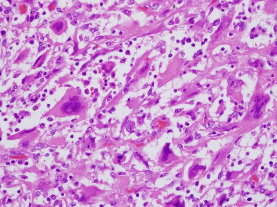
2 Case - 2 Clinical History: A 43 year old man with no significnt past history was seen for right chest pain and shortness of breath. A chest X-ray showed right pleural effusion and consolidation of the right lower lobe of lung. A CT scan showed an infiltrative mass extensively involving the diaphragmatic and costal margin of the pleura.

3

4

5

6

7

8
9

10 CK AE1/AE3

11 Calretinin SMA

12 MIB-1

13 Other Stains: CEA negative MOC31 negative TTF1 negative S-100 negative HMB45 negative Bcl-2 negative CD31 negative CD34 negative P63 negative WT1 negative EMA - negative CEA MOC31

14

15 Diagnosis: 1) Metastatic sarcomatoid renal cell carcinoma 2) Pleomorphic leiomyosarcoma 3) Sarcomatoid malignant mesothelioma 4) Pleural invasion by sarcomatoid carcinoma of lung 5) Pleuropulmonary blastoma 6) Malignant solitary fibrous tumor
 Pleuropulmonary blastoma 6) Malignant solitary")
16 Additional History: Extensive clinical studies and radiographic evaluation failed to identify any evidence of tumor elsewhere Additional history indicated that the patient had worked in his youth in a factory where he had been exposed to asbestos The patient died of progressive local disease 8 months after the operation

17 Diagnosis: SARCOMATOID MALIGNANT MESOTHELIOMA

18 Sarcomatoid Mesothelioma a New Paradigm? Diagnosis of exclusion Necessitates extensive clinical evaluation and imaging studies to rule out the possibility of a tumor elsewhere Immunohistochemical studies and electron microscopy are non-specific and serve mostly to rule out other diagnostic possibilities Often abused diagnosis in clinical practice

19 Sarcomatoid Mesothelioma Spindle cells arranged in fascicles or having a haphazard distribution that resemble fibrosarcoma or MFH The differentiation from sarcomatoid carcinoma of the lung or metastatic sarcomatoid renal cell carcinoma can be exceedingly difficult Immunostains do not reliably differentiate between these possibilities In such cases, gross and clinical features may be helpful. WHO Pathology and Genetics of Lung, Pleura, Thymus and Heart, 2004.

20 Assumptions Regarding the Diagnosis of Mesothelioma Any history of asbestos exposure is adequate proof that the patient has malignant mesothelioma Any diffuse plaque-like tumor growth involving the pleura is evidence of malignant mesothelioma There are specific and distinctive IHC markers that permit a reliable diagnosis of pleural mesothelioma

21 History of Asbestos Exposure Not all mesotheliomas are related to asbestos exposure Not all exposure to asbestos automatically leads to the development of malignant mesothelioma The vast majority of individuals exposed to asbestos do NOT develop mesothelioma Trivial or brief exposure to asbestos is highly unlikely to cause malignant mesothelioma An appropriate latency period (15-30 years) between exposure and development of mesothelioma should exist
22 Diffuse Pleural Spread of Tumor Malignant mesothelioma Metastatic adenocarcinoma Pseudomesotheliomatous adenocarcinoma of lung Synovial sarcoma Angiosarcoma Leiomyosarcoma Metastatic malignant melanoma Thymoma Others
23 IHC Diagnosis of Mesothelioma Numerous studies and markers reported in the literature during past 20 years There is no specific marker so far for malignant mesothelioma IHC represents a diagnosis of EXCLUSION Panel of positive and negative markers Many exceptions to the rules; diagnosis must be adapted to specific clinical and pathological circumstances
24 IHC Limitations in the DD with Adenocarcinoma The positive marker, CK5/6, is only of utility in the differential diagnosis between lung adenocarcinoma and epithelioid mesothelioma, but not for discriminating adenocarcinomas from other organs Calretinin can also be expressed in a large variety of other epithelial neoplasms, including lung, breast, ovarian and thymic carcinoma, in addition to spindle cell carcinoma, synovial sarcoma, sex-cord stromal tumors and malignant melanoma WT1 is also seen in ovarian serous carcinoma, renal cell carcinoma, ovarian stromal tumors, Sertoli cell tumor and malignant melanoma The negative markers can also be positive in mesothelioma
25 Role of IHC for the Diagnosis of Sarcomatoid Mesothelioma The percentage of reactivity of the currently available positive markers in sarcomatoid mesothelioma is significantly lower than for conventional epithelioid tumors, indicating very poor sensitivity There is a great deal of overlap in the expression of markers between mesothelioma and other primary and metastatic malignant spindle cell tumors of the pleura, such as synovial sarcoma, leiomyosarcoma, spindle cell carcinoma, etc.
26 IHC in Sarcomatoid Mesothelioma Calretinin was positive in 17% of spindle cell sarcomas and in 60% of sarcomatoid carcinomas Thrombomodulin was positive in 38% of sarcomas and in 40% of sarcomatoid mesotheliomas Smooth muscle actin was positive in 60% of sarcomatoid mesotheliomas (Lucas et al. Histopathology 42: , 2003)
27 IHC in Sarcomatoid Mesothelioma Study CK CALRET Thr-MOD HBME1 CK5/6 Oates & Edwards -- 4/8 -- 3/8 -- Miettinen et al -- 7/7 5/7 0/7 1/7 Lucas et al 7/10 7/10 7/ /10 Attanoos et al 24/31 12/31 9/ /31 Ordonez -- 7/8 1/8 -- 1/8 TOTAL 31/41 37/64 22/56 3/15 11/56 (75%) (58%) (39%) (20%) (19%)
28 IHC in Sarcomatoid Mesothelioma The same number of cases of sarcomatoid mesothelioma stained positive for calretinin than did sarcomatoid carcinomas metastatic to the pleura and sarcomas The same number of cases of sarcomatoid mesothelioma stained positive for pan-keratin and vimentin than other non-mesotheliomatous spindle cell tumors of the pleura In the pleura, cytokeratin staining is only of value in the setting of malignant solitary fibrous tumors, but of no help with the rest of the tumors involved in the differential diagnosis
29 Spindle Cell Tumors of the Pleura Metastatic spindle cell carcinoma Synovial sarcoma Malignant solitary fibrous tumor Other primary or metastatic spindle cell sarcomas of the pleura (leiomyosarcoma, angiosarcoma, etc) Spindle cell thymoma Metastatic spindle cell melanoma
30 Conclusions: Sarcomatoid mesothelioma is a diagnosis of exclusion and should not be automatically rendered on just any spindle cell tumor of the pleura Immunohistochemical stains are of limited value for the diagnosis of sarcomatoid mesothelioma, and the usual panel of stains used for conventional epithelioid mesothelioma cannot be applied with similar results Ruling out alternative conditions, such as sarcomatoid carcinoma, synovial sarcoma, and metastases of other spindle cell neoplasms, is always indicated.
31 Case 4 Clinical History: A 45 year old man with no significant past history was seen for the development of a palpable right thyroid nodule. An FNA showed follicular cells consistent with follicular process. A right thyroid lobectomy was done. The resected specimen showed a tan-brown, well-circumscribed nodule that measured 5.0 cm. in greatest diameter and was surrounded by a thin rim of normal thyroid parenchyma.
32
33
34
35
36
37
38
39
40
41 Diagnosis: 1. Follicular tumor with artifactual clearing of the nuclei 2. Follicular variant of papillary thyroid carcinoma 3. Macrofollicular variant of papillary carcinoma 4. Hyperplastic nodule with degenerative changes secondary to poor fixation 5. Benign follicular adenoma with pseudoclear nuclei of Haepke and Dehner
42 Diagnosis: THYROID FOLLICULAR ADENOMA WITH PSEUDOCLEAR NUCLEI
43
44
45
46
47 Diagnosis of PTC: Is based on a constellation of findings not on a single feature: - Typical nuclear features - Architectural changes, including papillation, invasion and stromal desmoplasia - Invasive properties - Other findings: psammoma bodies, multinucleated giant cells, scalloping of dense-staining colloid, etc.
48 Nuclear Features of Papillary Carcinoma Optically clear (Orphan Annie) nuclei Longitudinal nuclear grooves Cytoplasmic pseudonuclear inclusions Overlapping of nuclei ( shingling )
49 The Optically Clear Nucleus (a) Optically clear nucleus of papillary carcinoma (b) Pseudoclear nucleus of Hapke and Dehner. (c) Nucleus in follicular adenoma/carcinoma Hapke & Dehner, Am J Surg Pathol, Vol.3: 31-38, 1979
50 Other Thyroid Conditions with Clear Nuclear Changes Follicular adenoma Hyperplastic nodules Hashimoto s thyroiditis
51
52
53
54 Thyroid intrafollicular neoplasia: A spectrum of morphological appearances from benign cytologic precursors to microscopic papillary carcinoma N. Pennelli University of Padova, Italy J Postgrad Med 53:5-6, 2007
55 Follicular Variant of Papillary Thyroid Carcinoma (FVPTC) Definition: a tumor with a predominant or exclusive follicular growth pattern displaying the characteristic nuclear features of PTC First described by Lindsay in 1960 Same prognosis and behavior as conventional PTC Two histologic types: - Encapsulated FVPTC - Widely invasive FVPTC
56
57 Laryngoscope, Vol. 110: , 2000.
58 Follicular Variant of Papillary Carcinoma: The Diagnostic Limitations of Preoperative Fine-Needle Aspiration and Intraoperative Frozen Section Evaluation 47 patients with PTC studied who had both FNA and FS done 24/47 patients had a final diagnosis of FVPTC Sensitivity for FNA in cases of FVPTC was 25% Sensitivity of FS in cases of FVPTC was 29% Sensitivity of conventional PTC for FNA was 74% Sensitivity of conventional PTC for FS was 87% CONCLUSION: The thyroid surgeon needs to realize that, like follicular carcinoma, FVPTC is often diagnosed only on final pathological examination Laryngoscope, Vol.110: , 2000.
59 Problem Areas in the Diagnosis of Follicular Variant of PTC: Wide variation in pathologist s perception of what constitutes the nuclear features of PTC Tumors in which the nuclear features of PTC are only seen focally within the tumor Tumors in which multiple microscopic foci with nuclear features suggestive of PTC are present Tumors in which the nuclei adopt a pseudoclear appearance, but no other features of FVPTC are present
60
61
62
63
64 Am J Surg Pathol Vol.28: , 2004.
65 Percentage of Diagnoses by 10 Reviewers in 87 Follicular Tumors Reviewer FVPTC FA FCA Other 1 100% % 12.6% % % 13.8% 1.1% % 20.7% 1.1% 1.1% % 4.7% 0 3.5% 6 100% % 1.1% 0 6.9% % 0 1.1% % 37.9% 12.6% 3.5% % 11.5% 1.2% 26.4%
66 Observer Variation in the Diagnosis of Follicular Variant of Papillary Thyroid Carcinoma All 10 experts agreed on the diagnosis in only 39% of all cases All 10 experts agreed on the diagnosis in 66.7% of cases that were widely invasive and metastasized The lowest concordance was for well-circumscribed, non-invasive and encapsulated tumors CONCLUSION: Since most cases with metastatic disease had obvious invasion, caution should be used in making a diagnosis of FVPTC in the absence of the major histopathologic features of clearcut invasive growth Am J Surg Pathol 28: , 2004.
67 Chernobyl Pathologists Group (Int J Surg Pathol Vol.8: , 2000) There are a small number of follicular tumors in which there is no vascular invasion and genuine doubt exists about capsular invasion. Since follicular tumors with only capsular invasion carry an extremely good prognosis, as do encapsulated FVPTC, these cases should not be subjected to further treatment (i.e., total thyroidectomy + radioactive iodine); lobectomy with clear margins is generally curative The term Well-differentiated tumor of uncertain malignant potential or Follicular tumor of uncertain malignant potential is proposed for these lesions to avoid overtreatment and overdiagnosis
68 Nomenclature for Encapsulated Follicular Tumors by the Chernobyl Group Nuclear Features of PTC Obvious Equivocal Definite capsular or vasc. invasion None or questionable caps/vasc invasion PTC Well-differentiated Ca Not otherwise specified Well-differentiated tumor of uncertain malignant potential
69 Architectural and Stromal Changes in Papillary Thyroid Carcinoma Papillary architecture Stromal desmoplasia Infiltrative growth pattern Capsular invasion Vascular invasion Psammoma bodies These changes in and of themselves are not pathognomonic for papillary thyroid carcinoma but their absence in a thyroid process with clear nuclear features should challenge a diagnosis of PTC
70 CONCLUSIONS: It may be better to err on the benign side than to overdiagnose FVPTC, because lobectomy or nodulectomy is curative in most instances A diagnosis of carcinoma should be avoided in tumors with only focal, partial or equivocal features of invasion or nuclear features of PTC Well-differentiated tumor of uncertain malignant potential or Follicular tumor of uncertain malignant potential may be a preferable designations for such cases
71 Conclusions: Clear nuclear features in thyroid nodules per se and in isolation are not diagnostic of papillary thyroid carcinoma Focal clear nuclear changes can be seen in a variety of conditions other than papillary carcinoma, including nodular hyperplasia, follicular adenoma and Hashimoto s thyroiditis The diagnosis of papillary thyroid carcinoma is established based on a constellation of findings including nuclear features, architecture, and stromal changes, not on the basis of any given single feature
72 Case - 6 Clinical History: A 43 year old woman without any significant past medical history was seen for symptoms of abdominal pain and cramps. Upper endoscopy showed luminal obstruction of the duodenum by a mass lesion infiltrating the wall of the bowel. A biopsy was read as a high-grade malignant neoplasm. A small bowel resection was done.
73
74
75
76
77
78 Round/polygonal Spindle/oval
79
80 Diagnosis: 1) GIST 2) GANT 3) Metastatic malignant melanoma 4) Clear cell sarcoma-like tumor of the GI tract 5) Epithelioid sarcoma 6) Anaplastic carcinoma
81 S-100
82 SOX-10 HMB-45 Synapto Melan-A
83 Summary of IHC Stains: Positive: S-100 SOX-8 Synaptophysin Vimentin NB84-neuronal-assoc.p CD56 Neurofilament protein Negative: HMB45 Melan-A Tyrosinase MiTF C-kit (CD117) DOG-1 CD34 BCL-2 Ker AE1/AE3 CAM5.2 Desmin/SMA/Myo D1 CD99 Chromogranin-A
84 Ultrastructure
85 Splitting of fluorescent signal with break apart probe for chromosomal translocation
86 Additional Molecular Findings: Break-apart FISH for partner fusion genes showed involvement of the activating transcription factor (ATF1) gene
87 Diagnosis: Gastrointestinal Neuroectodermal Tumor (GNET)
88 Modern Pathology, Vol.24: Supplement 1, 2011, p.21a Presented at 100 th Annual Meeting of the United States and Canadian Academy of Pathology (USCAP), Feb.2011
89 GNET: Clinicopathologic, immunohistochemical, ultrastructural and molecular analysis of 16 cases of a distinctive gastrointestinal neoplasm showing features of neural differentiation. Stockman D, Miettinen M, Spagnolo D, Dominguez H, Hornick J, Adsay V, Chou P, Amanuel BH, Van Tuinen P, Suster S, Zambrano E Mod Pathol Vol.24: 21A; cases of primary malignant neoplasms of the GI tract with features reminiscent of so-called Clear cell sarcoma-like tumor of the GIT All cases were studied by histology, IHC, molecular pathology, and in 5 cases by electron microscopy A large panel of IHC stains was utilized: DOG1, SMA, desmin, vimentin, S- 100, CD34, CD56, CD99, CD117, HMB45, Melan-A, tyrosinase, MiTF, neurofilament, MSE, synaptophysin, chromogranin, NB84, NeuN, SOX10, GFAP, CK AE1/AE3, CAM5.2 and Ki-67. FISH for EWSR1 and FUS was done using dual-color break-apart probes FISH rearrangement of ATF1 on chromosome 12q13 and for CREB1 on chromosome 2q34 was performed on paraffin sections
90 Clinical Findings: 8 women and 8 men, aged years (mean: 42 years) Abdominal pain, obstruction and abdominal mass discovered on imaging studies No history or evidence of similar tumor elsewhere Location: small intestine (10), stomach (4), colon (2) Size: cm in greatest diameter (mean: 5 cm) Gross: solid, firm, tan white and lobulated Treatment: surgery; chemotherapy Follow-up: 6/12 died of tumor before 2 yrs; 4/12 are alive but with regional metastases; 2 pts A&W at 3 years
91 Histologic Findings: All tumors involved the wall of the GI tract (submucosa and muscularis propia) Solid population of uniform, small epithelioid or oval to spindled tumor cells forming sheets or nests Pseudoalveolar, pseudopapillary, pseudoglandular, trabecular, microcystic patterns of growth were observed focally 6/16 cases showed clear cytoplasmic features 12/16 cases showed osteoclast-type giant cells 2/3 of cases showed ulceration of the overlying mucosa and areas of necrosis Significant mitotic activity (average: 6 mitoses per 10 HPF)
92 Epithelioid Spindle
93
94 Clear cell features Pseudorosettes
95 Osteoclast-type giant cells
96
97 Immunohistochemical Findings: S-100, SOX10 and Vimentin + in ALL cases Melanocytic-associated markers (HMB45, Melan-A, MiTF) were negative in ALL cases GIST markers (DOG-1, CD34, C-Kit) were negative in ALL Epithelial markers (AE1/AE3, CAM5.2) were negative in ALL Glial, neuronal (NeuN) and muscle markers were negative CD99 negative in ALL cases Neural and neuroendocrine markers, including synaptophysin, chromogranin, CD56, NB84 and NSE showed variable degrees of positivity ranging from 45-62% of cases
98 Ultrastructural Findings: EM was done in 5 cases: Polygonal cells with multiple interdigitating cell processes joined by macula adherens type junctions Slender and bulbous cytoplasmic processes In 2 cases, scattered cytoplasmic dense-core neurosecretory granules could be identified No evidence of muscle, nerve sheath, or melanocytic differentiation could be identified on extensive search (no premelanosomes or intracysternal tubules seen)
99 Molecular Studies: Ewing sarcoma breakpoint region 1 (EWSR1) was studied in 14 cases by break-apart FISH 12 cases showed involvement of EWSR1 11 cases showed splitting of the fluorescent probe consistent with chromosomal translocation involving ESWR1 1 case showed extra copies of the ESWR1 gene 5/11 cases showed involvement of ATF1 partner fusion gene with EWSR1 by break-apart FISH 3/11 cases showed involvement of CREB1 partner fusion gene with EWRS1 by break-apart FISH All cases were negative for FUS gene rearrangements
100 Clear Cell Sarcoma-Like Tumors of the Gastrointestinal Tract Six cases reported by Zambrano et al in 2003 characterized by S-100+ and multinucleated osteoclastic giant cells Tumors have been felt to represent the GI counterpart of clear cell sarcoma of tendons and aponeurosis arising in soft tissues Unlike most other clear cell sarcoma-like tumors, our cases did not reveal any immunohistochemical or ultrastructural features of melanogenesis or melanocytic differentiation Ultrastructural and IHC features are more in keeping with a primitive neuroectodermal tumor with autonomic nerve differentiation (similar to GANT)
101 Molecular Profile: Precise significance of the molecular genetic abnormality is unknown The translocation EWSR1-ATF1 is shared with: > Clear cell sarcoma of tendons and aponeurosis > Small round blue cell tumor of the intraosseous membrane > Polyphenotypical round cell sarcoma of bone > Angiomatoid fibrous histiocytoma The sharing of this common rearrangement with other related and unrelated neoplasms may be an indication that the different phenotypes are not a direct result of the molecular alteration but rather of the cell types in which the chimeric gene is expressed
102 Molecular Promiscuity Translocation/Fusion Genes T(12,22) EWSR1-ATF1 T(X,17) TFE3-ASPS T(12,16) ETV6-TRK-C Tumor Clear cell carcinoma of salivary gland Clear cell sarcoma of tendons and aponeurosis Small round desmoplastic tumor of bone Angiomatoid fibrous histiocytoma Translocation-associated renal cell carcinoma Alveolar soft parts sarcoma Congenital fibrosarcoma Cellular mesoblastic nephroma Acute myeloid leukemia Secretory breast carcinoma Mammary-type secretory skin CA
103
104 Summary: We believe these tumors correspond to a distinct, previously unrecognized entity that is closely related to clear cell sarcoma of tendons and aponeurosis but that possess distinctive features that set them apart; namely, lack of melanocytic differentiation and evidence of autonomic nerve features The tumors may be part of a family of neurocristomas that are derived from neural crest progenitor cells and that may give rise to a spectrum of phenotypes, including melanocytic and neural (autonomic nerve differentiation) We propose the designation of gastrointestinal neuroectodermal tumor (GNET) to distinguish them from other similar and related lesions
105 David Stockman, M.D. Medical College of Wisconsin Eduardo Zambrano, MD Medical College of Wisconsin Markku Miettinen, M.D. Armed Forces Institute of Pathology Volkan Adsay, M.D. Emory University Jason Hornick, M.D. Harvard Medical School Pauline Chou, M.D. Univ. Chicago Children s Hospital Dominic Spagnolo, M.D. Univ. of Western Australia Saul Suster, M.D. Medical College of Wisconsin
Update on Mesothelioma
 November 8, 2012 Update on Mesothelioma Intro incidence and nomenclature Update on Classification Diagnostic specimens Morphologic features Epithelioid Histology Biphasic Histology Immunohistochemical
Diagnosis of Mesothelioma Pitfalls and Practical Information
 Diagnosis of Mesothelioma Pitfalls and Practical Information Mary Beth Beasley, M.D. Mt Sinai Medical Ctr Dept of Pathology One Gustave L Levy Place New York, NY 10029 (212) 241-5307 mbbeasleymd@yahoo.com
PATHOLOGY OF THE PLEURA: Mesothelioma and mimickers Necessity of Immunohistochemistry. M. Praet
 PATHOLOGY OF THE PLEURA: Mesothelioma and mimickers Necessity of Immunohistochemistry M. Praet Pathology of the Pleura Normal serosa: visceral and parietal layers Inflammation Neoplasia: Primary: mesothelioma
MALIGNANT MESOTHELIOMA UPDATE ON PATHOLOGY AND IMMUNOHISTOCHEMISTRY
 MALIGNANT MESOTHELIOMA CLASSIFICATION MALIGNANT MESOTHELIOMA UPDATE ON PATHOLOGY AND IMMUNOHISTOCHEMISTRY Sisko Anttila, MD, PhD Jorvi Hospital Laboratory of Pathology Helsinki University Hospital Espoo,
MALIGNANT MESOTHELIOMA UPDATE ON PATHOLOGY AND IMMUNOHISTOCHEMISTRY
 MALIGNANT MESOTHELIOMA UPDATE ON PATHOLOGY AND IMMUNOHISTOCHEMISTRY Sisko Anttila, MD, PhD Jorvi Hospital Laboratory of Pathology Helsinki University Hospital Espoo, Finland 2nd Nordic Conference on Applied
HKCPath Anatomical Pathology Peer Review and Scores : PDF version for download
 AP2003R1 http://hkcpath.org. Correspondence: pkhui@ha.org.hk 1of 10 07/08/2003 HKCPath Anatomical Pathology Peer Review and Scores : PDF version for download AP141 Bone Marrow: Metastatic Carcinoma from
Seattle. Case Presentations. Case 1. 76 year old female with a history of breast cancer 12 years ago. Now presents with a pleural effusion.
 Seattle Montreal IAP September 2006 Case Presentations Allen M. Gown, M.D. Medical Director and Chief Pathologist PhenoPath Laboratories Clinical Professor of Pathology University of British Columbia Case
Immunohistochemical differentiation of metastatic tumours
 Immunohistochemical differentiation of metastatic tumours Dr Abi Wheal ST1. TERA 3/2/14 Key points from a review article written by Daisuke Nonaka Intro Metastatic disease is the initial presentation in
Something Old, Something New.
 Something Old, Something New. Michelle A. Fajardo, D.O. Loma Linda University Medical Center Clinical Presentation 6 year old boy, presented with hematuria Renal mass demonstrated by ultrasound & CT scan
Notice of Faculty Disclosure
 The Diagnosis of Malignant Mesothelioma Andrew Churg, MD Department of Pathology University of British Columbia Vancouver, BC, Canada achurg@mail.ubc.ca Notice of Faculty Disclosure In accordance with
The develpemental origin of mesothelium
 Mesothelioma Tallinn 14.12.06 Henrik Wolff Finnish Institute of Occupational Health The develpemental origin of mesothelium Mesodermal cavities (pleura, peritoneum and pericardium ) are lined with mesenchymal
DESMOPLASTIC SMALL ROUND CELL TUMOR: A RARE PATHOLOGY PUZZLE
 DESMOPLASTIC SMALL ROUND CELL TUMOR: A RARE PATHOLOGY PUZZLE Ryan Granger University of Rhode Island Cytotechnology program May 2, 2015 ASCT Annual Meeting Nashville, Tennessee DESMOPLASTIC SMALL ROUND
Case of the. Month October, 2012
 Case of the Month October, 2012 Case The patient is a 47-year-old male with a 3-week history of abdominal pain. A CT scan of the abdomen revealed a suggestion of wall thickening at the tip of the appendix
The evolving pathology of solitary fibrous tumours. Luciane Dreher Irion MREH / CMFT / NSOPS
 The evolving pathology of solitary fibrous tumours Luciane Dreher Irion MREH / CMFT / NSOPS Historical review Haemangiopericytoma (HPC) first described primarily as a soft tissue vascular tumour of pericytic
Ovarian tumors Ancillary methods
 Ovarian tumors Ancillary methods Ovarian tumor course Oslo, 24-25/11/14 Prof. Ben Davidson, MD PhD Department of Pathology, Norwegian Radium Hospital, Oslo University Hospital, Oslo, Norway Division of
CASE OF THE MONTH AUGUST-2015 DR. GURUDUTT GUPTA HEAD HISTOPATHOLOGY
 CASE OF THE MONTH AUGUST-2015 DR. GURUDUTT GUPTA HEAD HISTOPATHOLOGY CASE HISTORY 52Y MALE RIGHT RADICAL NEPHERECTOMY Case of right renal mass with IVC thrombus. History of surgery and RT for right occipital
Today s Topics. Tumors of the Peritoneum in Women
 Today s Topics Tumors of the Peritoneum in Women Charles Zaloudek, M.D. Department of Pathology 505 Parnassus Ave., M563 University of California, San Francisco San Francisco, CA USA charles.zaloudek@ucsf.edu
Practical Effusion Cytology
 Practical Effusion Cytology A Community Pathologist s Approach to Immunocytochemistry in Body Fluid Cytology Emily E. Volk, MD William Beaumont Hospital Troy, MI College of American Pathologists 2004.
Diagnostic Challenge. Department of Pathology,
 Cytology of Pleural Fluid as a Diagnostic Challenge Paavo Pääkkö,, MD, PhD Chief Physician and Head of the Department Department of Pathology, Oulu University Hospital,, Finland Oulu University Hospital
Immunohistochemistry of soft tissue tumors
 Immunohistochemistry of soft tissue tumors Immunohistochemistry Major advances : antigen retrieval techniques (HIER) sensitive detection systems numerous antibodies of good quality Standardization : automated
Académie internationale de Pathologie - Division arabe XX ème congrès 24-26 novembre 2008 Alger. Immunohistochemistry in malignant mesotheliomas
 Académie internationale de Pathologie - Division arabe XX ème congrès 24-26 novembre 2008 Alger Immunohistochemistry in malignant mesotheliomas Françoise Thivolet-Béjui Groupement Hospitalier Est Lyon-Bron
Lessons from a consultation practice
 Pitfalls in the Application of Immunohistochemistry in Diagnostic Pathology Lessons from a consultation practice Kevin O. Leslie, MD Professor and Consultant Mayo Clinic Arizona Scottsdale, Arizona Presenter
Immunohistochemistry on cytology specimens from pleural and peritoneal fluid
 Immunohistochemistry on cytology specimens from pleural and peritoneal fluid Dr Naveena Singh Consultant Pathologist Bart health NHS Trust London United Kingdom Disclosures and Acknowledgements I have
ATLAS OF HEAD AND NECK PATHOLOGY THYROID PAPILLARY CARCINOMA
 Papillary carcinoma is the most common of thyroid malignancies and occurs in all age groups but particularly in women under 45 years of age. There is a high rate of cervical metastatic disease and yet
Pathology of lung cancer
 Pathology of lung cancer EASO COURSE ON LUNG CANCER AND MESOTHELIOMA DAMASCUS (SYRIA), MAY 3-4, 2007 Gérard ABADJIAN MD Pathologist Associate Professor, Saint Joseph University Pathology Dept. Hôtel-Dieu
Outline. Workup for metastatic breast cancer. Metastatic breast cancer
 Metastatic breast cancer Immunostain Update: Diagnosis of metastatic breast carcinoma, emphasizing distinction from GYN primary 1/3 of breast cancer patients will show metastasis 1 st presentation or 20-30
TUMORS OF THE TESTICULAR ADNEXA and SPERMATIC CORD
 TUMORS OF THE TESTICULAR ADNEXA and SPERMATIC CORD Victor E. Reuter, MD Memorial Sloan-Kettering Cancer Center reuterv@mskcc.org 66 th Annual Pathology Seminar California Society of Pathologists Short
Renal Cell Carcinoma: Advances in Diagnosis B. Iványi, MD
 Renal Cell Carcinoma: Advances in Diagnosis B. Iványi, MD Department of Pathology University of Szeged, Hungary ISUP Vancouver Classification of Renal Neoplasia Am J Surg Pathol 37:14691489, 2013 13 histologic
PRIMARY SEROUS CARCINOMA OF PERITONEUM: A CASE REPORT
 PRIMARY SEROUS CARCINOMA OF PERITONEUM: A CASE REPORT Dott. Francesco Pontieri (*) U.O. di Anatomia Patologica P.O. di Rossano (CS) Dott. Gian Franco Zannoni Anatomia Patologica Facoltà di Medicina e Chirurgia
How To Test For Cancer
 Diagnosis Of Serous Cavity Effusions - Beware The Mesothelial Cell! Effusion = Confusion Syed Z. Ali, M.D. Professor of Pathology and Radiology The Johns Hopkins Hospital Baltimore, Maryland Diagnostic
3-F. Pathology of Mesothelioma
 3-F. Pathology of Mesothelioma Kouki Inai Professor of Department of Pathology, Graduate School of Biomedical Science, Hiroshima University Introduction Mesothelioma is a peculiar type of malignancy, which
Effusions: Mesothelioma and Metastatic Cancers
 Effusions: Mesothelioma and Metastatic Cancers Malignant Mesothelioma Incidence: 2,500 cases/year ~60-80% pts with pleural MM relationship with asbestos exposure Other risk factors: radiation, other carcinogens,
Emerging Subtypes in Renal Cancer. Donna E. Hansel, MD PhD Professor of Pathology, UC San Diego Division Chief, Anatomic Pathology dhansel@ucsd.
 Emerging Subtypes in Renal Cancer Donna E. Hansel, MD PhD Professor of Pathology, UC San Diego Division Chief, Anatomic Pathology dhansel@ucsd.edu Some General Comments Fuhrman nuclear grading clear cell
Disclosures. Learning Objectives. Effusion = Confusion. Diagnosis Of Serous Cavity Effusions - Beware The Mesothelial Cell!
 Disclosures Diagnosis Of Serous Cavity Effusions - Beware The Mesothelial Cell! No Relevant Financial Relationships with Commercial Interests Syed Z. Ali, M.D. Syed Z. Ali, M.D. Associate Professor of
Index. F Factor VIII-related antigen, see VWF FactorXIIIa, for dermatofibroma, 272-275 5-HT, see Serotonin
 A Acantholytic squamous cell carcinoma vs epithelioid angiosarcoma, 56-57 Acinic cell carcinoma of pancreas, 76-77 vs ductal adenocarcinoma, 74-75 vs islet cell tumor, 78-81 Adenomatoid tumor vs hemangioma,
MODERN IMMUNOHISTOCHEMISTRY
 MODERN IMMUNOHISTOCHEMISTRY Cambridge Illustrated Surgical Pathology Peiguo G. Chu City of Hope National Medical Center, Duarte, California Lawrence M. Weiss City of Hope National Medical Center, Duarte,
Male. Female. Death rates from lung cancer in USA
 Male Female Death rates from lung cancer in USA Smoking represents an interesting combination of an entrenched industry and a clearly drug-induced cancer Tobacco Use in the US, 1900-2000 5000 100 Per Capita
The Role of Genetic Testing in the Evaluation of Thyroid Nodules. Thyroid Cancer and FNA. Thyroid Cancer. Pure Follicular Cancers.
 Where does Molecular Analysis of FNA Specimens fit into the evaluation of thyroid nodules? The Role of Genetic Testing in the Evaluation of Thyroid Nodules Ultrasound TSH Risk factors Jill E. Langer, MD
Renal Tumors with Eosinophilic Cytoplasm: 2013 Classification. Jesse K. McKenney, MD Associate Head, Surgical Pathology
 Renal Tumors with Eosinophilic Cytoplasm: 2013 Classification Jesse K. McKenney, MD Associate Head, Surgical Pathology Renal Epithelial Neoplasia History 1981: WHO Classification of Renal Neoplasms 1.
"Advances in Surgical Pathology: New Entities, Emerging Concepts and New Perspectives on Old Problems"
 123rd Semi-Annual Slide Seminar "Advances in Surgical Pathology: New Entities, Emerging Concepts and New Perspectives on Old Problems" Saul Suster, M.D. Chairman of Pathology Medical College of Wisconsin
Neoplasms of the LUNG and PLEURA
 Neoplasms of the LUNG and PLEURA 2015-2016 FCDS Educational Webcast Series Steven Peace, BS, CTR September 19, 2015 2015 Focus o Anatomy o SSS 2000 o MPH Rules o AJCC TNM 1 Case 1 Case Vignette HISTORY:
YOUR LUNG CANCER PATHOLOGY REPORT
 UNDERSTANDING YOUR LUNG CANCER PATHOLOGY REPORT 1-800-298-2436 LungCancerAlliance.org A GUIDE FOR THE PATIENT 1 CONTENTS What is a Pathology Report?...3 The Basics...4 Sections of a Pathology Report...7
Introduction: Tumor Swelling / new growth / mass. Two types of growth disorders: Non-Neoplastic. Secondary / adaptation due to other cause.
 Disorders of Growth Introduction: Tumor Swelling / new growth / mass Two types of growth disorders: Non-Neoplastic Secondary / adaptation due to other cause. Neoplastic. Primary growth abnormality. Non-Neoplastic
Tumor Patterns. Common Morphologic Patterns in Soft Tissue Tumors. Update on Lipomatous Tumors. MFH-like. Highly cellular spindle cell pattern
 Common Morphologic Patterns in Soft Tissue Tumors John R. Goldblum, M.D. Chairman, Department of Anatomic Pathology The Cleveland Clinic Professor of Pathology Cleveland Clinic Lerner College of Medicine
Cytology : first alert of mesothelioma? Professor B. Weynand, UCL Yvoir, Belgium
 Cytology : first alert of mesothelioma? Professor B. Weynand, UCL Yvoir, Belgium Introduction 3 cavities with the same embryologic origin the mesoderme Pleura Exudates Pleura Peritoneum Pericardium 22%
Neoplasia gastrica cistica: GIST o leiomiosarcoma? Sebastiano Cacciaguerra U. O. Chirurgia Pediatrica Ospedale Garibaldi Catania
 Neoplasia gastrica cistica: GIST o leiomiosarcoma? Sebastiano Cacciaguerra U. O. Chirurgia Pediatrica Ospedale Garibaldi Neoplasia gastrica cistica: GIST o leiomiosarcoma? Aims of presentation Atypical
Case based applications part III
 Case based applications part III Los Angeles Society Of Pathologists January 25, 2014 Sanja Dacic, MD, PhD University of Pittsburgh Medical Center 1 CASE 1 A 44-year-old woman with multiple lung nodules.
A 70-year old Man with Pleural Effusion
 Mesothelioma Diagnosis: Pitfalls and Latest Updates S Klebe and DW Henderson Recommendations Indisputable malignant cells on cytomorphological criteria which demonstrate a mesothelial phenotype, which
Original Article Differential diagnosis of sarcomatoid mesothelioma from true sarcoma and sarcomatoid carcinoma using immunohistochemistry
 Pathology International 2008; 58: 75 83 doi:10.1111/j.1440-1827.2007.02193.x Original Article Differential diagnosis of sarcomatoid mesothelioma from true sarcoma and sarcomatoid carcinoma using immunohistochemistry
Primary -Benign - Malignant Secondary
 TUMOURS OF THE LUNG Primary -Benign - Malignant Secondary The incidence of lung cancer has been increasing almost logarithmically and is now reaching epidemic levels. The overall cure rate is very low
Frozen Section Diagnosis
 Frozen Section Diagnosis Dr Catherine M Corbishley Honorary Consultant Histopathologist St George s Healthcare NHS Trust and lead examiner final FRCPath Practical 2008-2011 Frozen Section Diagnosis The
20 Diagnostic Cytopathology, Vol 36, No 1 ' 2007 WILEY-LISS, INC.
 Utility of WT-1, p63, MOC31, Mesothelin, and Cytokeratin (K903 and CK5/6) Immunostains in Differentiating Adenocarcinoma, Squamous Cell Carcinoma, and Malignant Mesothelioma in Effusions Robert T. Pu,
Molecular Diagnostics in Thyroid Cancer
 Disclosure Nothing to disclose Jonathan George, MD, MPH Assistant Professor Head and Neck Oncologic & Endocrine Surgery Molecular Diagnostics in Thyroid Cancer Current Practices & Future Trends UCSF Medical
J of Evidence Based Med & Hlthcare, pissn- 2349-2562, eissn- 2349-2570/ Vol. 2/Issue 33/Aug. 17, 2015 Page 5063
 PERITONEAL MALIGNANT MESOTHELIOMA: A RARE S. R. Dhamotharan 1, S. Shanthi Nirmala 2, F. Celine Foustina Mary 3, M. Arul Raj Kumar 4, R. Vinothprabhu 5 HOW TO CITE THIS ARTICLE: S. R. Dhamotharan, S. Shanthi
MALIGNANT MESOTHELIOMA: A TYPICAL PRESENTATION IN AN ATYPICAL PATIENT
 MALIGNANT MESOTHELIOMA: A TYPICAL PRESENTATION IN AN ATYPICAL PATIENT Written by: Karyn Varley MS, SCT(ASCP) The donating laboratory would like to remain anonymous. PATIENT HISTORY 28 year old female Lived
Plueral Malignancy: Radiologic-pathologic
 Plueral Malignancy: Radiologic-pathologic Correlation Ritu R. Gill, MD Pleural Malignancies: Radiologic-Pathologic Correlation Ritu R Gill MD Brigham and Women s Hospital Boston, Massachusetts Pleural
WORKPLACE SAFETY AND INSURANCE APPEALS TRIBUNAL DECISION NO. 1557/14
 WORKPLACE SAFETY AND INSURANCE APPEALS TRIBUNAL DECISION NO. 1557/14 BEFORE: M. Crystal: Vice-Chair HEARING: August 20, 2014 at Toronto Written DATE OF DECISION: December 4, 2014 NEUTRAL CITATION: 2014
Case presentation. Awatif Al-Nafussi
 Case presentation Awatif Al-Nafussi Case History 49 year old DVT & small PE June 08, Pelvic mass Ca125 33 Laparotomy-TAHBSO, drainage of ascites Ovarian carcinoma Clinical diagnosis Multiple specimens
Cytopathology Case Presentation #8
 Cytopathology Case Presentation #8 Emily E. Volk, MD William Beaumont Hospital, Troy, MI Jonathan H. Hughes, MD Laboratory Medicine Consultants, Las Vegas, Nevada Clinical History 44 year old woman presents
Diagnosis of Synovial Sarcoma of the Pleura and Differentiation from Malignant Mesothelioma
 36 Diagnosis of Synovial Sarcoma of the Pleura and Differentiation from Malignant Mesothelioma Amy Powers and Michele Carbone Synovial sarcomas (SSs) are soft tissue tumors that occur primarily in adolescents
Thymic carcinoma: update of current diagnostic criteria and histologic types
 Seminars in Diagnostic Pathology 22, 198-212 Thymic carcinoma: update of current diagnostic criteria and histologic types Saul Suster, MD From the Department of Pathology, The Ohio State University and
Mesothelioma. 1995-2013, The Patient Education Institute, Inc. www.x-plain.com ocft0101 Last reviewed: 03/21/2013 1
 Mesothelioma Introduction Mesothelioma is a type of cancer. It starts in the tissue that lines your lungs, stomach, heart, and other organs. This tissue is called mesothelium. Most people who get this
THYROID CANCER. I. Introduction
 THYROID CANCER I. Introduction There are over 11,000 new cases of thyroid cancer each year in the US. Females are more likely to have thyroid cancer than men by a ratio of 3:1, and it is more common in
The Value of Thyroid Transcription Factor-1 in Cytologic Preparations as a Marker for Metastatic Adenocarcinoma of Lung Origin
 Anatomic Pathology / TTF-1 IN CYTOLOGY OF BODY FLUIDS The Value of Thyroid Transcription Factor-1 in Cytologic Preparations as a Marker for Metastatic Adenocarcinoma of Lung Origin Jonathan L. Hecht, MD,
CHAPTER 2. Neoplasms (C00-D49) March 2014. 2014 MVP Health Care, Inc.
 March 2014. 2014 MVP Health Care, Inc.") Neoplasms (C00-D49) March 2014 2014 MVP Health Care, Inc. CHAPTER SPECIFIC CATEGORY CODE BLOCKS C00-C14 Malignant neoplasms of lip, oral cavity and pharynx C15-C26 Malignant neoplasms of digestive organs
Neoplasms (C00-D49) March 2014 2014 MVP Health Care, Inc. CHAPTER SPECIFIC CATEGORY CODE BLOCKS C00-C14 Malignant neoplasms of lip, oral cavity and pharynx C15-C26 Malignant neoplasms of digestive organs
Changes in Breast Cancer Reports After Second Opinion. Dr. Vicente Marco Department of Pathology Hospital Quiron Barcelona. Spain
 Changes in Breast Cancer Reports After Second Opinion Dr. Vicente Marco Department of Pathology Hospital Quiron Barcelona. Spain Second Opinion in Breast Pathology Usually requested when a patient is referred
Case presentation: Mesothelioma of the tunica vaginalis. Dr Ben Shepherd Pathology Queensland Princess Alexandra Hospital Brisbane
 Case presentation: Mesothelioma of the tunica vaginalis Dr Ben Shepherd Pathology Queensland Princess Alexandra Hospital Brisbane A 76 year old man presented June 2011 with a 6 month history of painless
Translocation Renal Cell Carcinomas
 Translocation Renal Cell Carcinomas Cora N. Sternberg, MD, FACP Chair, Department of Medical Oncology San Camillo and Forlanini Hospitals Rome, Italy Kidney cancer is not a single disease Clear cell (75%)
A Cytokeratin- and Calretinin-negative Staining Sarcomatoid Malignant Mesothelioma
 A Cytokeratin- and Calretinin-negative Staining Sarcomatoid Malignant Mesothelioma MICHAEL G. HURTUK and MICHELE CARBONE Cardinal Bernadin Cancer Center, Cancer Immunology Program, Department of Pathology,
Classificazioni citologiche: verso uno schema internazionale unificato?
 Cytology and molecular biology for thyroid nodules diagnos6c categories to clinical ac6ons From Classificazioni citologiche: verso uno schema internazionale unificato? A. Crescenzi Diagnostic categories
Lung Cancer. Ossama Tawfik, MD, PhD Professor, Vice Chairman Director of Anatomic &Surgical Pathology University of Kansas School of Medicine
 Lung Cancer Ossama Tawfik, MD, PhD Professor, Vice Chairman Director of Anatomic &Surgical Pathology University of Kansas School of Medicine Alexandria, Egypt July 1-1 3, 2008 OBJECTIVES Describe and
Diagnostic Pitfalls In Thoracic Tumors
 1376 Diagnostic Pitfalls In Thoracic Tumors Neda Kalhor, MD Cesar A. Moran, MD, FASCP WEEKEND OF PATHOLOGY AMERICAN SOCIETY FOR CLINICAL PATHOLOGY 33 W Monroe Ste 1600 Chicago, IL 60603 Program Content
Pleural Mesothelioma: An Institutional Experience of 66 Cases
 The Korean Journal of Pathology 2014; 48: 91-99 ORIGINAL ARTICLE Pleural Mesothelioma: An Institutional Experience of 66 Cases Soomin Ahn In Ho Choi Joungho Han Jhingook Kim 1 Myung-Ju Ahn 2 Departments
Renal Cell Carcinoma Associated with Xp11.2 Translocation: Clinicopathologic and Immunohistochemical Findings of 4 Cases
 The Korean Journal of Pathology 2005; 39: 406-11 Renal Cell Carcinoma Associated with Xp11.2 Translocation: Clinicopathologic and Immunohistochemical Findings of 4 Cases Sanghui Park Ji-Eun Kwon Yeon-Lim
MAJOR PARADIGM SHIFT IN EARLY 1990S IN UNDERSTANDING RENAL CANCER
 Renal tumours WHO 4 MAJOR PARADIGM SHIFT IN EARLY 1990S IN UNDERSTANDING RENAL CANCER Molecular differential pathology of renal cell tumours G. KOVACS A CLASSIFICATION BASED ON UNDERSTANDING THE GENETIC
PROTOCOL OF THE RITA DATA QUALITY STUDY
 PROTOCOL OF THE RITA DATA QUALITY STUDY INTRODUCTION The RITA project is aimed at estimating the burden of rare malignant tumours in Italy using the population based cancer registries (CRs) data. One of
Clinicopathological Study on Malignant Pleural Mesotheliomas
 Table of Contents Clinicopathological Study on Malignant Pleural Mesotheliomas PL-4-03 Kenzo Hiroshima Kenzo Hiroshima 1, Akira Iyota 1, Kiyoshi Sibuya 1, Toshikazu Yusa 2, Takehiko Fujisawa 1 and Yukio
METASTATIC CLEAR CELL RENAL CELL CARCINOMA TO THE SUBCUTANEOUS AREA IN ILLIAC FOSSA AND ADRENAL GLAND WITHOUT AN IDENTIFIABLE PRIMARY TUMOR
 Indian J.Sci.Res. 5(1) : 121-125, 2014 METASTATIC CLEAR CELL RENAL CELL CARCINOMA TO THE SUBCUTANEOUS AREA IN ILLIAC FOSSA AND ADRENAL GLAND WITHOUT AN IDENTIFIABLE PRIMARY TUMOR a1 b c d REETA DHAR, SHILPI
SEMESTER VI 3 RD YEAR PATHOLOGY KIDNEY TUMORS
 SEMESTER VI 3 RD YEAR PATHOLOGY KIDNEY TUMORS LEARNING OBJECTIVES At the end of the lecture, students should be able to: Know the pathology of renal tumors. RENAL TUMORS RENAL PAPILLARY ADENOMA Common
Epithelial Tumors of the Kidney Diagnostic Problems and Recently Described Entities
 Pathology of Renal Neoplasia Epithelial Tumors of the Kidney Diagnostic Problems and Recently Described Entities Wael A Sakr, MD Wayne State University School of Medicine CURRENT CLASSIFICATION = EPITHELIAL
Protocol applies to all primary borderline and malignant epithelial tumors, and malignant mesothelial neoplasms of the peritoneum.
 Peritoneum Protocol applies to all primary borderline and malignant epithelial tumors, and malignant mesothelial neoplasms of the peritoneum. Protocol revision date: January 2004 No AJCC/UICC staging system
BIOBANK LPCE-NICE CHEST
 BIOBANK LE-NIE HEST athologist :. BUTORI 12/09/2013 LE / HU Unit atient : N LH13-3603 N LB 13-0691 ID : RO A onsent : YES Age : 54 Diagnosis and staging : chronic pleuresia 5x1000µL BIOBANK LE-NIE HEST
Fredrik.Enlund@gu.se Sahlgrenska universitetssjukhuset
 Fredrik.Enlund@gu.se Sahlgrenska universitetssjukhuset 1 Techniques for sarcoma diagnostics Molecular Pathology of Solid Tumors Uppsala 120924 Klinisk Molekylär Patologi Klinisk Patologi och Cytologi Gene
ASBESTOS EXPOSURE AND SARCOMATOID MALIGNANT PLEURAL MESOTHELIOMA Gorantla Sambasivarao 1, Namballa Usharani 2, Tupakula Suresh Babu 3
 ASBESTOS EXPOSURE AND SARCOMATOID MALIGNANT PLEURAL MESOTHELIOMA Gorantla Sambasivarao 1, Namballa Usharani 2, Tupakula Suresh Babu 3 HOW TO CITE THIS ARTICLE: Gorantla Sambasivarao, Namballa Usharani,
Cardiac Masses and Tumors
 Cardiac Masses and Tumors Question: What is the diagnosis? A. Aortic valve myxoma B. Papillary fibroelastoma C. Vegetation from Infective endocarditis D. Thrombus in transit E. None of the above Answer:
INTERNATIONAL ASSOCIATION FOR THE STUDY OF LUNG CANCER Prospective Mesothelioma Staging Project
 INTERNATIONAL ASSOCIATION FOR THE STUDY OF LUNG CANCER Prospective Mesothelioma Staging Project Data Forms and Fields in CRAB Electronic Data Capture System - Reduced Set - Pivotal data elements for developing
Rotation Specific Goals & Objectives: University Health Network-Princess Margaret Hospital/ Sunnybrook Breast/Melanoma
 Rotation Specific Goals & Objectives: University Health Network-Princess Margaret Hospital/ Sunnybrook Breast/Melanoma Medical Expert: Breast Rotation Specific Competencies/Objectives 1.0 Medical History
Case Report Epithelioid malignant mesothelioma presenting with features of gastric tumor in a child
 Int J Clin Exp Pathol 2014;7(5):2636-2640 www.ijcep.com /ISSN:1936-2625/IJCEP0000187 Case Report Epithelioid malignant mesothelioma presenting with features of gastric tumor in a child Qihan You 1, Jing
Objectives. Mylene T. Truong, MD. Malignant Pleural Mesothelioma Background
 Imaging of Pleural Tumors Mylene T. Truong, MD Imaging of Pleural Tumours Mylene T. Truong, M. D. University of Texas M.D. Anderson Cancer Center, Houston, TX Objectives To review tumors involving the
- Slide Seminar - Endocrine pathology in non-endocrine organs. Case 11. Stefano La Rosa, Gioacchino D Ambrosio, Fausto Sessa
 - Slide Seminar - Endocrine pathology in non-endocrine organs Case 11 Stefano La Rosa, Gioacchino D Ambrosio, Fausto Sessa Dept. of Pathology, Multimedica, Milan, Italy Dept. of Surgical and Morphological
Recommendations for the Reporting of Pleural Mesothelioma
 Recommendations for the Reporting of Pleural Mesothelioma Association of Directors of Anatomic and Surgical Pathology * DOI: 10.1309/6A30YQHBMTHEJTEM It has been evident for decades that pathology reports
Ovarian mucinous lesions. Ovarian mucinous lesions: Common diagnostic dilemmas. Ovarian mucinous lesions: problematic issues
 Ovarian mucinous lesions Ovarian mucinous lesions: Common diagnostic dilemmas Karuna Garg, MD University of California San Francisco Intestinal or usual type Seromucinous (Endocervical mucinous or Mullerian
The Diagnosis of Cancer in the Pathology Laboratory
 The Diagnosis of Cancer in the Pathology Laboratory Dr Edward Sheffield Christmas Select 74 Meeting, Queen s Hotel Cheltenham, 3 rd December 2014 Agenda Overview of the pathology of cancer How specimens
RENAL CELL CARCINOMA EPIDEMIOLOGY
 RENAL CELL CARCINOMA: THE 2012 ISUP VANCOUVER CLASSIFICATION David Grignon MD, Indiana University, Indianapolis, IN RENAL CELL CARCINOMA EPIDEMIOLOGY Siegel et al. CaA Cancer J Clin 63:11-30, 2013 1 EPITHELIAL
A23: Oncologic Disease- Tumor Markers
 A23: Oncologic Disease- Tumor Markers Diagnosis Tumor Markers and Genetic Markers Use for Specific Malignancy The following information is from multiple guideline sources as recommendations for use of
بسم هللا الرحمن الرحيم
 بسم هللا الرحمن الرحيم Updates in Mesothelioma By Samieh Amer, MD Professor of Cardiothoracic Surgery Faculty of Medicine, Cairo University History Wagner and his colleagues (1960) 33 cases of mesothelioma
Cystic Neoplasms of the Pancreas: A multidisciplinary approach to the prevention and early detection of invasive pancreatic cancer.
 This lecture is drawn from the continuing medical education program Finding Hope: Prevention, Early Detection and Treatment of Pancreatic Cancer, Nov, 2011. Robert P. Jury, MD Cystic Neoplasms of the Pancreas:
Lung Cancer: Diagnosis, Staging and Treatment
 PATIENT EDUCATION patienteducation.osumc.edu Lung Cancer: Diagnosis, Staging and Treatment Cancer begins in our cells. Cells are the building blocks of our tissues. Tissues make up the organs of the body.
Advances in Differentiated Thyroid Cancer
 Advances in Differentiated Thyroid Cancer Steven A. De Jong, M.D., FACS, FACE Professor and Vice Chair Clinical Affairs Department of Surgery Loyola University Medical Center Thyroid Cancer classification