Immunohistochemical classification of the unknown primary tumour
|
|
|
- Kerry Chase
- 8 years ago
- Views:
Transcription

1 Immunohistochemical classification of the unknown primary tumour NordiQC Workshop 2014 Prof. Mogens Vyberg Institute of Pathology Aalborg, Denmark PART 2 Secondary panels for carcinoma identification/subclassification Cytokeratin subtypes Cell adhesion molecules Oncofetal proteins Transcription factors Neuroendocrine proteins Hormone receptors Secretory proteins... Breast markers Lung markers GI-markers Fem.gen.tract markers Urinary tract markers Prostate markers Squamous cell markers Mesothelial markers NE cell markers Liver cell markers Adrenal cortical markers Germinal cell markers Cell adhesion molecules Epithelial cell adhesion molecule (Ep-CAM) Claudin 4 (c) Mogens Vyberg

2 Ep-CAM Glycoproteins located on the cell membrane surface (preferentially basolaterally) and in the cytoplasm of virtually all epithelial cells with the exception of squamous epithelia (mucosae focal pos.) hepatocytes (pos. with inflammation and fibrosis) renal proximal tubular cells gastric parietal cells some endocrine cells Also negative mesothelial cells (reactive pos.) mesenchymal cells germ cells neural crest cells (except. olfactory neurons) Liver Ep-CAM +(-) Adenocarcinomas (of most types) Neuroenocrine neoplasms Olfactory neuroblastoma Seminoma/Dysgerminoma Synovial sarcoma Desmoplastic small cell tumour Brenner tumour Large cell lung carcinoma Ep-CAM -/+ Lobular breast carcinoma Hepatocellular carcinoma Squamous cell carcinoma Renal cell carcinoma Malignant mesothelioma Embryonal carcinoma - Adrenal cortical carcinoma Sarcomas (apart from synovial sarcoma) Lymphomas HCC (c) Mogens Vyberg
 Liver Ep-CAM +(-) Adenocarcinomas (of most types) Neuroenocrine neoplasms Olfactory neuroblastoma Seminoma/Dysgerminoma Synovial sarcoma Desmoplastic small cell tumour Brenner")
3 Ep-CAM Undiff carcinoma Ep-CAM Choroid plexus pap. Malignant mesothelioma Yolk sac tumour EpCAM EMA (c) Mogens Vyberg
")
4 EpCAM protocol Colon adenocarcinoma Lab A Lab B Claudin 4 Integral membrane protein, which belongs to the claudin family. The protein is a component of tight junction strands and may play a role in internal organ development and function. Tonsil Claudin 4 Ep-CAM Claudin 4 vs. Ep-CAM Mesothelioma Claudin 4 Ep-CAM (c) Mogens Vyberg

5 Claudin 4 vs. Ep-CAM Renal clear cell carc Claudin 4 Ep-CAM Breast cancer markers Estrogen receptor GCDFP-15 Mammaglobin GATA3 Estrogen receptor alpha Estrogen sensitive tissues: Breast Ovarium Fallopian tube Endo- and myometrium Uterine cervix Thyroid gland Pituitary gland Pancreas Sweat gland Salivary gland Prostate, Liver (c) Mogens Vyberg


6 Estrogen receptor alpha Adenocarcinomas Breast carcinoma +/ Ovarian (non-muc.) +/ Endometrial carc. +/ Cervix, lung, kidney, endocrine... /+ ERα Sica et al. APLM 132,1889,2008 Estrogen receptor alpha Lung adenocarcinomas ERα Sica et al. APLM 132,1889,2008 Estrogen receptor alpha: The influence of antibody selection 92 lung adenocarcinomas ER+ ( 1% pos. cells) ERα 1D5: 8% 6F11: 14% SP1: 27% Gomez-Fernandez et al. AIMM 2010;18:137 (c) Mogens Vyberg

7 Serial sections stained for Estrogen receptor Lab. A Lab. B 19 ER Run B17/2013: 281 labs 60% optimal 12% insufficient Lab. A Lab. B 20 Serial sections stained for Estrogen receptor Control: uterine cervix Lab. A Lab. B 21 (c) Mogens Vyberg

8 Serial sections stained for Estrogen receptor Control: uterine cervix All Abs but 6F11 6F11 in 15/36 labs Lab. A Lab. B 22 GCDFP-15 (gross cystic disease fluid protein 15) Prolactin induced glycoprotein of apocrine cells Adenocarcinomas Breast +/ Sweat gland +/ Salivary gland +/ Lung (+) Renal cell (+) Ovary (+) GCDFP-15 in sweat glands GCDFP-15 Breast carcinomas (c) Mogens Vyberg
 Renal cell (+) Ovary")
9 Gastric biopsies, 43 y ER GCDFP-15 Mammaglobin Mammary-specific member of the uteroglobin family Adenocarcinomas Breast +/ Sweat gland +/ Salivary gland +/ Lung /+ Stomach, biliary, thyroid, ovary, endom. (+) Mammaglobin in breast Mammaglobin Breast ductal carcinomas (c) Mogens Vyberg
 Mammaglobin in breast Mammaglobin Breast ductal carcinomas (c)")
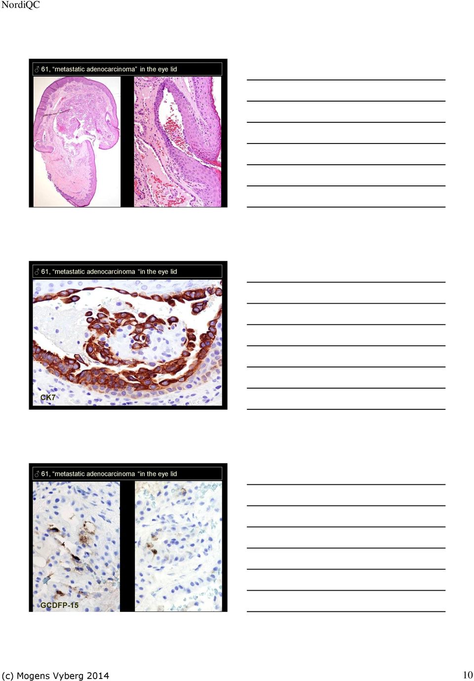
10 61, metastatic adenocarcinoma in the eye lid 61, metastatic adenocarcinoma in the eye lid CK7 61, metastatic adenocarcinoma in the eye lid GCDFP-15 (c) Mogens Vyberg
11 61, metastatic adenocarcinoma in the eye lid: Moll gland ca. MAMMAGLOBIN GATA3 Transcription factor recognizing the G-A-T-A nucleotide sequences in target gene promoters, regulating genes involved in the development/maintenance of T-cells breast epithelium urothelium skin trophoblast endothelia GATA3 Transcription factor recognizing the G-A-T-A nucleotide sequences in target gene promoters, regulating genes involved in the development/maintenance of T-cells breast epithelium urothelium skin trophoblast endothelia (c) Mogens Vyberg

12 GATA3 Transcription factor recognizing the G-A-T-A nucleotide sequences in target gene promoters, regulating genes involved in the development/maintenance of T-cells breast epithelium urothelium skin trophoblast endothelia GATA3 + Breast carcinoma Urothelial carcinoma Skin carcinoma (squam., bas., adnex) Yolk sac tumour Lobular breast carcinoma Urothelial carcinoma Miettinen et al. Am J Surg Pathol. 2014, 38: GATA3 +/- Malignant mesothelioma Pancreas adenocarcinoma Chromophobe renal cell carcinoma Paraganglioma -/+ Non-skin squamous cell carc. Salivary gland carcinomas Adrenal cortical carcinoma Embryonal carcinoma (c) Mogens Vyberg

13 GATA3 -(+) Serous and endometrioid adenocarcinoma Colon and gastric adenocarcinoma Prostate adenocarcinoma Renal cell carcinoma (non-chromophobic) Thyroid carcinoma Sarcomas - Endocrine neoplasms Malignant melanoma Seminoma Lung markers TTF-1 Napsin Thyroid transcription factor-1 Thyroid gland: regulating thyroglobulin, thyroperoxidase and thyrotropin receptor. Lung: regulating surfactant proteins and Clara cell secretory protein. Thyroid follicles and C-cells Lung pneumocytes II and Clara cells Brain Pituitary Parathyroid Normal lung (c) Mogens Vyberg

14 TTF-1 Run 39/2013: 227 labs Optimal: 51% Insufficient: 29% Thyroid transcription factor-1 % pos.: SPT24 8G7G3/1 Lung adenocarcinoma large cell carcinoma small cell carcinoma carcinoid squamous cell carcinoma 15 0 Non-lung small cell carcinoma 20-40? Thyroid carc. (non-anaplastic) Non-lung carcinomas Non-lung carcinoids ~ 0 ~ 0 Matoso et al, AIMM 2010,18: Napsin A Aspartic proteinase Type II pneumocytes Proximal and convoluted renal tubules (Pancreatic acini and ducts) (c) Mogens Vyberg
 100 100 Non-lung carcinomas 0-5 0-5 Non-lung carcinoids ~ 0 ~ 0 Matoso et al, AIMM 2010,18:142-149 Napsin A Aspartic")
15 Napsin A Aspartic proteinase Type II pneumocytes Proximal and convoluted renal tubules (Pancreatic acini and ducts) Napsin A: lung adenocarcinoma ~ 80% Napsin A: Lung squamous carcinoma 0 30% (c) Mogens Vyberg
 Mogens")
16 Napsin A: Renal cell carcinoma 0 70% 49 Liver adenocarcinoma of unknown primary CK20 49 Liver adenocarcinoma of unknown primary: Lung TTF Napsin A (c) Mogens Vyberg

17 Napsin Run 39/2013: 104 labs 30% optimal 42% insufficient GI markers CDX2 CAD17 CEA SMAD4 CDX-2 protein Drosophila caudal related homeobox gene 2 product: Nuclear transcription factor for intestinal differentiation Intestine all cell types incl. endocrine Intestinal metaplasia chronic gastritis Barrett s esophagus Pancreas/bil.tract colon pancreas (c) Mogens Vyberg

18 CDX-2 protein in adenocarcinoma Colorectum +( ) Mucinous ovar. +( ) Esoph./Stom. +/ Mucinous lung +/ Pancr./biliary /+ Prostate (+) Urothelial (+) Endometrioid (+) Endocrine midgut + Yolk sac tumour + Colon adenocarcinoma CDX-2 protein in adenocarcinoma Colorectum +( ) Mucinous ovar. +( ) Esoph./Stom. +/ Mucinous lung +/ Pancr./biliary /+ Prostate (+) Urothelial (+) Endometrioid (+) Endocrine midgut + Yolk sac tumour + Colon adenoc. medullary adenoc. Colon adenosquamous carc. Endometrioid carcinoma: ER & CDX-2 ER CDX-2 (c) Mogens Vyberg
 Mogens Vyberg 2014 18")
19 CDX2 Normal colon 1 EPRCON EPRRTU DAK AMT 88 CDX2 Normal pancreas EPRCON EPRRTU DAK AMT 88 CDX2 Colon adenocarc EPRCON EPRRTU DAK AMT 88 (c) Mogens Vyberg
 Mogens")
20 Quality assurance of CDX2 detection Mean H-score and proportion of positives N EPR* CONC EPR RTU DAK- CDX2 AMT- 28 CDX2-88 High Ex % 100% 100% 98% 96% Low Ex rmab EPR2764Y (Ventana, CellMarque) 95% 48% 58% 19% 13% Borrisholt M, Nielsen S, Vyberg M. Appl Immunohistochem Mol Morphol Jan;21(1): Cadherin 17 Calcium dependent adhesion molecules CAD17 = Liver-Intestine (LI-) Cadherin Regulated by CDX2 Intestine, pancreas Cadherin 17 Adenocarcinoma, colon + (100%) Adenocarc., esophagus, stomach, pancreas biliary tract +/ (often heterogenous) Adenocarc., lung, endometrium, ovary, breast (+) (focal) Endocrine tumour, small intest + (100%) Endocrine tumour, lung, pancr. /+ Squamous cell carc. (+) (c) Mogens Vyberg
 Adenocarc.")
21 CAD17 Appendix Cadherin 17: rmab SP183, CM, 1:50, CC1M/16M/UV CAD17 and CDX2 Colon adenocarcinoma Cadherin 17: rmab SP183, CM, 1:50, CC1M/16M/UV CDX2: rmab EPR2764Y, CM Carcinoembryonic antigen (CD66e) Adhesion molecule espc. associated with intestine (c) Mogens Vyberg
22 Carcinoembryonic antigen (CD66e) in adenocarcinomas Colorectal + Medull. thyroid + Pancreas/biliary tract +/ Stomach +/ Lung +/ Ovary, mucinous +/ Ovary, non-muc. /+ Prostate Kidney Liver metast. colon adenoc Carcinoembryonic antigen Medul. thyroid carc. Breast ductal carc. Carcinoembryonic antigen which antibody? Mal. mesothelioma Lab A Lab B (c) Mogens Vyberg
23 SMAD4 Ep-CAM Similar to Mothers Against Drosophila 4 = Deleted in pancreatic cancer-4 (DPC4) Nuclear transcription activator in all normal cells Pancreatico-biliary carcinomas ~50% Colorectal carcinomas ~10% Other carcinmas: rare 53 liver with ACUP 53 liver with ACUP CDX2 SMAD4 (c) Mogens Vyberg
24 SMAD4 loss in pancreatic and ampullary carcinomas Ep-CAM Female genital tract markers WT1 PAX8 ER HNFB1 Wilms tumour-1 (WT1) protein Transcription factor for development of the genitourinary system Mesothelium Ovary (surface / inclusion cysts) Fallopian tube Female genital tract: stromal cells Bone marrow stem cells Fallopian tube (c) Mogens Vyberg
25 Wilms tumour-1 (WT1) protein in adenocarcinomas Serous carcinoma + Endometrioid carcinoma +/ Other carcinomas (+) Mal. mesothelioma +/ Serous carcinoma PAX8 Transcription factor crucial to the organogenesis and development of Urogenital tract Adult genital tract Thyroid (Neuroendocrine system) PAX8 Ovary - serous carcinoma LG + - serous carcinoma HG / clear cell + - endometrioid carcinoma +/- - mucinous carcinoma -(+) Kidney - clear cell / papillary carcinoma + - chromophobe / collect. duct carc. + - RCC, sarcomatoid +/- Thyroid carcinoma + Other carcinomas -(+) Malignant mesothelioma - (c) Mogens Vyberg
26 PAX8 Run 34/2012: 36 labs Tube RCC Optimal: 26% Insufficient: 36% 48 mediastinal lymph node metastasis Case 2 mediastinal lymph node metastasis Pan-CK CK7 (c) Mogens Vyberg
27 Case 2 Metastatic clear cell renal cell carcinoma VIM PAX8 Markers for endocrine tumours Neuroendocrine Synaptophysin Chromogranin A Thyroid Thyroglobulin Liver Arginase CD66a/CD10 Glypican 3 Adrenal cortex, sex cord Calretinin Inhibin Melan A (A 103) Hormones... Unspecific NE: (PGP 9.5) (neuron specific enolase) (CD56) Synaptophysin An integral-membrane glycoprotein of presynaptic vesicles Neurons Neuroendocrine cells (Adrenal cortical cells) SYP 81 pancreas (c) Mogens Vyberg
28 Synaptophysin in neoplasias Neural tumours NE tumours NE carcinomas Adrenal cortical tumours Lung NE large cell carcinoma 82 Synaptophysin in neoplasias SCLC in skin 83 Adrenal cortical carcinoma Synaptophysin Run 37/2013: 214 labs. NE gr. 2 Optimal: 19% Insufficient: 42% SCLC (c) Mogens Vyberg
29 Mesothelial markers Calretinin Podoplanin Calretinin Calcium-binding protein related to S-100 proteins Neurons Mesothelial cells Steroid producing cells adrenal cortical cells testicular Leydig and Sertoli cells ovarian theca interna cells and surface epithelium Neuroendocrine cells Breast glands... Calretinin Mesothelium Appendix (c) Mogens Vyberg
30 Calretinin in neoplasms Malignant mesothelioma Malignant mesothelioma Calretinin Adrenal gland Granulosa cell tumour Podoplanin Lymphatic endothelium Fibroblasts, osteocytes Smooth and striated muscle cells Myoepithelial cells, Cajal cells Basal squamous epithelial cells Gastric crypt c., prostatic basal c. Immature Sertoli c. and gonocytes Renal glomerular podocytes Mesothelium (reactive) Follicular dendritic cells Some lymphocytes Glial/Schwann cells (c) Mogens Vyberg
31 Podoplanin Mesothelioma Many other tumours but rarely adenocarcinomas Malignant mesothelioma vs. lung carcinoma Claudin Ep- P63 WT1 CR PDP VIM 4 CAM CEA NAPA TTF-1 p40 CK5 Ep. malignant mesothelioma (f) Lung adenocarcinoma 0 10 (f) 10 (f ) 30 5? (5) Lung squamous cell carcinoma 0 50? 10 10? ? 10f Liver cell markers Glypican 3 Arginase cancd66a (biliary glycoprotein) Hepatocyte antigen Glutamine synthetase cancd10 canvillin AFP (c) Mogens Vyberg
32 Glypican 3 Heparan sulphate proteoglycan modifying cell signaling Yolk sac tumour + Choriocarcinoma + Embryonal carc. /+ Hepatoblastoma + Hepatocellular carc. +/ Ovarian clear cell c. /+ Carcinoid a.o. /+ HCC Glypican 3 Heparan sulphate proteoglycan modifying cell signaling Yolk sac tumour + Choriocarcinoma + Embryonal carc. /+ Hepatoblastoma + Hepatocellular carc. +/ Ovarian clear cell c. /+ Carcinoid a.o. /+ Teratoma with Emb. carc. Arginase I The final enzyme of the urea cycle: arginine + H2O ornithine + urea Predominantly located in liver cells Hepatocellular carcinoma +/ (80-90%) Adenocarcinoma, biliary tract, pancreas /+ Adencarcinoma breast, colorectum (+) (c) Mogens Vyberg
33 Arg and hepa Hepatocellular carcinoma I rmab SP156 1:25 CM 16M/CC1S/UV mab OCH1E5 1:400 Dako 32M/CC1S/UV+amp Arg and Hepa Pancreas adenocarcinoma rmab SP156 1:25 CM 16M/CC1S/UV mab OCH1E5 1:400 Dako 32M/CC1S/UV+amp CD66a Biliary glycoprotein CEA-like adhesion molecule Canalicular staining! HCC (c) Mogens Vyberg
Cytokeratins and generel epithelial markers in tumour classification
 Cytokeratins and generel epithelial markers in tumour classification 42, liver adenocarcinoma of unknown primary Colonrectum? Pancreas/biliary tract? Stomach? Lung? Ovary/endometrium? Mogens Vyberg, NordiQC
Immunohistochemical differentiation of metastatic tumours
 Immunohistochemical differentiation of metastatic tumours Dr Abi Wheal ST1. TERA 3/2/14 Key points from a review article written by Daisuke Nonaka Intro Metastatic disease is the initial presentation in
Ovarian tumors Ancillary methods
 Ovarian tumors Ancillary methods Ovarian tumor course Oslo, 24-25/11/14 Prof. Ben Davidson, MD PhD Department of Pathology, Norwegian Radium Hospital, Oslo University Hospital, Oslo, Norway Division of
Outline. Workup for metastatic breast cancer. Metastatic breast cancer
 Metastatic breast cancer Immunostain Update: Diagnosis of metastatic breast carcinoma, emphasizing distinction from GYN primary 1/3 of breast cancer patients will show metastasis 1 st presentation or 20-30
Tumour Markers. What are Tumour Markers? How Are Tumour Markers Used?
 Dr. Anthony C.H. YING What are? Tumour markers are substances that can be found in the body when cancer is present. They are usually found in the blood or urine. They can be products of cancer cells or
Practical Effusion Cytology
 Practical Effusion Cytology A Community Pathologist s Approach to Immunocytochemistry in Body Fluid Cytology Emily E. Volk, MD William Beaumont Hospital Troy, MI College of American Pathologists 2004.
Index. F Factor VIII-related antigen, see VWF FactorXIIIa, for dermatofibroma, 272-275 5-HT, see Serotonin
 A Acantholytic squamous cell carcinoma vs epithelioid angiosarcoma, 56-57 Acinic cell carcinoma of pancreas, 76-77 vs ductal adenocarcinoma, 74-75 vs islet cell tumor, 78-81 Adenomatoid tumor vs hemangioma,
MALIGNANT MESOTHELIOMA UPDATE ON PATHOLOGY AND IMMUNOHISTOCHEMISTRY
 MALIGNANT MESOTHELIOMA UPDATE ON PATHOLOGY AND IMMUNOHISTOCHEMISTRY Sisko Anttila, MD, PhD Jorvi Hospital Laboratory of Pathology Helsinki University Hospital Espoo, Finland 2nd Nordic Conference on Applied
MALIGNANT MESOTHELIOMA UPDATE ON PATHOLOGY AND IMMUNOHISTOCHEMISTRY
 MALIGNANT MESOTHELIOMA CLASSIFICATION MALIGNANT MESOTHELIOMA UPDATE ON PATHOLOGY AND IMMUNOHISTOCHEMISTRY Sisko Anttila, MD, PhD Jorvi Hospital Laboratory of Pathology Helsinki University Hospital Espoo,
How To Test For Cancer With A Blood Test
 Histolab Products AB Eva Alströmer Jonas Falgén Helsingborg 7-8/10 2010 Table 1 Formalin Fixation Times and Estrogen Receptor Staining With 25 Minutes Antigen Retrieval Pretreatment Formalin Q-Score Difference
Académie internationale de Pathologie - Division arabe XX ème congrès 24-26 novembre 2008 Alger. Immunohistochemistry in malignant mesotheliomas
 Académie internationale de Pathologie - Division arabe XX ème congrès 24-26 novembre 2008 Alger Immunohistochemistry in malignant mesotheliomas Françoise Thivolet-Béjui Groupement Hospitalier Est Lyon-Bron
Immunohistochemistry in the Diagnosis of Metastatic Carcinoma of Unknown Primary Origin
 Immunohistochemistry in the Diagnosis of Metastatic Carcinoma of Unknown Primary Origin Rodney T. Miller, M.D. Director of Immunohistochemistry ProPath Laboratory 1355 River Bend Drive Dallas, TX 75247-4915
A 70-year old Man with Pleural Effusion
 Mesothelioma Diagnosis: Pitfalls and Latest Updates S Klebe and DW Henderson Recommendations Indisputable malignant cells on cytomorphological criteria which demonstrate a mesothelial phenotype, which
MODERN IMMUNOHISTOCHEMISTRY
 MODERN IMMUNOHISTOCHEMISTRY Cambridge Illustrated Surgical Pathology Peiguo G. Chu City of Hope National Medical Center, Duarte, California Lawrence M. Weiss City of Hope National Medical Center, Duarte,
Effusions: Mesothelioma and Metastatic Cancers
 Effusions: Mesothelioma and Metastatic Cancers Malignant Mesothelioma Incidence: 2,500 cases/year ~60-80% pts with pleural MM relationship with asbestos exposure Other risk factors: radiation, other carcinogens,
Update on Mesothelioma
 November 8, 2012 Update on Mesothelioma Intro incidence and nomenclature Update on Classification Diagnostic specimens Morphologic features Epithelioid Histology Biphasic Histology Immunohistochemical
Immunohistochemistry in diagnostic pathology of tumors. Approach, benefits, limits and pitfalls
 Aus dem Institut für Pathologie der Medizinischen Fakultät Charité Universitätsmedizin Berlin DISSERTATION Immunohistochemistry in diagnostic pathology of tumors. Approach, benefits, limits and pitfalls
page antibody Adipophilin (polyclonal) ALDH1A1 (44) c-myc (EP121) * Cadherin-17 (SP183) Cathepsin K (3F9)
 ALDH1A1 (44) c-myc (EP121) * Cadherin-17 (SP183) Cathepsin K (3F9)") New IHC Antibodies 2 Table of Contents page antibody 1 2 3 4 5 6 7 8 Adipophilin (polyclonal) ALDH1A1 (44) c-myc (EP121) * Cadherin-17 (SP183) Cathepsin K (3F9) Caveolin-1 (2297) CD13 (SP187) CD16 (SP175)
New IHC Antibodies 2 Table of Contents page antibody 1 2 3 4 5 6 7 8 Adipophilin (polyclonal) ALDH1A1 (44) c-myc (EP121) * Cadherin-17 (SP183) Cathepsin K (3F9) Caveolin-1 (2297) CD13 (SP187) CD16 (SP175)
Efficient Tumor Immunohistochemistry A Differential Diagnosis-Driven Approach
 Efficient Tumor Immunohistochemistry A Differential Diagnosis-Driven Approach Publishing Team Erik Tanck (production manager/designer) Joshua Weikersheimer (publisher) Copyright 2006 by the American Society
Immunohistochemistry of clear cell tumours. What are clear cell tumours? Jan Klos 1
 Immunohistochemistry of clear cell tumours J. Klos MD Department of Pathology Stavanger University Hospital Norway What are clear cell tumours? Multiple factors in etiology of clear cell changes: - technical
A23: Oncologic Disease- Tumor Markers
 A23: Oncologic Disease- Tumor Markers Diagnosis Tumor Markers and Genetic Markers Use for Specific Malignancy The following information is from multiple guideline sources as recommendations for use of
Diagnosis of Mesothelioma Pitfalls and Practical Information
 Diagnosis of Mesothelioma Pitfalls and Practical Information Mary Beth Beasley, M.D. Mt Sinai Medical Ctr Dept of Pathology One Gustave L Levy Place New York, NY 10029 (212) 241-5307 mbbeasleymd@yahoo.com
How To Test For Cancer
 Diagnosis Of Serous Cavity Effusions - Beware The Mesothelial Cell! Effusion = Confusion Syed Z. Ali, M.D. Professor of Pathology and Radiology The Johns Hopkins Hospital Baltimore, Maryland Diagnostic
The term undifferentiated tumor has been used in reference
 Undifferentiated Tumor True Identity by Immunohistochemistry Armita Bahrami, MD; Luan D. Truong, MD; Jae Y. Ro, MD, PhD Context. Undifferentiated tumor refers to a heterogeneous group of neoplasms with
HKCPath Anatomical Pathology Peer Review and Scores : PDF version for download
 AP2003R1 http://hkcpath.org. Correspondence: pkhui@ha.org.hk 1of 10 07/08/2003 HKCPath Anatomical Pathology Peer Review and Scores : PDF version for download AP141 Bone Marrow: Metastatic Carcinoma from
Renal Cell Carcinoma: Advances in Diagnosis B. Iványi, MD
 Renal Cell Carcinoma: Advances in Diagnosis B. Iványi, MD Department of Pathology University of Szeged, Hungary ISUP Vancouver Classification of Renal Neoplasia Am J Surg Pathol 37:14691489, 2013 13 histologic
Disclosures. Learning Objectives. Effusion = Confusion. Diagnosis Of Serous Cavity Effusions - Beware The Mesothelial Cell!
 Disclosures Diagnosis Of Serous Cavity Effusions - Beware The Mesothelial Cell! No Relevant Financial Relationships with Commercial Interests Syed Z. Ali, M.D. Syed Z. Ali, M.D. Associate Professor of
Contents. 1. Introduction and Approach to Fine Needle Aspiration Cytology... 1. 2. Head, Neck, Orbit and Salivary Glands... 12
 Contents 1. Introduction and Approach to Fine Needle Aspiration Cytology... 1 Complications 1 Fine Needle Aspiration Technique 1 Evaluation of FNAC Smear 4 Cell Morphology 4 Nucleus 4 Cytoplasm 6 Background
PRIMARY SEROUS CARCINOMA OF PERITONEUM: A CASE REPORT
 PRIMARY SEROUS CARCINOMA OF PERITONEUM: A CASE REPORT Dott. Francesco Pontieri (*) U.O. di Anatomia Patologica P.O. di Rossano (CS) Dott. Gian Franco Zannoni Anatomia Patologica Facoltà di Medicina e Chirurgia
The Value of Thyroid Transcription Factor-1 in Cytologic Preparations as a Marker for Metastatic Adenocarcinoma of Lung Origin
 Anatomic Pathology / TTF-1 IN CYTOLOGY OF BODY FLUIDS The Value of Thyroid Transcription Factor-1 in Cytologic Preparations as a Marker for Metastatic Adenocarcinoma of Lung Origin Jonathan L. Hecht, MD,
Thyroid Transcription Factor-1 (TTF 1): protein expression is not exclusive to lung and thyroid tissue.
: protein expression is not exclusive to lung and thyroid tissue.") Thyroid Transcription Factor-1 (TTF 1): protein expression is not exclusive to lung and thyroid tissue. Scorer PW, Pinkney M, McIntosh GG. Leica Biosystems Newcastle Ltd, Balliol Business Park West, Benton
Thyroid Transcription Factor-1 (TTF 1): protein expression is not exclusive to lung and thyroid tissue. Scorer PW, Pinkney M, McIntosh GG. Leica Biosystems Newcastle Ltd, Balliol Business Park West, Benton
Determination of the type and origin of metastatic
 Diagnosis of Metastatic Neoplasms An Immunohistochemical Approach Murli Krishna, MD N Context. It is important to determine the type and/or site of origin of metastatic tumors for optimal clinical management.
PATHOLOGY OF THE PLEURA: Mesothelioma and mimickers Necessity of Immunohistochemistry. M. Praet
 PATHOLOGY OF THE PLEURA: Mesothelioma and mimickers Necessity of Immunohistochemistry M. Praet Pathology of the Pleura Normal serosa: visceral and parietal layers Inflammation Neoplasia: Primary: mesothelioma
TUMORS OF THE TESTICULAR ADNEXA and SPERMATIC CORD
 TUMORS OF THE TESTICULAR ADNEXA and SPERMATIC CORD Victor E. Reuter, MD Memorial Sloan-Kettering Cancer Center reuterv@mskcc.org 66 th Annual Pathology Seminar California Society of Pathologists Short
Immunostain Update: Diagnosis of Metastatic Breast Carcinoma, Emphasizing the Distinction from Gynecologic Cancers
 Immunostain Update: Diagnosis of Metastatic Breast Carcinoma, Emphasizing the Distinction from Gynecologic Cancers Yunn-Yi Chen, M.D., Ph.D. UCSF Pathology Department yunn-yi.chen@ucsf.edu June 3, 2010
Immunohistochemistry on cytology specimens from pleural and peritoneal fluid
 Immunohistochemistry on cytology specimens from pleural and peritoneal fluid Dr Naveena Singh Consultant Pathologist Bart health NHS Trust London United Kingdom Disclosures and Acknowledgements I have
Chapter I Overview Chapter Contents
 Chapter I Overview Chapter Contents Table Number Contents I-1 Estimated New Cancer Cases and Deaths for 2005 I-2 53-Year Trends in US Cancer Death Rates I-3 Summary of Changes in Cancer Incidence and Mortality
Pathology of lung cancer
 Pathology of lung cancer EASO COURSE ON LUNG CANCER AND MESOTHELIOMA DAMASCUS (SYRIA), MAY 3-4, 2007 Gérard ABADJIAN MD Pathologist Associate Professor, Saint Joseph University Pathology Dept. Hôtel-Dieu
Cytology of Effusion Fluids. Cytology of Effusion Fluids. Types of Effusion Fluids. Anatomy. Causes of Effusions. Sampling of Effusion Fluids
 Cytology of Effusion Fluids John W. Wong, MD, FRCPC Sunnybrook Health Sciences Centre Assistant Professor, Laboratory Medicine and Pathobiology Faculty of Medicine, University of Toronto November 10, 2012
R-16: Chronic nonspecific cervisit
 R-16: Chronic nonspecific cervisit Ectoservikal squamous epithelium Endoservical columnar epithelium Dilated cystic endoservical glands lymphoplasmocytes R18:Squamous cell carcinoma insitu Neoplastic epithelium
The develpemental origin of mesothelium
 Mesothelioma Tallinn 14.12.06 Henrik Wolff Finnish Institute of Occupational Health The develpemental origin of mesothelium Mesodermal cavities (pleura, peritoneum and pericardium ) are lined with mesenchymal
Applications of immunohistology to non-heme tumor differential diagnosis R V Rouse 7/22/2014 http://surgpathcriteria.stanford.edu
 Applications of immunohistology to non-heme tumor differential diagnosis R V Rouse 7/22/2014 http://surgpathcriteria.stanford.edu Table of Contents Page Undifferentiated panel 1 CK7/20 table 2 Breast carcinoma
Frozen Section Diagnosis
 Frozen Section Diagnosis Dr Catherine M Corbishley Honorary Consultant Histopathologist St George s Healthcare NHS Trust and lead examiner final FRCPath Practical 2008-2011 Frozen Section Diagnosis The
Articles. Nordic Immunohistochemical Quality Control (NordiQC) - An Organization for External Quality Assurance. Mogens Vyberg, MD
 - An Organization for External Quality Assurance. Mogens Vyberg, MD") Articles Nordic Immunohistochemical Quality Control (NordiQC) - An Organization for External Quality Assurance Mogens Vyberg, MD Institute of Pathology. Aalborg Hospital Aarhus University Hospital Denmark
Articles Nordic Immunohistochemical Quality Control (NordiQC) - An Organization for External Quality Assurance Mogens Vyberg, MD Institute of Pathology. Aalborg Hospital Aarhus University Hospital Denmark
MOC-31 Exhibits Superior Reactivity Compared With Ber-EP4 in Invasive Lobular and Ductal Carcinoma of the Breast. A Tissue Microarray Study
 RESEARCH ARTICLE MOC-31 Exhibits Superior Reactivity Compared With Ber-EP4 in Invasive Lobular and Ductal Carcinoma of the Breast A Tissue Microarray Study Reetesh K. Pai, MD and Robert B. West, MD Abstract:
Schedule of Accreditation issued by United Kingdom Accreditation Service 2 Pine Trees, Chertsey Lane, Staines-upon-Thames, TW18 3HR, UK
 Schedule of ccreditation United Kingdom ccreditation Service 2 Pine Trees, Chertsey Lane, Staines-upon-Thames, TW18 3HR, UK University College London, operating UK NEQS for ccredited to UK NEQS ICC & ISH
Changes in Breast Cancer Reports After Second Opinion. Dr. Vicente Marco Department of Pathology Hospital Quiron Barcelona. Spain
 Changes in Breast Cancer Reports After Second Opinion Dr. Vicente Marco Department of Pathology Hospital Quiron Barcelona. Spain Second Opinion in Breast Pathology Usually requested when a patient is referred
Seattle. Case Presentations. Case 1. 76 year old female with a history of breast cancer 12 years ago. Now presents with a pleural effusion.
 Seattle Montreal IAP September 2006 Case Presentations Allen M. Gown, M.D. Medical Director and Chief Pathologist PhenoPath Laboratories Clinical Professor of Pathology University of British Columbia Case
Lessons from a consultation practice
 Pitfalls in the Application of Immunohistochemistry in Diagnostic Pathology Lessons from a consultation practice Kevin O. Leslie, MD Professor and Consultant Mayo Clinic Arizona Scottsdale, Arizona Presenter
Introduction: Tumor Swelling / new growth / mass. Two types of growth disorders: Non-Neoplastic. Secondary / adaptation due to other cause.
 Disorders of Growth Introduction: Tumor Swelling / new growth / mass Two types of growth disorders: Non-Neoplastic Secondary / adaptation due to other cause. Neoplastic. Primary growth abnormality. Non-Neoplastic
A Better Path for Cancer Diagnostics
 INTERPRETATION FLEX Ready-to-Use Atlas of Stains - 4 th Edition A Better Path for Cancer Diagnostics Committed to raising the bar for higher quality During the last decades, organizations such as CAP,
The Diagnosis of Cancer in the Pathology Laboratory
 The Diagnosis of Cancer in the Pathology Laboratory Dr Edward Sheffield Christmas Select 74 Meeting, Queen s Hotel Cheltenham, 3 rd December 2014 Agenda Overview of the pathology of cancer How specimens
4/15/2013. bi/o carcin/ chem/o immun/o onc/o radi/o sarc/o. anabrachydysectoendoneo- -ectomy -genesis -oma -plasia -sarcoma
 Chapter Sixteen Oncology bi/o carcin/ chem/o immun/o onc/o radi/o sarc/o Combining Forms Prefixes and Suffixes Carcinogenesis anabrachydysectoendoneo- -ectomy -genesis -oma -plasia -sarcoma Causes of cancer
Today s Topics. Tumors of the Peritoneum in Women
 Today s Topics Tumors of the Peritoneum in Women Charles Zaloudek, M.D. Department of Pathology 505 Parnassus Ave., M563 University of California, San Francisco San Francisco, CA USA charles.zaloudek@ucsf.edu
ORGAN SYSTEMS OF THE BODY
 ORGAN SYSTEMS OF THE BODY DEFINITIONS AND CONCEPTS A. Organ a structure made up of two or more kinds of tissues organized in such a way that they can together perform a more complex function that can any
This module consists of four units which will provide the user a basic knowledge of cancer as a disease.
 Module 5: What is Cancer? This module consists of four units which will provide the user a basic knowledge of cancer as a disease. After completing this module, cancer abstractors will be able to: Define
- Slide Seminar - Endocrine pathology in non-endocrine organs. Case 11. Stefano La Rosa, Gioacchino D Ambrosio, Fausto Sessa
 - Slide Seminar - Endocrine pathology in non-endocrine organs Case 11 Stefano La Rosa, Gioacchino D Ambrosio, Fausto Sessa Dept. of Pathology, Multimedica, Milan, Italy Dept. of Surgical and Morphological
Introduction to Anatomy and Physiology: Tissues and Integumentary System. Biology 105 Lecture 7 Chapter 4
 Introduction to Anatomy and Physiology: Tissues and Integumentary System Biology 105 Lecture 7 Chapter 4 Outline I. Tissues A. Epithelial B. Connective C. Muscle D. Nervous tissues II. Cell-to-cell contact
Cytopathology Case Presentation #8
 Cytopathology Case Presentation #8 Emily E. Volk, MD William Beaumont Hospital, Troy, MI Jonathan H. Hughes, MD Laboratory Medicine Consultants, Las Vegas, Nevada Clinical History 44 year old woman presents
Ep-CAM/Epithelial Specific Antigen (Ber-EP4)
") Ep-CAM/Epithelial Specific Antigen (Ber- Product Identification Cat. No. Description 45614 Ep-CAM 0,1 M (Ber- 45615 Ep-CAM 1 M (Ber- 45635 Ep-CAM RTU M (Ber- Symbol Definitions P C A E S DIL DOC# DIS ready-to-use
Ep-CAM/Epithelial Specific Antigen (Ber- Product Identification Cat. No. Description 45614 Ep-CAM 0,1 M (Ber- 45615 Ep-CAM 1 M (Ber- 45635 Ep-CAM RTU M (Ber- Symbol Definitions P C A E S DIL DOC# DIS ready-to-use
MAJOR PARADIGM SHIFT IN EARLY 1990S IN UNDERSTANDING RENAL CANCER
 Renal tumours WHO 4 MAJOR PARADIGM SHIFT IN EARLY 1990S IN UNDERSTANDING RENAL CANCER Molecular differential pathology of renal cell tumours G. KOVACS A CLASSIFICATION BASED ON UNDERSTANDING THE GENETIC
FRCPath Part 2 Histopathology Short Cases, autumn 2014
 FRCPath Part 2 Histopathology Short Cases, autumn 2014 Commentary Case 1 Female age 52: palpable lesion, left breast Breast, fat necrosis. Average: 2.6/5 This case was chosen as a good example of fat necrosis
Report series: General cancer information
 Fighting cancer with information Report series: General cancer information Eastern Cancer Registration and Information Centre ECRIC report series: General cancer information Cancer is a general term for
BRIEF REPORTS. Introduction
 BRIEF REPORTS WT1, Monoclonal CEA, TTF1, and CA125 Antibodies in the Differential Diagnosis of Lung, Breast, and Ovarian Adenocarcinomas in Serous Effusions Weijian Zhu, M.D., Ph.D. and Claire W. Michael,
Abstracts and References
 Abstracts and References Soft Tissue Pathology Professor Cyril Fisher, Royal Marsden Hospital Learning points CD34 and CK positivity coexist in epithelioid sarcoma and epithelioid endothelial tumours.
Excellent FFPE. Staining
 Outstanding Markers Excellent FFPE Staining Superior Staining Quality High Titer Best Price O P T I S TA I N Anti-PD-1 (clone NAT105) - a marker for follicular helper T cells (T FH ) and T-cell lymphomas
190.25 - Alpha-fetoprotein
 Other Names/Abbreviations AFP 190.25 - Alpha-fetoprotein Alpha-fetoprotein (AFP) is a polysaccharide found in some carcinomas. It is effective as a biochemical marker for monitoring the response of certain
BIO 137: CHAPTER 1 OBJECTIVES
 BIO 137: CHAPTER 1 OBJECTIVES 1. Define the terms anatomy and physiology, and explain their relationship using an example of a human structure with its corresponding function. A. ANATOMY = the study of
20 Diagnostic Cytopathology, Vol 36, No 1 ' 2007 WILEY-LISS, INC.
 Utility of WT-1, p63, MOC31, Mesothelin, and Cytokeratin (K903 and CK5/6) Immunostains in Differentiating Adenocarcinoma, Squamous Cell Carcinoma, and Malignant Mesothelioma in Effusions Robert T. Pu,
Common Cancers & Hereditary Syndromes
 Common Cancers & Hereditary Syndromes Elizabeth Hoodfar, MS, LCGC Regional Cancer Genetics Coordinator Kaiser Permanente Northern California Detect clinical characteristics of hereditary cancer syndromes.
Immunohistochemistry of soft tissue tumors
 Immunohistochemistry of soft tissue tumors Immunohistochemistry Major advances : antigen retrieval techniques (HIER) sensitive detection systems numerous antibodies of good quality Standardization : automated
Diagnostic Challenge. Department of Pathology,
 Cytology of Pleural Fluid as a Diagnostic Challenge Paavo Pääkkö,, MD, PhD Chief Physician and Head of the Department Department of Pathology, Oulu University Hospital,, Finland Oulu University Hospital
Cytology : first alert of mesothelioma? Professor B. Weynand, UCL Yvoir, Belgium
 Cytology : first alert of mesothelioma? Professor B. Weynand, UCL Yvoir, Belgium Introduction 3 cavities with the same embryologic origin the mesoderme Pleura Exudates Pleura Peritoneum Pericardium 22%
Metastasis. Brookdale Hospital, Brooklyn, New York 11212, USA; 2 Cambridge, MA 02138, USA ma8080@gmail.com
 Metastasis Ma Hongbao 1, Margaret Ma 2, Yang Yan 1 1 Brookdale Hospital, Brooklyn, New York 11212, USA; 2 Cambridge, MA 02138, USA ma8080@gmail.com Abstract: Cancer begins when cells in a part of the body
Case presentation. Awatif Al-Nafussi
 Case presentation Awatif Al-Nafussi Case History 49 year old DVT & small PE June 08, Pelvic mass Ca125 33 Laparotomy-TAHBSO, drainage of ascites Ovarian carcinoma Clinical diagnosis Multiple specimens
FNA Cytology of Mediastinal Lesions. Presenters: Xiaoqi Lin, M.D., Ph.D. Ritu Nayar, M.D.
 Disclosure information The speakers have no relationship that represents a possible conflict of interest with respect to the content of this presentation. FNA Cytology of Mediastinal Lesions Presenters:
Case of the. Month October, 2012
 Case of the Month October, 2012 Case The patient is a 47-year-old male with a 3-week history of abdominal pain. A CT scan of the abdomen revealed a suggestion of wall thickening at the tip of the appendix
The Tissue Level of Organization
 The Tissue Level of Organization Tissues A groups of similar cells, usually having similar embryonic origin and specialized function Histology: the study of tissues Four general types Epithelial Muscle
Ovarian mucinous lesions. Ovarian mucinous lesions: Common diagnostic dilemmas. Ovarian mucinous lesions: problematic issues
 Ovarian mucinous lesions Ovarian mucinous lesions: Common diagnostic dilemmas Karuna Garg, MD University of California San Francisco Intestinal or usual type Seromucinous (Endocervical mucinous or Mullerian
NEOPLASMS C00 D49. Presented by Jan Halloran CCS
 NEOPLASMS C00 D49 Presented by Jan Halloran CCS 1 INTRODUCTION A neoplasm is a new or abnormal growth. In the ICD-10-CM classification system, neoplastic disease is classified in categories C00 through
Something Old, Something New.
 Something Old, Something New. Michelle A. Fajardo, D.O. Loma Linda University Medical Center Clinical Presentation 6 year old boy, presented with hematuria Renal mass demonstrated by ultrasound & CT scan
NEOPLASIA (4) Tumor-Host Interactions and Systemic Effects of Neoplasms
 Tumor-Host Interactions and Systemic Effects of Neoplasms") NEOPLASIA (4) Tumor-Host Interactions and Systemic Effects of Neoplasms Rex Bentley, M.D. Department of Pathology M216A, Duke South Green Zone Rex.Bentley@duke.edu 684-6423 NEOPLASIA (IV) Goals and Objectives
NEOPLASIA (4) Tumor-Host Interactions and Systemic Effects of Neoplasms Rex Bentley, M.D. Department of Pathology M216A, Duke South Green Zone Rex.Bentley@duke.edu 684-6423 NEOPLASIA (IV) Goals and Objectives
Table 2.2. Cohort studies of consumption of alcoholic beverages and cancer in special populations
 North America Canada Canadian 1951 Schmidt & Popham (1981) 1951 70 9 889 alcoholic men, aged 15 years, admitted to the clinical service of the Addiction Research Foundation of Ontario between Death records
How To Diagnose And Treat A Tumour In An Effusion
 Effusions of the Serous Cavities Annika Dejmek Professor/Consultant in Cytopathology Clinical Pathology; Department of Laboratory Medicine, Malmö, Lund University 5th EFCS Tutorial Trondheim 2012 Pleura
Biology 321. Mammalian Histology. Fall, 2012
 Biology 321. Mammalian Histology. Fall, 2012 Instructor: Dr. Elaine Chapman. Parker 130. Text/Atlas: Junqueira and Carneiro. Basic Histology: Text and Atlas. 12th edition. McGraw Hill, 2010. ISBN 978-0-07-163020-7
CHAPTER 2. Neoplasms (C00-D49) March 2014. 2014 MVP Health Care, Inc.
 March 2014. 2014 MVP Health Care, Inc.") Neoplasms (C00-D49) March 2014 2014 MVP Health Care, Inc. CHAPTER SPECIFIC CATEGORY CODE BLOCKS C00-C14 Malignant neoplasms of lip, oral cavity and pharynx C15-C26 Malignant neoplasms of digestive organs
Neoplasms (C00-D49) March 2014 2014 MVP Health Care, Inc. CHAPTER SPECIFIC CATEGORY CODE BLOCKS C00-C14 Malignant neoplasms of lip, oral cavity and pharynx C15-C26 Malignant neoplasms of digestive organs
Renal Tumors with Eosinophilic Cytoplasm: 2013 Classification. Jesse K. McKenney, MD Associate Head, Surgical Pathology
 Renal Tumors with Eosinophilic Cytoplasm: 2013 Classification Jesse K. McKenney, MD Associate Head, Surgical Pathology Renal Epithelial Neoplasia History 1981: WHO Classification of Renal Neoplasms 1.
THE HUMAN BODY SYSTEMS
 Name Period Date THE HUMAN BODY SYSTEMS System Function Diagram Major Organs Digestive 1. take in food (ingestion) 2. digest food into smaller molecules and absorb nutrients 3. remove undigestable food
Oncology. Topic-Bile Duct Carcinoma. Topic-Adenocarcinoma, Lung. Topic-Hemangiosarcoma, Spleen and Liver
 Topic-Adenocarcinoma, Lung Figure 1. Pulmonary adenocarcinoma. Figure 2. Pulmonary bronchiolar carcinoma. Figure 3. Pulmonary fibrosarcoma. Topic-Adenocarcinoma, Skin (Sweat Gland, Sebaceous) Figure 1.
Hospital-Based Tumor Registry. Srinagarind Hospital, Khon Kaen University
 Hospital-Based Tumor Registry Srinagarind Hospital, Khon Kaen University Statistical Report 2012 Cancer Unit, Faculty of Medicine Khon Kaen University Khon Kaen, Thailand Tel & Fax:+66(43)-202485 E-mail:
THE CANCER CENTER 2013 ANNUAL REPORT CONTAINING 2012 STATISTICS
 THE CANCER CENTER 2013 ANNUAL REPORT CONTAINING 2012 STATISTICS Northside Medical Center Cancer Committee Mission Statement It is the mission of the Cancer Committee to evaluate and monitor the care of
Carbohydrate antigen 19 9 (CA 19 9) (serum, plasma)
 (serum, plasma)") Carbohydrate antigen 19 9 (CA 19 9) (serum, plasma) 1 Name and description of analyte 1.1 Name of analyte Carbohydrate antigen 19 9 (CA 19 9) 1.2 Alternative names Cancer antigen 19 9, cancer antigen GI
Carbohydrate antigen 19 9 (CA 19 9) (serum, plasma) 1 Name and description of analyte 1.1 Name of analyte Carbohydrate antigen 19 9 (CA 19 9) 1.2 Alternative names Cancer antigen 19 9, cancer antigen GI
Biochemistry of Cancer Cell
 Biochemistry of Cancer Cell Prof. Taha Kumosani Prof. Taha Kumosani http://biochemistry4all.com/taha/5.htm Cancer: an Overview Paleopathologists Dinosaur bones Egyptians Papyrus Autopsis Hippocrates Carcinoma
PROTOCOL OF THE RITA DATA QUALITY STUDY
 PROTOCOL OF THE RITA DATA QUALITY STUDY INTRODUCTION The RITA project is aimed at estimating the burden of rare malignant tumours in Italy using the population based cancer registries (CRs) data. One of
Gynecology Abnormal Physiology of the ovaries. Simple Cystic Masses
 Gynecology Abnormal Physiology of the ovaries (Effective February 2007) pediatric, reproductive, and perimenopausal/postmenopausal (24-28 %) Simple Cystic Masses ovary s function is to mature oocytes until
Pediatric Oncology for Otolaryngologists
 Pediatric Oncology for Otolaryngologists Frederick S. Huang, M.D. Division of Hematology/Oncology Department of Pediatrics The University of Texas Medical Branch Grand Rounds Presentation to Department
ProSono Copyright 2006. Ovarian Pathology
 Ovarian Pathology Physiologic cysts: Functional cysts Pathology: A simple cyst is a sac containing fluid or semi-solid material. Physiologic cysts are generic types of hormonally active cysts that result
New strategies in anticancer therapy
 癌 症 診 療 指 引 簡 介 及 臨 床 應 用 New strategies in anticancer therapy 中 山 醫 學 大 學 附 設 醫 院 腫 瘤 內 科 蔡 明 宏 醫 師 2014/3/29 Anti-Cancer Therapy Surgical Treatment Radiotherapy Chemotherapy Target Therapy Supportive
Tumor Markers. What are tumor markers? How are tumor markers used? Screening and early detection of cancer
 Tumor Markers What are tumor markers? Tumor markers are substances that can be found in the body when cancer is present. Ideally, a tumor marker would always be found in the blood in higher-than-normal
Avastin: Glossary of key terms
 Avastin: Glossary of key terms Adenocarcinoma Adenoma Adjuvant therapy Angiogenesis Anti-angiogenics Antibody Antigen Avastin (bevacizumab) Benign A form of carcinoma that originates in glandular tissue.