Longitudinal diffusion tensor imaging shows progressive changes in white matter in Huntington s disease
|
|
|
- Amber Lindsey
- 5 years ago
- Views:
Transcription
1 Longitudinal diffusion tensor imaging shows progressive changes in white matter in Huntington s disease Sarah Gregory a *, James H. Cole b, c *, Ruth E. Farmer d, Elin M. Rees b, Raymund A.C. Roos e, Reiner Sprengelmeyer f, Alexandra Durr g, Bernhard Landwehrmeyer f, Hui Zhang h, Rachael I. Scahill b, Sarah J. Tabrizi b, Chris Frost d, Nicola Z. Hobbs b, i *Equal first author a Wellcome Trust Centre for Neuroimaging, UCL, London, WC1N 3BG, UK b UCL Institute of Neurology, University College London, UK c Computational, Cognitive & Clinical Neuroimaging Laboratory, Department of Medicine, Imperial College London, UK d Department of Medical Statistics, London School of Hygiene & Tropical Medicine e Department of Neurology, Leiden University Medical Centre, 2300RC Leiden, The Netherlands f Department of Neurology, Ulm University, Ulm, Germany g Department of Genetics and Cytogenetics, INSERM UMR S679, APHP Hôpital de la Salpêtrière, Paris, France h Centre for Medical Image Computing, University College London, UK i IXICO Plc., London, UK Corresponding author Sarah Gregory Wellcome Trust Centre for Neuroimaging at University College London 12 Queen Square, London, WC1N 3BG s.gregory@ucl.ac.uk Telephone: Running Title: Longitudinal DTI in Huntington s disease 1
2 Abstract BACKGROUND: Huntington s disease is marked by progressive neuroanatomical changes, assumed to underlie the development of the disease s characteristic symptoms. Previous work has demonstrated longitudinal macrostructural white-matter atrophy, with some evidence of microstructural change focused in the corpus callosum. OBJECTIVE: To more accurately characterise longitudinal patterns, we examined white matter microstructural change using Diffusion Tensor Imaging (DTI) data from three timepoints over a 15 month period. METHODS: In 48 early-stage HD patients and 36 controls from the multi-site PADDINGTON project, diffusion tensor imaging (DTI) was employed to measure changes in fractional anisotropy (FA) and axial (AD) and radial diffusivity (RD) in 24 white matter regions-of-interest (ROIs). RESULTS: Cross-sectional analysis indicated widespread baseline group differences, with significantly decreased FA and increased AD and RD found in HD patients across multiple ROIs. Longitudinal rates of change differed between HD patients and controls in the genu and body of corpus callosum, corona radiata and anterior limb of internal capsule. Change in RD in the body of the corpus callosum was associated with baseline disease burden, but other clinical associations were not significant. CONCLUSIONS: We detected subtle longitudinal white matter changes in early HD patients. Progressive white matter abnormalities in HD may not be uniform throughout the brain, with some areas remaining static in the early symptomatic phase. Longer assessment periods across disease stages will help map this progressive trajectory. Keywords: Huntington s disease, Diffusion Tensor Imaging, Longitudinal, Symptomatic 2
3 Introduction Huntington s disease (HD) causes progressive neurodegeneration, leading to debilitating motor, cognitive and psychiatric symptoms. Despite identification of the gene responsible for HD [1] and subsequent years of concerted clinical and research efforts, the search for disease-modifying treatments is still in its infancy. Studying HD in vivo using magnetic resonance imaging (MRI) is a powerful approach for improving our understanding of the disease s progressive nature. Used to elucidate aspects of the neural pathogenesis of HD in the striatum, studies using volumetric approaches have also shown that HD pathology extends to macrostructural abnormalities within cortical and white matter regions [2-6]. By employing diffusion tensor imaging (DTI), we can similarly investigate microstructural changes in white matter with several studies already having identified abnormalities in manifest HD genecarriers [7-11], pre-manifest HD (prehd) gene carriers [12-17] and across multiple disease stages [5, 18-30]. Cross-sectional DTI studies which have included both prehd and early-hd gene-carriers have attempted to characterise disease progression, but this is better achieved by conducting longitudinal analyses which can map individual patterns of change. There is strong evidence of progressive macrostructural white matter degeneration [31-39] and current evidence suggests that longitudinal change in caudate nucleus volume is a particularly promising candidate neuroimaging biomarker for future clinical trials [36]. If the microstructural properties captured by DTI are more sensitive to the cellular processes underlying neurodegeneration [40], DTI may potentially be a better index of HD progression than macrostructural volumetric methods. Thus, further longitudinal DTI analysis of white matter in HD is warranted. However, only a limited number of studies have examined DTI in HD longitudinally (see Rees et al., for review [41]) and these have generally either been restricted to striatal grey matter regions [42, 43], or limited in sample size and scope. For example, white matter change was investigated in a small combined group (N = 7) of early HD and prehd gene carriers 3
4 using whole-brain tract-based spatial statistics (TBSS) [44, 45]. This preliminary research identified longitudinal decreases in fractional anisotropy (FA) and axial diffusivity (AD) after one year in the HD patients when compared to a matched control group. Conversely, Sritharan and colleagues [46] were unable to detect similar changes over the same interval, using a region-of-interest (ROI) approach in a larger sample of 18 HD patients and 17 controls. However, this analysis only examined mean diffusivity (MD) of the corpus callosum. A recent study has investigated longitudinal change over 18 months using data from two timepoints in a larger group of prehd and early HD genecarriers [29]. Again, using TBSS, they have shown that when compared to both prehd and control groups, early HD gene-carriers have reduced FA in the genu, body and splenium of the corpus callosum and mid cingulum and that callosal FA levels are predicted by the Unified Huntington s Disease Rating Scale Total Motor Score (UHDRS-TMS)[47]. Furthermore, there was evidence of increased radial diffusivity (RD) in the corpus callosum, cingulum, internal capsule and striatal projections when compared to the premanifest, but not the control group. Another recent study applying histogram analysis to DTI data over 24 months using data from two timepoints, assessed microstructural properties of whole-brain gray matter, white matter and the striatum in different stages of HD [30]. In that study evidence was found for alterations in crosssectional diffusion profiles between early manifest HD and prehd subjects compared to healthy controls, without evidence for longitudinal differences in the degree of diffusivity change between the groups. Investigating the structural connectome using the same dataset by applying graph theoretical analysis, Odish and colleagues revealed longitudinal differences between early manifest HD, prehd subjects and controls in various network measures[48]. These results further emphasise the importance of exploring different analytical approached to study the biomarker potential of diffusion MRI. The aim of the current study was to investigate progressive alterations in white matter microstructure during the early stages of symptomatic HD using multi-site DTI data combined with an ROI approach. To more clearly characterise longitudinal change, we used three timepoints over a 4
5 15 month interval to track the subtle, non-linear changes in progression across this period. We focussed on FA as a measure of the direction of diffusivity, in addition to both AD and RD which respectively index water movement parallel and perpendicular to the main fibre. We investigated white matter changes with respect to genetic factors (i.e. CAG repeat length) and disease burden, as these markers of HD show associations with cross-sectional white matter abnormalities [8, 23, 24] and in using the ROI approach, this analysis was designed to highlight specific brain regions for future evaluation as DTI-derived biomarkers for potential therapeutic studies. Materials and Methods Participants 61 HD patients and 40 control participants were recruited as part of the PADDINGTON study [11] across four study sites (Leiden, Netherlands; London, UK; Paris, France; Ulm, Germany). Participants attended three separate study visits, with an initial baseline and subsequent 6-month and 15-month follow-ups. Due to a combination of participant dropout, missing data and quality control failure, 17 participants (13 HD patients, 4 controls) did not have complete diffusion MRI datasets for all three visits (two missing 6-month assessment; 15 missing 15-month); hence 84 participants (48 HD patients and 36 controls see Table 1) were included in the final analysis. These exclusions were necessary as the longitudinal image registration scheme outlined below cannot account for missing image data. Excluded HD patients had marginally higher disease burden and Total Motor Score (TMS) and lower Total Functional Capacity (TFC) at baseline but overall the demographic differences were minimal (see Supplementary Table 1). In addition to MRI data, behavioural, cognitive and clinical assessments were performed at each visit. All patients had received a diagnosis of manifest HD, were at disease stage I [49] and had a baseline UHDRS TFC score between One patient had a TFC score of 9 and was classified as disease stage II (this score remained stable across all study visits). CAG repeat length was assessed to confirm diagnosis and all patients had between repeats. Control participants were spouses, 5
6 partners or gene-negative siblings of the HD patients. Inclusion criteria were: years of age, no diagnosis of psychiatric or other neurological disorders, not currently taking part in other pharmacological research and no contraindication to MRI scanning. The respective local ethical committees for each study site approved the study and written informed consent was obtained from each participant. Data acquisition 3T MRI Diffusion-weighted T1 structural and DTI images were acquired at all sites. T1 Imaging parameters: For the Siemens Tim Trio and Verio scanners (London and Paris respectively) the T1 MP-RAGE scans were acquired with the following parameters: TR = 2200ms, TE = 2.2ms, flip angle = 10, FOV = 28cm, matrix size = 256x256, yielding 208 sagittal slices with a slice thickness of 1.0 mm with no inter-slice gap. For the Siemens Allegra (Ulm), parameters were as above except TE = 2.81ms, flip angle = 9 and a slice thickness of 1.1 mm For the Philips Achieva scanner (Leiden) T1- weighted magnetisation-prepared rapid gradient echo (MP-RAGE) scans were acquired with the following parameters; TR = 7.7ms, TE = 3.5ms, flip angle = 8, FOV = 24cm, matrix size = 224x224, yielding 164 sagittal slices to cover the entire brain with a slice thickness of 1.0 mm with no interslice gap. DTI Imaging Parameters: For the Siemens Tim Trio (London) DTI data were acquired using an EPI sequence with the following parameters: 65 axial slices of 2 mm thickness, with no inter-slice gaps, acquisition matrix = 96 x 128, in-plane resolution of 2 mm 2, resulting in isotropic voxels (TR = 7600 ms, TE = 84 ms). For the Siemens Verio (Paris) DTI data were acquired using an EPI sequence with the following parameters; 75 axial slices of 2 mm thickness, with no inter-slice gaps, acquisition matrix = 128 x 128, in-plane resolution of 2 mm 2, resulting in isotropic voxels (TR = ms, TE = 86 ms). For both, data were acquired in 42 different encoding directions with b = 1000 s/mm 2, along with 7 b = 0 images. For the Siemens Allegra (Ulm), each data volume consisted of 52 axial slices of 2.2 mm thickness, with no inter-slice gaps, acquisition matrix = 96 x 128, in-plane resolution of 2.2 6
7 mm 2, resulting in isotropic voxels (TR = 7600 ms, TE = 85 ms). Diffusion data were acquired in 47 different encoding directions with b = 1000 s/mm 2, along with three b = 0 images. For the Philips Achieva scanner (Leiden) DTI data were acquired using the following parameters; 55 axial slices of 2 mm thickness, with no inter-slice gaps, acquisition matrix = 112 x 112, in-plane resolution of 2 mm 2, resulting in isotropic voxels (TR = 8062 ms, TE = 56 ms). Diffusion data were acquired in 42 different encoding directions with b = 1000 s/mm 2, along with one b = 0 image. For further details on acquisition parameters and pooling of multi-site site data see our previous work on this sample [11, 50]. Visual quality control was performed to check for acquisition protocol compliance, artefacts and head positioning. Rescans were requested for four participants and scans were excluded due to failure to meet the required quality criteria, leaving 84 participants for the analysis. All image processing and analysis was performed blinded to participant diagnosis. All site effects were modelled within the analysis. Data pre-processing Diffusion-weighted images were initially registered to the corresponding b0 reference image to correct for motion and eddy current distortions, with updates applied to the gradient scheme accordingly. Subsequently, a non-linear least-squares method was used to fit the tensor at each voxel, using Camino [51]. Tensor-based registration was then performed using DTI-TK ( [52]. This offers improvements over conventional scalar-based registration as it incorporates information from local fibre orientations and has been shown to improve the sensitivity of DTI metrics to microstructural group differences [53, 54] and the performance of study-specific atlas generation [55]. We employed a well-established specific longitudinal registration procedure, which registers average within-subject templates to a study-specific group template; this avoids biases caused by asymmetric image registration or imbalanced application of interpolation schemes [56] (see Figure 1). In short, for every participant, tensor images for each visit were co-registered together to form a bootstrap template, then each image was non-linearly registered to the 7
8 template using an iterative approach. The resulting affine and non-linear registrations were then combined so that the input images could be mapped to subject-template space in one interpolation step [57]. A group template was then created using this same iterative affine and non-linear procedure; with participant average images as input. Again, transformations were combined to take within-participant template images to group template space with one interpolation. This resulted in all participants tensor images being spatially normalised to a study-specific group template, from which individual subject maps for FA, AD and RD could be generated for further processing and statistical analysis. Atlas-based ROI analysis To define our ROIs, we used the 2mm 3 ICBM-DTI-81 atlas [58] employed within FSL software. Based on our previous work [59], the number of regions was reduced from 48 to 24 to include only those with high test-retest reliability for FA, AD and RD (i.e. intraclass correlation coefficient > 0.8) (Tables 2 and 3). It should be noted that the current sample includes the subsample reported in [59]. However, the analysis in that study used sequentially acquired data from a single visit, not multiple timepoints, and therefore presents no confound when using that data to inform the selection of ROIs for the present study. The DTI-81 atlas was registered to the group FA template using the Nifty- Toolkit ( components Aladin, for affine registration [60] and F3D, for non-linear refinement [61]. The computed transformation was then used to warp the label files to match the group FA template. The group FA template was thresholded at FA > 0.2 and belowthreshold voxels removed from DTI-81 label images in group-template space. The mean value of each metric (FA, AD and RD) was then calculated across each ROI for each participant at each visit. Statistical analysis All analysis was conducted in STATA version 13. The repeated measures of each ROI/metric combination were separately analysed using generalised least squares regression models, with outcome variance (and correlations between pairs of measures) allowed to differ both by disease 8
9 group and by visit. The models included a group factor (HD patient or Control), time from baseline (in days) and a quadratic term that modelled non-linear change over the three visits. Also, baseline covariates of age, sex and study site were included. Interaction terms between time (both linear and quadratic) and all other variables were included. The primary measures of interest were group differences at baseline and in change over 15 months. Due to the large number of ROIs and ensuing increased risk of false positives, a correction for multiple testing was carried out using a procedure to control the False Discovery Rate (FDR) [62] at 5%. It was considered appropriate to do this for each metric separately, thus corrections were based on analysing 24 ROIs. Due to a priori concern of between-site differences, two interaction terms were tested using the same multiple comparison procedure as above: baseline interaction between group and study site, three-way site by group by time (both linear and quadratic) interaction. If the three-way interaction was significant, then both the baseline group differences and the between-group difference in 15-month change were reported by site. If only the baseline interaction between site and group was significant, this was retained in the model and a single between-group difference in 15-month change reported (nonsignificant three-way interactions were removed for clarity). If neither interaction was statistically significant they were both removed from the model. FDR correction for the primary measures of interest was conducted after removal of any measures that needed to be reported as site-specific. Clinical associations Longitudinal imaging measures were tested for associations with baseline CAG and disease burden score DBS) in the HD participant group only. This was restricted to those measures identified as having shown significant between-group differences in 15-month change. For simplicity, a linear regression was used to model 15-month change with an adjustment for the baseline diffusion value. Additional adjustments were also made for age, sex and study site. Again, interaction between group and site was allowed for, and if significant, site-specific associations were presented. If not, the interaction was removed from the model. As above, p-values were adjusted using FDR 9
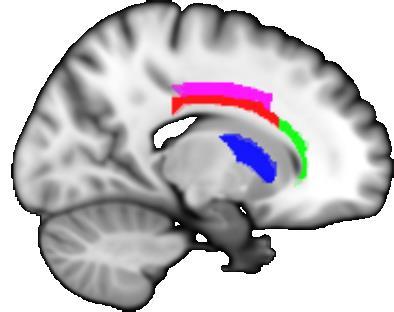
10 correction at 5%, this time based on the number of measures carried through to the secondary analysis. Results Baseline comparison of early HD patients and controls To provide context for understanding longitudinal changes, we investigated cross-sectional differences between early HD patients and controls at baseline. All 24 regions showed statistically significant group differences (corrected for multiple comparisons) for at least one metric, with the HD group demonstrating either decreases in FA or increases in AD or RD when compared with controls (Table 2). Regions where all three DTI metrics showed significant differences were the three subdivisions of the corpus callosum (genu, body and splenium), the left external capsule and left bilateral sagittal stratum. There were no regions where increased FA was evident in the HD group, nor where decreased AD or RD was found. Comparison of longitudinal changes in early HD patients and controls DTI metrics showed significant differences in rates of change between early HD patients and controls in nine regions (see Figure 2 and Table 3). The genu and body of the corpus callosum showed reduced FA and increased RD, with increased AD in the genu only. There was increased RD and AD in the left anterior corona radiata; and increases in AD were also evident in both the right superior corona radiate and anterior limb of internal capsule. All differences were significant following multiple correction. Four metrics showed evidence of interaction between group differences in 15- month change and site (see Supplementary Table 2). If between-group differences were driven by disease, we would expect to see a relatively flat trajectory of DTI measures over time for the control group, coupled with a deterioration in HD subjects to reflect disease progression. As an informal check, we plotted the model-estimated trajectories of diffusivity measures throughout the study period in controls and HD groups 10
11 separately. Fitted values are shown for hypothetical participants age 50, with a 50/50 male-female split and a 25% contribution from each site (Figure 3). Examination of the example trajectories highlighted FA in the genu of corpus callosum, AD in the anterior limb of internal capsule and right superior corona radiata and RD in the left anterior corona radiata as matching this pattern. The longitudinal trajectory in other regions indicated a possible regression to the mean in controls influencing the detected group differences in rates of change over time. Clinical associations with longitudinal neuroimaging measures The nine measures that reached statistical significance for longitudinal change (Table 3) were examined for associations between 15-month change and baseline CAG and disease burden in the HD subjects. Estimates and 95% confidence intervals are displayed in Table 4. After correction for multiple comparisons, only RD in the body of the corpus callosum showed a significant association with disease burden, with other measures showing directionally consistent but only borderline statistically significant results. After adjustment for a FDR of 5% there was no evidence that the associations differed between study sites. Discussion Early-stage HD gene-carriers show progressive changes in white matter microstructure in a number of key tracts when compared with healthy, age-matched controls. Using multi-site data, we have demonstrated widespread cross-sectional differences in diffusivity at baseline between controls and early stage HD, with longitudinal change in a number of key tracts including the genu and body of corpus callosum, left anterior corona radiata, right superior corona radiata and right anterior limb of internal capsule. Our cross-sectional findings at baseline concur with previous work using the current cohort [11] and much of the previous DTI research examining white matter in early-stage HD, which demonstrated abnormalities in the corpus callosum [18, 19, 24, 27, 63] and more widely throughout the brain [5, 7-11
12 10, 20-23, 26, 28, 30]. We have also shown longitudinal change in the corpus callosum, corona radiate, and right anterior internal capsule across a series of DTI metrics, which supports evidence from previous longitudinal studies [29, 45]. Weaver et al., for example, identified FA decreases at follow-up in the corpus callosum, internal capsule and corona radiate. Most significantly, however, the recent longitudinal study by Poudel et al. demonstrated that the most robust white matter changes in manifest HD occur within the corpus callosum [29]. Both the current study and that of Poudel have shown that in early HD there are FA increases and RD decreases in the corpus callosum when compared with controls. Although this previous study used only two timepoints, incorporating a longer period of change, and a whole-brain rather than an a priori hypothesis driven method, findings are still highly consistent regarding the corpus callosum. These three studies highlight a consistent disturbance within the corpus callosum above all other white matter tracts. This is compatible with the aforementioned extant evidence at the cross-sectional level which robustly demonstrates white matter abnormalities within the corpus callosum. Despite concordance concerning measures of RD and FA, there is far greater inconsistency in terms of AD measures. We saw increased AD not only in the corpus callosum, but also the anterior and superior corona radiate and the anterior internal capsule. Weaver, however, reported decreased AD in a small set of voxels [45], these differences likely due to low statistical power and variability in terms of disease progression; while Poudel et al. did not show any significant differences in AD at all [29]. Crosssectional studies of early HD patients have previously shown increases in AD compared with controls [7, 11, 18, 27, 30] and there is evidence that the negative correlation between AD and RD acts as an independent predictor of HD disease progression (see unpublished data Mike O). However, given the limited number of studies examining longitudinal change in white matter, further replication of our findings would be required to confirm the increases in AD in early HD gene-carriers. It is important to note that some longitudinal differences may potentially be driven by a regression to the mean in controls, rather than being explicitly disease-related. Nevertheless, some regions do appear to show a consistent trajectory of change in early HD patients, while those of the controls 12
13 remaining static (e.g. genu FA, right anterior limb of internal capsule AD, right superior corona radiata AD, left anterior corona radiata RD). Inferring specific biological processes underlying alterations in DTI metrics can be contentious [64]. Animal studies combining DTI and histology to model white matter damage [65, 66] and HD specifically [67] have indicated that differing pathological processes may underlie AD and RD measurements. Under this assumption, RD increases reflect demyelination [68], while changes in AD are more complex and time-dependent. Decreases in AD are thought to occur in response to acute axonal damage and inflammation [69]; conversely AD increases reflect expanded extra-cellular spaces resulting from Wallerian degeneration and other more gross atrophic processes [70, 71]. However, we would caution against the over-interpretation of instances where, for example, AD was significant and RD was not. Cellular modifications caused by HD pathology undoubtedly affect white matter, but the mechanisms underlying this are unclear. Elements of intrinsic axonal degeneration, altered fibre coherence or configuration due to inflammation, decreased axonal density or demyelination all occur during the progression of HD and current DTI methods lack such aetiological specificity. More recent developments in diffusion MRI [72-76], may be more aetiologicallyinformative regarding microstructural white matter changes occurring during HD progression. Analysis of clinical associations found a relationship between DBS and change in RD in the body of corpus callosum, with greater disease burden at baseline related to greater increases in RD over 15 months during the early stages of manifest HD. Again, it is the corpus callosum that is most significantly targeted and, notwithstanding the aforementioned caveats, this may reflect on-going demyelination in the corpus callosum of the more severe patients, in line with previous evidence for corpus callosum abnormalities in early HD [18, 19, 24, 27, 63]. The number of associations between DTI metrics and clinical measures that did not reach statistical significance was however, unexpectedly high. Poudel et al also found that when correlating FA with a series of clinical measures, only UHDRS-TMS scores were significant predictors of white matter microstructural 13
14 changes [29], illustrating the potential difficulty in demonstrating clear relationships between clinical measures and longitudinal neuroimaging metrics. While this has been achieved using robust volumetric methods [36-38], issues regarding signal-to-noise ratio, reliability and validity hinder both DTI and many clinical measures and detecting associations may be better achieved by adopting novel diffusion-imaging techniques or by combining multiple imaging modalities with clinical data in a multivariate framework. Combining data from multiple scanners can present limitations, however our previous work has demonstrated that the cross-site pooling of data does not introduce bias in DTI analyses [50]. Furthermore, longitudinal within-participant change measures were not confounded by scanner differences. Our study did not model any potentially confounding effects of medication on white matter metrics. However, given the considerable heterogeneity in medication usage among the participants, it would not have been possible to test medication effect systematically. In conclusion, patients in the early stages of clinically-manifest HD show robust abnormalities in white matter structure throughout the brain, most robustly in the corpus callosum. Moreover, some of those regions appear to be undergoing microstructural degeneration as the disease progresses during the early diagnosed stages, with some evidence for a relationship between disease burden and structure of the corpus callosum. Despite these findings, our results demonstrate the difficulties in mapping effects of clinical variables onto a dynamically and heterogeneous pattern of brain changes that take place during early HD. Novel diffusion imaging methods which provide more information regarding the biological underpinnings of changes in white matter microstructure are required to further understand the heterogeneity of such alterations within HD. Furthermore, the use of multivariate modelling, which incorporates both imaging and clinical data, will also help to understand how white matter changes are related to clinical symptoms. Acknowledgements 14
15 The authors would like to thank Shiva Keihaninejad for help with the tensor based registration methods. The authors would also like to thank the patients and controls who took part in this study and acknowledge the efforts of all the PADDINGTON investigators in collecting the data used in the study. This work has been supported by the European Union PADDINGTON project and all authors, with the exception of RS, SG and HZ receive funding from this project. RS, SG and HZ are supported by the CHDI/High Q Foundation, a not-for-profit organization dedicated to finding treatments for Huntington's disease. This work was undertaken at UCLH/UCL supported by the National Institute for Health Research [NIHR] University College London Hospitals [UCLH] Biomedical Research Centre [BRC]. Conflict of interest The authors have no conflict of interest to report. 15
16 References [1] The Huntington's Disease Collaborative Research Group. A novel gene containing a trinucleotide repeat that is expanded and unstable on Huntington's disease chromosomes. The Huntington's Disease Collaborative Research Group. Cell. 1993;72(6): [2] Rosas HD, Koroshetz WJ, Chen YI, Skeuse C, Vangel M, Cudkowicz ME, et al. Evidence for more widespread cerebral pathology in early HD: An MRI-based morphometric analysis. Neurology. 2003;60(10): [3] Tabrizi SJ, Langbehn DR, Leavitt BR, Roos RA, Durr A, Craufurd D, et al. Biological and clinical manifestations of Huntington's disease in the longitudinal TRACK-HD study: cross-sectional analysis of baseline data. Lancet neurology. 2009;8(9): [4] Aylward EH, Codori AM, Rosenblatt A, Sherr M, Brandt J, Stine OC, et al. Rate of caudate atrophy in presymptomatic and symptomatic stages of Huntington's disease. Movement disorders : official journal of the Movement Disorder Society. 2000;15(3): [5] Georgiou-Karistianis N, Gray MA, Dominguez DJ, Dymowski AR, Bohanna I, Johnston LA, et al. Automated differentiation of pre-diagnosis Huntington's disease from healthy control individuals based on quadratic discriminant analysis of the basal ganglia: the IMAGE-HD study. Neurobiology of disease. 2013;51: [6] Georgiou-Karistianis N, Scahill R, Tabrizi SJ, Squitieri F, Aylward E. Structural MRI in Huntington's disease and recommendations for its potential use in clinical trials. Neuroscience and biobehavioral reviews. 2013;37(3): [7] Bohanna I, Georgiou-Karistianis N, Sritharan A, Asadi H, Johnston L, Churchyard A, et al. Diffusion Tensor Imaging in Huntington's disease reveals distinct patterns of white matter degeneration associated with motor and cognitive deficits. Brain Imaging and Behavior. 2011;5(3):
17 [8] Della Nave R, Ginestroni A, Tessa C, Giannelli M, Piacentini S, Filippi M, et al. Regional distribution and clinical correlates of white matter structural damage in Huntington disease: A tractbased spatial statistics study. American Journal of Neuroradiology. 2010;31(9): [9] Delmaire C, Dumas EM, Sharman MA, van den Bogaard SJ, Valabregue R, Jauffret C, et al. The structural correlates of functional deficits in early huntington's disease. Hum Brain Mapp. 2013;34(9): [10] Douaud G, Behrens TE, Poupon C, Cointepas Y, Jbabdi S, Gaura V, et al. In vivo evidence for the selective subcortical degeneration in Huntington's disease. NeuroImage. 2009;46(4): [11] Hobbs NZ, Cole JH, Farmer RE, Rees EM, Crawford HE, Malone IB, et al. Evaluation of multimodal, multi-site neuroimaging measures in Huntington's disease: Baseline results from the PADDINGTON study. NeuroImage: Clinical. 2013;2(1): [12] Klöppel S, Draganski B, Golding CV, Chu C, Nagy Z, Cook PA, et al. White matter connections reflect changes in voluntary-guided saccades in pre-symptomatic Huntington's disease. Brain. 2008;131(1): [13] Magnotta VA, Kim J, Koscik T, Beglinger LJ, Espinso D, Langbehn D, et al. Diffusion Tensor Imaging in Preclinical Huntington's Disease. Brain imaging and behavior. 2009;3(1): [14] Matsui JT, Vaidya JG, Johnson HJ, Magnotta VA, Long JD, Mills JA, et al. Diffusion weighted imaging of prefrontal cortex in prodromal huntington's disease. Human brain mapping. 2014;35(4): [15] Reading SAJ, Yassa MA, Bakker A, Dziorny AC, Gourley LM, Yallapragada V, et al. Regional white matter change in pre-symptomatic Huntington's disease: A diffusion tensor imaging study. Psychiatry Research - Neuroimaging. 2005;140(1): [16] Rizk-Jackson A, Stoffers D, Sheldon S, Kuperman J, Dale A, Goldstein J, et al. Evaluating imaging biomarkers for neurodegeneration in pre-symptomatic Huntington's disease using machine learning techniques. NeuroImage. 2011;56(2):
18 [17] Stoffers D, Sheldon S, Kuperman JM, Goldstein J, Corey-Bloom J, Aron AR. Contrasting gray and white matter changes in preclinical Huntington disease: An MRI study. Neurology. 2010;74(15): [18] Di Paola M, Luders E, Cherubini A, Sanchez-Castaneda C, Thompson PM, Toga AW, et al. Multimodal MRI Analysis of the Corpus Callosum Reveals White Matter Differences in Presymptomatic and Early Huntington's Disease. Cereb Cort. 2012; /cercor/bhr360. [19] Di Paola M, Phillips OR, Sanchez-Castaneda C, Di Pardo A, Maglione V, Caltagirone C, et al. MRI measures of corpus callosum iron and myelin in early Huntington's disease. Human brain mapping [20] Dumas EM, van den Bogaard SJ, Ruber ME, Reilman RR, Stout JC, Craufurd D, et al. Early changes in white matter pathways of the sensorimotor cortex in premanifest Huntington's disease. Hum Brain Mapp. 2012;33(1): [21] Mascalchi M, Lolli F, Della Nave R, Tessa C, Petralli R, Gavazzi C, et al. Huntington disease: volumetric, diffusion-weighted, and magnetization transfer MR imaging of brain. Radiology. 2004;232(3): [22] Muller HP, Glauche V, Novak MJ, Nguyen-Thanh T, Unrath A, Lahiri N, et al. Stability of white matter changes related to Huntington's disease in the presence of imaging noise: a DTI study. PLoS currents. 2011;3:RRN1232. [23] Novak MJU, Seunarine KK, Gibbard CR, Hobbs NZ, Scahill RI, Clark CA, et al. White matter integrity in premanifest and early Huntington's disease is related to caudate loss and disease progression. Cortex; a journal devoted to the study of the nervous system and behavior. 2014;52(1): [24] Phillips O, Sanchez-Castaneda C, Elifani F, Maglione V, Di Pardo A, Caltagirone C, et al. Tractography of the Corpus Callosum in Huntington's Disease. PLoS One. 2013;8(9). [25] Phillips O, Squitieri F, Sanchez-Castaneda C, Elifani F, Caltagirone C, Sabatini U, et al. Deep white matter in Huntington's disease. PloS one. 2014;9(10). 18
19 [26] Poudel GR, Stout JC, Domínguez D JF, Salmon L, Churchyard A, Chua P, et al. White matter connectivity reflects clinical and cognitive status in Huntington's disease. Neurobiology of disease. 2014;65: [27] Rosas HD, Lee SY, Bender AC, Zaleta AK, Vangel M, Yu P, et al. Altered white matter microstructure in the corpus callosum in Huntington's disease: Implications for cortical "disconnection". NeuroImage. 2010;49(4): [28] Rosas HD, Tuch DS, Hevelone ND, Zaleta AK, Vangel M, Hersch SM, et al. Diffusion tensor imaging in presymptomatic and early Huntington's disease: Selective white matter pathology and its relationship to clinical measures. Movement Disorders. 2006;21(9): [29] Poudel GR, Stout JC, Dominguez DJ, Churchyard A, Chua P, Egan GF, et al. Longitudinal change in white matter microstructure in Huntington's disease: The IMAGE-HD study. Neurobiology of disease. 2015;74: [30] Odish OF, Leemans A, Reijntjes RH, van den Bogaard SJ, Dumas EM, Wolterbeek R, et al. Microstructural brain abnormalities in Huntington's disease: A two-year follow-up. Human brain mapping. 2015;36(6): [31] Aylward EH, Nopoulos PC, Ross CA, Langbehn DR, Pierson RK, Mills JA, et al. Longitudinal change in regional brain volumes in prodromal Huntington disease. Journal of Neurology, Neurosurgery and Psychiatry. 2011;82(4): [32] Ciarmiello A, Cannella M, Lastoria S, Simonelli M, Frati L, Rubinsztein DC, et al. Brain whitematter volume loss and glucose hypometabolism precede the clinical symptoms of Huntington's disease. Journal of Nuclear Medicine. 2006;47(2): [33] Hobbs NZ, Henley SM, Ridgway GR, Wild EJ, Barker RA, Scahill RI, et al. The progression of regional atrophy in premanifest and early Huntington's disease: a longitudinal voxel-based morphometry study. J Neurol, Neurosurg & Psych. 2010;81(7):
20 [34] Rosas HD, Reuter M, Doros G, Lee SY, Triggs T, Malarick K, et al. A tale of two factors: What determines the rate of progression in Huntington's disease? A longitudinal MRI study. Movement Disorders. 2011;26(9): [35] Squitieri F, Cannella M, Simonelli M, Sassone J, Martino T, Venditti E, et al. Distinct brain volume changes correlating with clinical stage, disease progression rate, mutation size, and age at onset prediction as early biomarkers of brain atrophy in Huntington's disease. CNS neuroscience & therapeutics. 2009;15(1):1-11. [36] Tabrizi SJ, Reilmann R, Roos RA, Durr A, Leavitt B, Owen G, et al. Potential endpoints for clinical trials in premanifest and early Huntington's disease in the TRACK-HD study: analysis of 24 month observational data. Lancet neurology. 2012;11(1): [37] Tabrizi SJ, Scahill RI, Durr A, Roos RA, Leavitt BR, Jones R, et al. Biological and clinical changes in premanifest and early stage Huntington's disease in the TRACK-HD study: the 12-month longitudinal analysis. Lancet neurology. 2011;10(1): [38] Tabrizi SJ, Scahill RI, Owen G, Durr A, Leavitt BR, Roos RA, et al. Predictors of phenotypic progression and disease onset in premanifest and early-stage Huntington's disease in the TRACK-HD study: analysis of 36-month observational data. The Lancet Neurology [39] Hobbs NZ, Farmer RE, Rees EM, Cole JH, Haider S, Malone IB, et al. Short-interval observational data to inform clinical trial design in Huntington's disease. Journal of neurology, neurosurgery, and psychiatry. 2015; /jnnp [40] Beaulieu C. The basis of anisotropic water diffusion in the nervous system - A technical review. NMR Biomed. 2002;15(7-8): [41] Rees EM, Scahill RI, Hobbs NZ. Longitudinal Neuroimaging Biomarkers in Huntington's Disease. Journal of Huntington's Disease. 2013;2(1): [42] Domínguez DJF, Egan GF, Gray MA, Poudel GR, Churchyard A, Chua P, et al. Multi-Modal Neuroimaging in Premanifest and Early Huntington's Disease: 18 Month Longitudinal Data from the IMAGE-HD Study. PloS one. 2013;8(9). 20
21 [43] Vandenberghe W, Demaerel P, Dom R, Maes F. Diffusion-weighted versus volumetric imaging of the striatum in early symptomatic Huntington disease. Journal of neurology. 2009;256(1): [44] Smith SM, Jenkinson M, Johansen-Berg H, Rueckert D, Nichols TE, Mackay CE, et al. Tractbased spatial statistics: Voxelwise analysis of multi-subject diffusion data. NeuroImage. 2006;31(4): [45] Weaver KE, Richards TL, Liang O, Laurino MY, Samii A, Aylward EH. Longitudinal diffusion tensor imaging in Huntington's Disease. Experimental neurology. 2009;216(2): [46] Sritharan A, Egan GF, Johnston L, Horne M, Bradshaw JL, Bohanna I, et al. A longitudinal diffusion tensor imaging study in symptomatic Huntington's disease. Journal of Neurology, Neurosurgery and Psychiatry. 2010;81(3): [47] Unified Huntington's Disease Rating Scale: reliability and consistency. Huntington Study Group. Movement disorders : official journal of the Movement Disorder Society. 1996;11(2): [48] Odish OF, Caeyenberghs K, Hosseini H, van den Bogaard SJ, Roos RA, Leemans A. Dynamics of the connectome in Huntington's disease: A longitudinal diffusion MRI study. NeuroImage Clinical. 2015;9: [49] Shoulson I, Fahn S. Huntington disease: clinical care and evaluation. Neurology. 1979;29(1):1-3. [50] Müller HP, Grön G, Sprengelmeyer R, Kassubek J, Ludolph AC, Hobbs N, et al. Evaluating multicenter DTI data in Huntington's disease on site specific effects: An ex post facto approach. NeuroImage: Clinical. 2013;2(1): [51] Cook PA, Bai Y, Nedjati-Gilani S, Seunarine KK, Hall MG, Parker GJ, et al., editors. Camino: Open-Source Diffusion-MRI Reconstruction and Processing. 14th Scientific Meeting of the International Society for Magnetic Resonance in Medicine; 2006; Seattle, WA, USA. [52] Zhang H, Yushkevich PA, Alexander DC, Gee JC. Deformable registration of diffusion tensor MR images with explicit orientation optimization. Medical image analysis. 2006;10(5):
22 [53] Zhang H, Avants BB, Yushkevich PA, Woo JH, Wang S, McCluskey LF, et al. High-dimensional spatial normalization of diffusion tensor images improves the detection of white matter differences: An example study using amyotrophic lateral sclerosis. IEEE Transactions on Medical Imaging. 2007;26(11): [54] Wang Y, Gupta A, Liu Z, Zhang H, Escolar ML, Gilmore JH, et al. DTI registration in atlas based fiber analysis of infantile Krabbe disease. NeuroImage. 2011;55(4): [55] Muller MJ, Mazanek M, Weibrich C, Dellani PR, Stoeter P, Fellgiebel A. Distribution characteristics, reproducibility, and precision of region of interest-based hippocampal diffusion tensor imaging measures. AJNR American journal of neuroradiology. 2006;27(2): [56] Keihaninejad S, Zhang H, Ryan N, Malone I, Frost C, Cardoso M, et al. Longitudinal changes in fiber tract integrity in Alzheimer's disease: A serial diffusion tensor imaging study. Alzheimer's & Dementia. 2012;8(S4):30-1. [57] Lee CE, Danielian LE, Thomasson D, Baker EH. Normal regional fractional anisotropy and apparent diffusion coefficient of the brain measured on a 3 T MR scanner. Neuroradiology. 2009;51(1):3-9. [58] Mori S, Oishi K, Jiang H, Jiang L, Li X, Akhter K, et al. Stereotaxic white matter atlas based on diffusion tensor imaging in an ICBM template. NeuroImage. 2008;40(2): [59] Cole JH, Farmer RE, Rees EM, Johnson HJ, Frost C, Scahill RI, et al. Test-Retest Reliability of Diffusion Tensor Imaging in Huntington's Disease. PLoS Curr. 2014;6. [60] Ourselin S, Roche A, Subsol G, Pennec X, Ayache N. Reconstructing a 3D structure from serial histological sections. Image and Vision Computing. 2001;19(1-2): [61] Modat M, Ridgway GR, Taylor ZA, Lehmann M, Barnes J, Hawkes DJ, et al. Fast free-form deformation using graphics processing units. Comput Methods Programs Biomed. 2010;98(3): [62] Benjamini Y, Hochberg Y. Controlling the False Discovery Rate - a Practical and Powerful Approach to Multiple Testing. J Roy Stat Soc B Met. 1995;57(1):
23 [63] Crawford HE, Hobbs NZ, Keogh R, Langbehn DR, Frost C, Johnson H, et al. Corpus callosal atrophy in premanifest and early Huntington's disease. J Huntingtons Dis. 2013;2(4): [64] Jones DK, Knosche TR, Turner R. White matter integrity, fiber count, and other fallacies: the do's and don'ts of diffusion MRI. NeuroImage. 2013;73: [65] Song SK, Sun SW, Ju WK, Lin SJ, Cross AH, Neufeld AH. Diffusion tensor imaging detects and differentiates axon and myelin degeneration in mouse optic nerve after retinal ischemia. NeuroImage. 2003;20(3): [66] Sun SW, Liang HF, Cross AH, Song SK. Evolving Wallerian degeneration after transient retinal ischemia in mice characterized by diffusion tensor imaging. NeuroImage. 2008;40(1):1-10. [67] Van Camp N, Blockx I, Camón L, De Vera N, Verhoye M, Veraart J, et al. A complementary diffusion tensor imaging (DTI)-histological study in a model of Huntington's disease. Neurobiology of Aging. 2012;33(5): [68] Song SK, Yoshino J, Le TQ, Lin SJ, Sun SW, Cross AH, et al. Demyelination increases radial diffusivity in corpus callosum of mouse brain. NeuroImage. 2005;26(1): [69] Budde MD, Xie M, Cross AH, Song SK. Axial Diffusivity Is the Primary Correlate of Axonal Injury in the Experimental Autoimmune Encephalomyelitis Spinal Cord: A Quantitative Pixelwise Analysis. J Neurosci. 2009;29(9): [70] Concha L, Gross DW, Wheatley BM, Beaulieu C. Diffusion tensor imaging of time-dependent axonal and myelin degradation after corpus callosotomy in epilepsy patients. NeuroImage. 2006;32(3): [71] Della Nave R, Ginestroni A, Diciotti S, Salvatore E, Soricelli A, Mascalchi M. Axial diffusivity is increased in the degenerating superior cerebellar peduncles of Friedreich's ataxia. Neuroradiology. 2011;53(5): [72] Jensen JH, Helpern JA. MRI quantification of non-gaussian water diffusion by kurtosis analysis. NMR Biomed. 2010;23(7):
24 [73] Zhang H, Schneider T, Wheeler-Kingshott CA, Alexander DC. NODDI: Practical in vivo neurite orientation dispersion and density imaging of the human brain. NeuroImage. 2012;61(4): [74] Zhang H, Hubbard PL, Parker GJM, Alexander DC. Axon diameter mapping in the presence of orientation dispersion with diffusion MRI. NeuroImage. 2011;56(3): [75] Assaf Y, Blumenfeld-Katzir T, Yovel Y, Basser PJ. AxCaliber: A method for measuring axon diameter distribution from diffusion MRI. Magnetic Resonance in Medicine. 2008;59(6): [76] Tournier JD, Mori S, Leemans A. Diffusion tensor imaging and beyond. Magnetic resonance in medicine : official journal of the Society of Magnetic Resonance in Medicine / Society of Magnetic Resonance in Medicine. 2011;65(6): [77] Penney JB, Vonsattel JP, MacDonald ME, Gusella JF, Myers RH. CAG repeat number governs the development rate of pathology in huntington's disease. Annals of Neurology. 1997;41(5): [78] Papadakis NG, Xing D, Huang CL, Hall LD, Carpenter TA. A comparative study of acquisition schemes for diffusion tensor imaging using MRI. Journal of magnetic resonance. 1999;137(1): [79] Jones DK. The effect of gradient sampling schemes on measures derived from diffusion tensor MRI: a Monte Carlo study. Magnetic resonance in medicine : official journal of the Society of Magnetic Resonance in Medicine / Society of Magnetic Resonance in Medicine. 2004;51(4): [80] Poonawalla AH, Zhou XJ. Analytical error propagation in diffusion anisotropy calculations. Journal of magnetic resonance imaging : JMRI. 2004;19(4): [81] Barrio-Arranz G, de Luis-Garcia R, Tristan-Vega A, Martin-Fernandez M, Aja-Fernandez S. Impact of MR Acquisition Parameters on DTI Scalar Indexes: A Tractography Based Approach. PloS one. 2015;10(10):e [82] Muller HP, Sussmuth SD, Landwehrmeyer GB, Ludolph A, Tabrizi SJ, Kloppel S, et al. Stability effects on results of diffusion tensor imaging analysis by reduction of the number of gradient directions due to motion artifacts: an application to presymptomatic Huntington's disease. PLoS Curr. 2011;3:RRN
25 Tables Table 1. HD patient and control characteristics Baseline characteristics Controls Early HD patients N Age (Years) [Mean (SD) Range] (8.75) (9.38) Sex (Female/Male N (%)) 20 / 16 (55.6%) / (44.4%) 29 / 19 (60.4%) / (39.6%) Education Level (ISCED) [Mean (SD) Range] Baseline-15 month interval (Days) [Mean (SD) Range] 3.97 (1.23) (1.27) (32.27) (43.44) Centre Leiden N (%) 9 (25%) 11 (22.92%) London N (%) 10 (27.78%) 13 (27.08%) Paris N (%) 8 (22.22%) 9 (18.75%) Ulm N (%) 9 (25%) 15 (31.25%) CAG [Mean (SD) Range] (2.61) Disease Burden Score [Mean (SD) Range] Total Functional Capacity [Mean (SD) Range] (85.66) (0.17) (0.93) 9-13 Total Motor Score [Mean (SD) Range] 1.44 (1.89) (9.19) 6-45 Disease burden score [77] calculated as [age x (cag-35.5)]. SD = standard deviation. ISCED = International Standard Classification of Education. CAG = tri-nucleotide repeat length. 25
26 Table 2. Baseline comparison of diffusion metrics in white matter regions between early HD patient and controls Brain region Fractional Anisotropy P Axial Diffusivity P Radial Diffusivity P Cingulum (cingulate gyrus) - L [-0.055, ] [-0.009, 0.047] [0.025, 0.085] <0.001 Cingulum (cingulate gyrus) - R [-0.050, ] 0.002* [-0.026, 0.027] [0.015, 0.069] 0.002* Corona radiata - Anterior - L [-0.019, 0.005] [0.019, 0.055] <0.001* [0.007, 0.044] 0.006* Corona radiata - Anterior - R [-0.024, 0.006] [0.017, 0.060] <0.001* [0.004, 0.050] 0.023* Corona radiata - Posterior - L [-0.021, 0.008] [0.018, 0.056] <0.001* [0.007, 0.042] 0.007* Corona radiata - Posterior - R [-0.023, 0.006] [0.016, 0.061] 0.001* [0.011, 0.050] 0.002* Corona radiata - Superior - L [-0.019, 0.007] [0.022, 0.059] <0.001* [0.010, 0.038] 0.001* Corona radiata - Superior - R [-0.020, 0.007] [0.015, 0.056] 0.001* [0.007, 0.035] 0.003* Corpus callosum - Body [-0.036, ] <0.001* [0.038, 0.081] <0.001* [0.032, 0.076] <0.001* Corpus callosum - Genu [-0.041, ] 0.001* [0.021, 0.070] <0.001* [0.022, 0.080] 0.001* Corpus callosum - Splenium [-0.034, ] <0.001* [0.050, 0.114] <0.001* [0.032, 0.084] <0.001* External capsule - L [-0.028, ] <0.001* [0.027, 0.058] <0.001* [0.028, 0.064] <0.001* External capsule - R [-0.023, ] [0.038, 0.075] <0.001* [0.024, 0.066] <0.001* Internal capsule - Anterior limb - L [-0.012, 0.014] (0.046, 0.083) <0.001* [0.014, 0.047] <0.001* Internal capsule - Anterior limb - R [-0.012, 0.013] [0.040, 0.080] <0.001* [0.021, 0.057] <0.001* Internal capsule - Retrolenticular - L [-0.011, 0.017] [0.034, 0.077] <0.001* [-0.001, 0.037] Internal capsule - Retrolenticular - R [-0.014, 0.014] [0.028, 0.067] <0.001* [0.002, 0.037] 0.025* Pontine crossing tract [-0.041, ] 0.012* [-0.012, 0.051] [0.006, 0.060] 0.016* Posterior thalamic radiation - L [-0.015, 0.015] [0.004, 0.059] 0.023* [-0.007, 0.039] Posterior thalamic radiation - R [-0.025, 0.009] [0.002, 0.067] 0.035* [-0.005, 0.055] Sagittal stratum - L [-0.036, ] 0.001* [0.028, 0.090] <0.001* [0.037, 0.088] <0.001* Sagittal stratum - R [-0.031, ] [0.002, 0.067] 0.039* [0.008, 0.061] Superior longitudinal fasciculus L [-0.024, 0.001] [0.011, 0.040] 0.001* [0.010, 0.044] 0.002* Superior longitudinal fasciculus - R [-0.026, ] [0.017, 0.045] <0.001* [0.014, 0.049] <0.001* Values are in the form of adjusted group difference estimates, with 95% confidence intervals presented in brackets. *Denotes statistically significant group differences after FDR correction L = Left, R = Right 26
27 Table 3. Early HD and control longitudinal rate of change comparison Brain Region Fractional Anisotropy P Axial Diffusivity P Radial Diffusivity P Cingulum (cingulate gyrus) - L [-0.006, 0.010] [-0.013, 0.008] [0.015, 0.007] Cingulum (cingulate gyrus) - R [-0.010, 0.002] [-0.008, 0.015] [-0.003, 0.017] Corona radiata - Anterior - L [-0.012, 0.000] [0.005, 0.028] 0.003* [0.004, 0.026] 0.006* Corona radiata - Anterior - R [-0.014, ] [-0.004, 0.018] [0.000, 0.021] Corona radiata - Posterior - L [-0.004, 0.008] [-0.007, 0.012] [-0.008, 0.010] Corona radiata - Posterior - R [-0.007, 0.003] [0.000, 0.015] [-0.002, 0.011] Corona radiata - Superior - L [-0.005, 0.003] [0.001, 0.015] [-0.001, 0.011] Corona radiata - Superior - R [-0.008, 0.002] [0.002, 0.016] 0.006* [-0.001, 0.012] Corpus callosum - Body [-0.017, ] <0.001* [0.001, 0.022] [0.011, 0.028] <0.001* Corpus callosum - Genu [-0.010, ] 0.004* [0.014, 0.033] <0.001* [0.010, 0.026] <0.001* Corpus callosum - Splenium [-0.005, 0.004] [-0.007, 0.016] [-0.008, 0.010] External capsule - L [-0.009, 0.000] [-0.003, 0.015] [0.000, 0.015] External capsule - R [-0.010, 0.001] [-0.002, 0.019] [-0.001, 0.018] Internal capsule - Anterior limb - L [-0.003, 0.010] [0.000, 0.022] [-0.009, 0.009] Internal capsule - Anterior limb - R [-0.012, 0.004] [0.012, 0.041] <0.001* [0.003, 0.030] Internal capsule - Retrolenticular - L [-0.006, 0.013] [-0.009, 0.016] [-0.014, 0.012] Internal capsule - Retrolenticular - R [-0.006, 0.009] [-0.011, 0.014] [-0.011, 0.008] Pontine crossing tract [0.002, 0.034] [-0.027, 0.023] [-0.040, 0.007] Posterior thalamic radiation - L [-0.014, 0.003] [-0.005, 0.028] [-0.002, 0.024] Posterior thalamic radiation - R [-0.007, 0.007] [-0.015, 0.011] [-0.010, 0.011] Sagittal stratum - L [-0.006, 0.012] [0.000, 0.043] [-0.007, 0.025] Sagittal stratum - R [-0.012, 0.001] [-0.009, 0.021] [0.001, 0.023] Superior longitudinal fasciculus - L [-0.005, 0.003] [-0.005, 0.010] [-0.003, 0.009] Superior longitudinal fasciculus - R [-0.006, 0.001] [-0.002, 0.010] [-0.002, 0.008] Values are in the form of adjusted group difference estimates, with 95% confidence intervals presented in brackets. *Denotes statistically significant group differences after FDR correction L = Left, R = Right 27
28 Table 4. Association between 15-month change and CAG repeat length or baseline Disease Burden Score CAG Disease Burden Estimate [95% CI] P Estimate [95% CI] P Corpus callosum - Genu AD 4.07 [-4.58, 12.73] [-0.12, 0.29] 0.41 Corpus callosum - Genu FA [-6.08, 1.22] [-0.15, 0.02] 0.11 Corpus callosum - Genu RD 3.88 [-3.84, 11.6] [-0.07, 0.29] 0.23 Corona radiata - Anterior - L AD 0.92 [-10.83, 12.68] [-0.27, 0.28] 0.97 Corona radiata - Anterior - L RD [ , ] [-13.47, 11.27] 0.86 Internal capsule - Anterior limb - R AD [-0.41, 27.87] [0.05, 0.69] 0.03 Corona radiata - Superior-R AD 5.42 [0.06, 10.79] [0.01, 0.26] 0.03 Corpus callosum - Body FA [-8.07, -0.25] [-0.19, -0.01] 0.04 Corpus callosum - Body RD 8.70 [2.02, 15.39] [0.07, 0.38] <0.01* All analyses adjusted for age, sex and study site. x 10-6 * Statistically significant after FDR correction FA = Fractional anisotropy AD = Axial diffusivity RD = Radial diffusivity L = Left, R = Right 28
29 Figure legends Figure 1. Overview of the unbiased longitudinal registration methods. (A) Within-subject registration performed on tensor images using an initial affine and subsequent non-linear step in DTI-TK to warp images from each visit to a withinsubject mid-space. A within-subject average template was then generated. (B) These within-subject average images were registered together, again using DTI-TK, to define a longitudinal between-subject space. The initial input images were then warped to this between-subject space and averaged to form a longitudinal group template, specific to the study. (C) Using NiftyReg to calculate affine and non-linear registrations, the FA image from the ICBM-DTI-81 atlas was then warped to match the group template. The parameters of this registration were then applied to ICBM-DTI-81 labels. (D) Finally, subject and visit specific metrics for each ROI were calculated. Figure 2. The figure depicts the regions-of-interest (ROIs) that showed significant differences in rates of change in DTI metrics over 15 months, when comparing early HD patients and controls. The body (red) and genu (green) of the corpus callosum, along with the left anterior corona radiata (yellow) and right superior corona radiata (magenta) and anterior limb of internal capsule (blue) are shown. A) Coronal, B) sagittal and C) axial 2D slices, along with D) 3D representation are displayed in order to illustrate the spatial relatedness of the five significant regions. Figure 3. Longitudinal trajectories of DTI metrics for early HD patients and controls for all significant metric-roi combinations. The fitted values depicted were calculated for hypothetical participants age 50, with a 50/50 male-female split and a 25% contribution from each site. Points are plotted at each assessment (baseline, 6 months, 15 months), with the dashed line representing early HD patients and the whole line representing controls. 29


30 Figures Figure 1. 30


31 Figure 2. 31
Diffusione e perfusione in risonanza magnetica. E. Pagani, M. Filippi
 Diffusione e perfusione in risonanza magnetica E. Pagani, M. Filippi DW-MRI DIFFUSION-WEIGHTED MRI Principles Diffusion results from a microspic random motion known as Brownian motion THE RANDOM WALK How
Neuroimaging module I: Modern neuroimaging methods of investigation of the human brain in health and disease
 1 Neuroimaging module I: Modern neuroimaging methods of investigation of the human brain in health and disease The following contains a summary of the content of the neuroimaging module I on the postgraduate
DTI Fiber Tract-Oriented Quantitative and Visual Analysis of White Matter Integrity
 DTI Fiber Tract-Oriented Quantitative and Visual Analysis of White Matter Integrity Xuwei Liang, Ning Cao, and Jun Zhang Department of Computer Science, University of Kentucky, USA, jzhang@cs.uky.edu.
Software Packages The following data analysis software packages will be showcased:
 Analyze This! Practicalities of fmri and Diffusion Data Analysis Data Download Instructions Weekday Educational Course, ISMRM 23 rd Annual Meeting and Exhibition Tuesday 2 nd June 2015, 10:00-12:00, Room
Adolescent Brain Development and Effects of Alcohol Use
 Adolescent Brain Development and Effects of Alcohol Use Monica Luciana, Ph.D. Professor and Chair Department of Psychology and Center for Neurobehavioral Development University of Minnesota (lucia003@umn.edu)
SITE IMAGING MANUAL ACRIN 6698
 SITE IMAGING MANUAL ACRIN 6698 Diffusion Weighted MR Imaging Biomarkers for Assessment of Breast Cancer Response to Neoadjuvant Treatment: A sub-study of the I-SPY 2 TRIAL Version: 1.0 Date: May 28, 2012
MRI DATA PROCESSING. Compiled by: Nicolas F. Lori and Carlos Ferreira. Introduction
 MRI DATA PROCESSING Compiled by: Nicolas F. Lori and Carlos Ferreira Introduction Magnetic Resonance Imaging (MRI) is a clinical exam that is safe to the patient. Nevertheless, it s very important to attend
Subjects: Fourteen Princeton undergraduate and graduate students were recruited to
 Supplementary Methods Subjects: Fourteen Princeton undergraduate and graduate students were recruited to participate in the study, including 9 females and 5 males. The mean age was 21.4 years, with standard
MEDIMAGE A Multimedia Database Management System for Alzheimer s Disease Patients
 MEDIMAGE A Multimedia Database Management System for Alzheimer s Disease Patients Peter L. Stanchev 1, Farshad Fotouhi 2 1 Kettering University, Flint, Michigan, 48504 USA pstanche@kettering.edu http://www.kettering.edu/~pstanche
Chapter 10. Summary & Future perspectives
 Summary & Future perspectives 123 Multiple sclerosis is a chronic disorder of the central nervous system, characterized by inflammation and axonal degeneration. All current therapies modulate the peripheral
Advanced MRI methods in diagnostics of spinal cord pathology
 Advanced MRI methods in diagnostics of spinal cord pathology Stanisław Kwieciński Department of Magnetic Resonance MR IMAGING LAB MRI /MRS IN BIOMEDICAL RESEARCH ON HUMANS AND ANIMAL MODELS IN VIVO Equipment:
32-Channel Head Coil Imaging at 3T
 32-Channel Head Coil Imaging at 3T Thomas Benner Athinoula A. Martinos Center for Biomedical Imaging, Department of Radiology, Massachusetts General Hospital and Harvard Medical School, Boston, MA, USA
University of California San Francisco, CA, USA 4 Department of Veterans Affairs Medical Center, San Francisco, CA, USA
 Disrupted Brain Connectivity in Alzheimer s Disease: Effects of Network Thresholding Madelaine Daianu 1, Emily L. Dennis 1, Neda Jahanshad 1, Talia M. Nir 1, Arthur W. Toga 1, Clifford R. Jack, Jr. 2,
Integration and Visualization of Multimodality Brain Data for Language Mapping
 Integration and Visualization of Multimodality Brain Data for Language Mapping Andrew V. Poliakov, PhD, Kevin P. Hinshaw, MS, Cornelius Rosse, MD, DSc and James F. Brinkley, MD, PhD Structural Informatics
UMHS-PUHSC JOINT INSTITUTE
 Imaging Biomarkers for Staging and Assessing Response to Therapy in Multiple Myeloma Qian Dong, MD. Radiology University of Michigan Wei Guo, MD. Orthopedic Oncology Peking University Second Hospital Team
Myelinization. THOMAS P. NAIDICH, MD FACR Mt. Sinai Medical Center New York, NY USA
 Myelinization THOMAS P. NAIDICH, MD FACR Mt. Sinai Medical Center New York, NY USA ALTERS BRAIN WATER LOCALLY MYELIN CONTAINS: GLYCOLIPIDS PHOSPHOLIPIDS & CHOLESTEROL Maturation of the White Matter Maturation
Corso Integrato di Metodi per Immagini bio-mediche e Chirurgia Assistita MIMCAS. Metodi per bioimmagini Prof. G. Baselli Seminario 24/04/2012
 Corso Integrato di Metodi per Immagini bio-mediche e Chirurgia Assistita MIMCAS Metodi per bioimmagini Prof. G. Baselli Seminario 24/04/2012 DTI Applications Maria Giulia Preti maria.preti@mail.polimi.it
Update: MRI in Multiple sclerosis
 Nyt indenfor MS ved MR Update: MRI in Multiple sclerosis Hartwig Roman Siebner Danish Research Centre for Magnetic Resonance (DRCMR) Copenhagen University Hospital Hvidovre Dansk Radiologisk Selskabs 10.
advancing magnetic resonance imaging and medical data analysis
 advancing magnetic resonance imaging and medical data analysis INFN - CSN5 CdS - Luglio 2015 Sezione di Genova 2015-2017 objectives Image reconstruction, artifacts and noise management O.1 Components RF
ONLINE SUPPLEMENTARY DATA. Potential effect of skull thickening on the associations between cognition and brain atrophy in ageing
 ONLINE SUPPLEMENTARY DATA Potential effect of skull thickening on the associations between cognition and brain atrophy in ageing Benjamin S. Aribisala 1,2,3, Natalie A. Royle 1,2,3, Maria C. Valdés Hernández
MRC Autism Research Forum Interventions in Autism
 MRC Autism Research Forum Interventions in Autism Date: 10 July 2003 Location: Aim: Commonwealth Institute, London The aim of the forum was to bring academics in relevant disciplines together, to discuss
An Interactive Visualization Tool for Nipype Medical Image Computing Pipelines
 An Interactive Visualization Tool for Nipype Medical Image Computing Pipelines Ramesh Sridharan, Adrian V. Dalca, and Polina Golland Computer Science and Artificial Intelligence Lab, MIT Abstract. We present
Medical Image Processing on the GPU. Past, Present and Future. Anders Eklund, PhD Virginia Tech Carilion Research Institute andek@vtc.vt.
 Medical Image Processing on the GPU Past, Present and Future Anders Eklund, PhD Virginia Tech Carilion Research Institute andek@vtc.vt.edu Outline Motivation why do we need GPUs? Past - how was GPU programming
Adaptively Constrained Convex Optimization for Accurate Fiber Orientation Estimation with High Order Spherical Harmonics
 Adaptively Constrained Convex Optimization for Accurate Fiber Orientation Estimation with High Order Spherical Harmonics Giang Tran 1 and Yonggang Shi 2, 1 Dept. of Mathematics, UCLA, Los Angeles, CA,
Imaging Markers of Brain Network Dysfunction in Multiple Sclerosis
 Faculty of Medicine & Health Sciences School of Medicine Radiological Sciences Research Group The University of Nottinham University Park Nottingham NG7 2RD t: +44 (0)115 823 0018 f: +44 (0)115 823 0004
Diffusion MRI imaging for brain connectivity analysis
 Diffusion MRI imaging for brain connectivity analysis principles and challenges Prof. Jean-Philippe Thiran EPFL - http://lts5www.epfl.ch - jp.thiran@epfl.ch From Brownian motion to Diffusion MRI - Motivation
Award Number: W81XWH-10-1-0739
 AD Award Number: W81XWH-10-1-0739 TITLE: Voxel-Wise Time-Series Analysis of Quantitative MRI in Relapsing-Remitting MS: Dynamic Imaging Metrics of Disease Activity Including Prelesional Changes PRINCIPAL
3/30/2013. Disclosure. Advanced Neuro MRI: Imaging Techniques and Protocol Optimization. MRI, 35 year ago. MRI Today. Outlines
 http://www.magnet.fsu.edu Disclosure Advanced Neuro MRI: Imaging Techniques and Protocol Optimization Research funding provided by Siemens Healthcare. Chen Lin, PhD DABR Indiana University School of Medicine
Standardized MRI Protocol for Brain Tumor Clinical Trials. Benjamin M. Ellingson, Ph.D. Assistant Professor of Radiology at UCLA
 Standardized MRI Protocol for Brain Tumor Clinical Trials Benjamin M. Ellingson, Ph.D. Assistant Professor of Radiology at UCLA Standardized MRI Protocol for Therapeutic Studies FDA Meeting in January
Video-Based Eye Tracking
 Video-Based Eye Tracking Our Experience with Advanced Stimuli Design for Eye Tracking Software A. RUFA, a G.L. MARIOTTINI, b D. PRATTICHIZZO, b D. ALESSANDRINI, b A. VICINO, b AND A. FEDERICO a a Department
ParaVision 6. Innovation with Integrity. The Next Generation of MR Acquisition and Processing for Preclinical and Material Research.
 ParaVision 6 The Next Generation of MR Acquisition and Processing for Preclinical and Material Research Innovation with Integrity Preclinical MRI A new standard in Preclinical Imaging ParaVision sets a
Structural Brain Changes in remitted Major Depression
 Structural Brain Changes in remitted Major Depression Andreas Berger unter der Anleitung von Assoc.Prof. Priv.Doz. Dr. Lukas Pezawas Universitätsklinik für Psychiatrie und Psychotherapie Medizinische Universität
Victims Compensation Claim Status of All Pending Claims and Claims Decided Within the Last Three Years
 Claim#:021914-174 Initials: J.T. Last4SSN: 6996 DOB: 5/3/1970 Crime Date: 4/30/2013 Status: Claim is currently under review. Decision expected within 7 days Claim#:041715-334 Initials: M.S. Last4SSN: 2957
Machine Learning for Medical Image Analysis. A. Criminisi & the InnerEye team @ MSRC
 Machine Learning for Medical Image Analysis A. Criminisi & the InnerEye team @ MSRC Medical image analysis the goal Automatic, semantic analysis and quantification of what observed in medical scans Brain
Morphological analysis on structural MRI for the early diagnosis of neurodegenerative diseases. Marco Aiello On behalf of MAGIC-5 collaboration
 Morphological analysis on structural MRI for the early diagnosis of neurodegenerative diseases Marco Aiello On behalf of MAGIC-5 collaboration Index Motivations of morphological analysis Segmentation of
runl I IUI%I/\L Magnetic Resonance Imaging
 runl I IUI%I/\L Magnetic Resonance Imaging SECOND EDITION Scott A. HuetteS Brain Imaging and Analysis Center, Duke University Allen W. Song Brain Imaging and Analysis Center, Duke University Gregory McCarthy
Mapping Brain Changes Over Time during Development: Challenges, Limits and Potential
 Mapping Brain Changes Over Time during Development: Challenges, Limits and Potential Guido Gerig University of Utah Scientific Computing and Imaging (SCI) Institute Gerig 09-2008 Outline 1. Imaging Technology
Why do we have so many brain coordinate systems? Lilla ZölleiZ WhyNHow seminar 12/04/08
 Why do we have so many brain coordinate systems? Lilla ZölleiZ WhyNHow seminar 12/04/08 About brain atlases What are they? What do we use them for? Who creates them? Which one shall I use? Brain atlas
The Prospect of Stem Cell Therapy in Multiple Sclerosis. Multiple sclerosis is a multifocal inflammatory disease of the central
 The Prospect of Stem Cell Therapy in Multiple Sclerosis Multiple sclerosis is a multifocal inflammatory disease of the central nervous system that generally affects young individuals, causing paralysis
GE Medical Systems Training in Partnership. Module 8: IQ: Acquisition Time
 Module 8: IQ: Acquisition Time IQ : Acquisition Time Objectives...Describe types of data acquisition modes....compute acquisition times for 2D and 3D scans. 2D Acquisitions The 2D mode acquires and reconstructs
Between session reproducibility and between subject variability of diffusion MR and tractography measures
 www.elsevier.com/locate/ynimg NeuroImage 33 (2006) 867 877 Between session reproducibility and between subject variability of diffusion MR and tractography measures E. Heiervang, a,b T.E.J. Behrens, a
Predict the Popularity of YouTube Videos Using Early View Data
 000 001 002 003 004 005 006 007 008 009 010 011 012 013 014 015 016 017 018 019 020 021 022 023 024 025 026 027 028 029 030 031 032 033 034 035 036 037 038 039 040 041 042 043 044 045 046 047 048 049 050
Reversibility of Acute Demyelinating Lesions in relapsingremitting
 Reversibility of Acute Demyelinating Lesions in relapsingremitting Multiple Sclerosis Omar A. Khan ( Division of Neuroimmunology, Department of Neurology, Neurology and Research Services. Veterans Affairs
OHTAC Recommendation
 OHTAC Recommendation Multiple Sclerosis and Chronic Cerebrospinal Venous Insufficiency Presented to the Ontario Health Technology Advisory Committee in May 2010 May 2010 Issue Background A review on the
Current Industry Neuroimaging Experience in Clinical Trials Jerome Barakos, M.D.
 Current Industry Neuroimaging Experience in Clinical Trials Jerome Barakos, M.D. Melbourne Australia March 28, 2012 Synarc Experience and Expertise Largest imaging service provider dedicated to clinical
NEUROIMAGING in Parkinsonian Syndromes
 NEUROIMAGING in Parkinsonian Syndromes (Focus on Structural Techniques: CT and MRI) Dr. Roberto Cilia Parkinson Institute, ICP, Milan, Italy OUTLINE Primary Parkinsonism Idiopathic Parkinson s Disease
Unwarping Echo Planar Images Using CMTK 1 Release 1.2
 Unwarping Echo Planar Images Using CMTK 1 Release 1.2 Torsten Rohlfing October 1, 2012 Neuroscience Program, SRI International, Menlo Park, CA Abstract This document describes the workflow for unwarping
5 Factors Affecting the Signal-to-Noise Ratio
 5 Factors Affecting the Signal-to-Noise Ratio 29 5 Factors Affecting the Signal-to-Noise Ratio In the preceding chapters we have learned how an MR signal is generated and how the collected signal is processed
Internal Medicine Residency, University of Colorado Health Sciences Center. Psychiatry Residency, University of California at San Francisco
 Internal Medicine Residency, University of Colorado Health Sciences Center Psychiatry Residency, University of California at San Francisco Addiction Psychiatry Fellowship, University of New Mexico E-mail:
Primary Endpoints in Alzheimer s Dementia
 Primary Endpoints in Alzheimer s Dementia Dr. Karl Broich Federal Institute for Drugs and Medical Devices (BfArM) Kurt-Georg-Kiesinger-Allee 38, D-53175 Bonn Germany Critique on Regulatory Decisions in
Translational research facilitating experimental medicine in dementia in the UK
 Translational research facilitating experimental medicine in dementia in the UK Simon Lovestone Director Biomedical Research Unit for dementia at Maudsley and King s Route Map for Dementia Research June
2007 WHO Research Agenda for Extremely Low Frequency Fields
 2007 WHO Research Agenda for Extremely Low Frequency Fields Introduction In 1997, the WHO International EMF Project developed a Research Agenda in order to facilitate and coordinate research worldwide
1.0 Abstract. Title: Real Life Evaluation of Rheumatoid Arthritis in Canadians taking HUMIRA. Keywords. Rationale and Background:
 1.0 Abstract Title: Real Life Evaluation of Rheumatoid Arthritis in Canadians taking HUMIRA Keywords Rationale and Background: This abbreviated clinical study report is based on a clinical surveillance
DKE Fiber Tractography Module: User s Guide Version 1 Release Date: July 2015
 DKE Fiber Tractography Module: User s Guide Version 1 Release Date: July 2015 Author: G. Russell Glenn, B.S., B.A. Medical Scientist Training Program (MD / PhD) Department of Neurosciences Department of
CCNY. BME I5100: Biomedical Signal Processing. Linear Discrimination. Lucas C. Parra Biomedical Engineering Department City College of New York
 BME I5100: Biomedical Signal Processing Linear Discrimination Lucas C. Parra Biomedical Engineering Department CCNY 1 Schedule Week 1: Introduction Linear, stationary, normal - the stuff biology is not
Retinal Imaging Biomarkers for Early Diagnosis of Alzheimer s Disease
 Retinal Imaging Biomarkers for Early Diagnosis of Alzheimer s Disease Eleonora (Nora) Lad, MD, PhD Assistant Professor of Ophthalmology, Vitreoretinal diseases Duke Center for Macular Diseases Duke University
The File-Card-Browser View for Breast DCE-MRI Data
 The File-Card-Browser View for Breast DCE-MRI Data Sylvia Glaßer 1, Kathrin Scheil 1, Uta Preim 2, Bernhard Preim 1 1 Department of Simulation and Graphics, University of Magdeburg 2 Department of Radiology,
ALLEN Mouse Brain Atlas
 TECHNICAL WHITE PAPER: QUALITY CONTROL STANDARDS FOR HIGH-THROUGHPUT RNA IN SITU HYBRIDIZATION DATA GENERATION Consistent data quality and internal reproducibility are critical concerns for high-throughput
Technological Innovation: High Field Open Magnetic Resonance Spectroscopy
 Technological Innovation: High Field Open Magnetic Resonance Spectroscopy Student Investigator: Thomas R. Pace UVM COM Class of 2012 Faculty Mentor: Christopher Filippi Department of Radiology Fletcher
2. MATERIALS AND METHODS
 Difficulties of T1 brain MRI segmentation techniques M S. Atkins *a, K. Siu a, B. Law a, J. Orchard a, W. Rosenbaum a a School of Computing Science, Simon Fraser University ABSTRACT This paper looks at
1: Motor neurone disease (MND)
") 1: Motor neurone disease (MND) This section provides basic facts about motor neurone disease (MND) and its diagnosis. The following information is an extracted section from our full guide Living with motor
1: Motor neurone disease (MND) This section provides basic facts about motor neurone disease (MND) and its diagnosis. The following information is an extracted section from our full guide Living with motor
placebo-controlledcontrolled double-blind, blind,
 Clinical Potential of Minocycline for Depression with Psychotic Features Tsuyoshi Miyaoka Department of Psychiatry Shimane University School of Medicine Minocycline 1. Second-generation tetracycline which
Margaret Rosenbloom, M.A., Edith V. Sullivan, Ph.D., and Adolf Pfefferbaum, M.D.
 Using Magnetic Resonance Imaging and Diffusion Tensor Imaging to Assess Brain Damage in Alcoholics Margaret Rosenbloom, M.A., Edith V. Sullivan, Ph.D., and Adolf Pfefferbaum, M.D. Brain imaging using conventional
Magnetic Resonance Imaging
 Magnetic Resonance Imaging What are the uses of MRI? To begin, not only are there a variety of scanning methodologies available, but there are also a variety of MRI methodologies available which provide
Lecture 14. Point Spread Function (PSF)
") Lecture 14 Point Spread Function (PSF), Modulation Transfer Function (MTF), Signal-to-noise Ratio (SNR), Contrast-to-noise Ratio (CNR), and Receiver Operating Curves (ROC) Point Spread Function (PSF) Recollect
Lecture 14 Point Spread Function (PSF), Modulation Transfer Function (MTF), Signal-to-noise Ratio (SNR), Contrast-to-noise Ratio (CNR), and Receiver Operating Curves (ROC) Point Spread Function (PSF) Recollect
Handling attrition and non-response in longitudinal data
 Longitudinal and Life Course Studies 2009 Volume 1 Issue 1 Pp 63-72 Handling attrition and non-response in longitudinal data Harvey Goldstein University of Bristol Correspondence. Professor H. Goldstein
Spinal Muscular Atrophy
 Maryam Oskoui, MD, MSc, FRCPC Pediatric Neurologist Spinal Muscular Atrophy Elise Historical Timeline In vitro and animal studies Werdnig and Hoffmann describe SMA 1 (Arch Psych Nervenkrankheiten) Disease
Multivariate Analysis of Variance. The general purpose of multivariate analysis of variance (MANOVA) is to determine
 is to determine") 2 - Manova 4.3.05 25 Multivariate Analysis of Variance What Multivariate Analysis of Variance is The general purpose of multivariate analysis of variance (MANOVA) is to determine whether multiple levels
2 - Manova 4.3.05 25 Multivariate Analysis of Variance What Multivariate Analysis of Variance is The general purpose of multivariate analysis of variance (MANOVA) is to determine whether multiple levels
Motor dysfunction 2: Spinal cord injury and subcortical motor disorders ANATOMY REVIEW: Basal Ganglia
 Motor dysfunction 2: Spinal cord injury and subcortical motor disorders ANATOMY REVIEW: Basal Ganglia A group of subcortical nuclei caudate, putamen, globus pallidus Caudate & Putamen = Neostriatum caudate
Cognitive function, inflammation and neurodegeneration after traumatic brain injury!
 Cognitive function, inflammation and neurodegeneration after traumatic brain injury! Professor David Sharp, NIHR Research Professor, Computational Clinical and Cognitive Neuroimaging Laboratory, Imperial
GUIDE TO SETTING UP AN MRI RESEARCH PROJECT
 GUIDE TO SETTING UP AN MRI RESEARCH PROJECT Formal requirements and procedures OVERVIEW This document is intended to help a principle investigator set up a research project using magnetic resonance imaging
The Genetic Basis of Neurological Disorders
 The Genetic Basis of Neurological Disorders A complete advanced undergraduate/graduate course with: 22 online lectures by leading authorities Resources for workshops, tutorials, journal clubs, projects
Hacking Brain Disease for a Cure
 Hacking Brain Disease for a Cure Magali Haas, CEO & Founder #P4C2014 Innovator Presentation 2 Brain Disease is Personal The Reasons We Fail in CNS Major challenges hindering CNS drug development include:
Quantitative Analysis of White Matter Fiber Properties along Geodesic Paths
 Quantitative Analysis of White Matter Fiber Properties along Geodesic Paths 1,3 Pierre Fillard, 2 John Gilmore, 2 Joseph Piven, 4 Weili Lin, 1,2 Guido Gerig 1 Department of Computer Science, 2 Department
Cirrus 0.2T. MRI for Everyone. North America, Asia, Europe. contact: kturek@mri-tech.pl
 Cirrus 0.2T MRI for Everyone North America, Asia, Europe contact: kturek@mri-tech.pl MRI-TECH inc. Cirrus MRI system for all your needs: Low costs Low maintenance High quality Open geometry Imaging of
TOWARD BIG DATA ANALYSIS WORKSHOP
 TOWARD BIG DATA ANALYSIS WORKSHOP 邁 向 巨 量 資 料 分 析 研 討 會 摘 要 集 2015.06.05-06 巨 量 資 料 之 矩 陣 視 覺 化 陳 君 厚 中 央 研 究 院 統 計 科 學 研 究 所 摘 要 視 覺 化 (Visualization) 與 探 索 式 資 料 分 析 (Exploratory Data Analysis, EDA)
Neurobiology of Depression in Relation to ECT. PJ Cowen Department of Psychiatry, University of Oxford
 Neurobiology of Depression in Relation to ECT PJ Cowen Department of Psychiatry, University of Oxford Causes of Depression Genetic Childhood experience Life Events (particularly losses) Life Difficulties
John E. O Toole, Marjorie C. Wang, and Michael G. Kaiser
 Hypothermia and Human Spinal Cord Injury: Updated Position Statement and Evidence Based Recommendations from the AANS/CNS Joint Sections on Disorders of the Spine & Peripheral Nerves and Neurotrauma &
Sensitive and reproducible clinical rating
 CLINICAL AND MRI MARKERS OF MS DISEASE PROGRESSION * Richard A. Rudick, MD ABSTRACT Sensitive and reproducible measures of multiple sclerosis (MS) severity and progression are important in the treatment
Is there a Distinct Phenotype to Memory Loss in Alzheimer's Disease?
 Is there a Distinct Phenotype to Memory Loss in Alzheimer's Disease? David A. Wolk, M.D. Assistant Director Penn Memory Center Assistant Professor of Neurology University of Pennsylvania 5 Million Clinical
National Disability Authority Resource Allocation Feasibility Study Final Report January 2013
 National Disability Authority Resource Allocation Feasibility Study January 2013 The National Disability Authority (NDA) has commissioned and funded this evaluation. Responsibility for the evaluation (including
Sample Size and Power in Clinical Trials
 Sample Size and Power in Clinical Trials Version 1.0 May 011 1. Power of a Test. Factors affecting Power 3. Required Sample Size RELATED ISSUES 1. Effect Size. Test Statistics 3. Variation 4. Significance
Statistical Analysis. NBAF-B Metabolomics Masterclass. Mark Viant
 Statistical Analysis NBAF-B Metabolomics Masterclass Mark Viant 1. Introduction 2. Univariate analysis Overview of lecture 3. Unsupervised multivariate analysis Principal components analysis (PCA) Interpreting
Biomarkers for Alzheimer's Disease in Down Syndrome
 Biomarkers for Alzheimer's Disease in Down Syndrome Brad Christian, Ph.D. Waisman Laboratory for Brain Imaging Outline Rationale for Studying AD in Down Syndrome Background of Alzheimer s Disease Biomarkers
E-ADNI (PharmaCog WP5) Italian ADNI. E-ADNI / PharmaCog WP5
 Italian ADNI. E-ADNI / PharmaCog WP5") E-NI (PharmaCog WP5) Italian NI E-NI / PharmaCog WP5 DESIGN 1 E-NI / PharmaCog WP5 5 HV, T0 T1, scan rescan Within-site reproducibility (volume, thickness) Siemens Allegra 3T Average vol. change = 4.0
E-NI (PharmaCog WP5) Italian NI E-NI / PharmaCog WP5 DESIGN 1 E-NI / PharmaCog WP5 5 HV, T0 T1, scan rescan Within-site reproducibility (volume, thickness) Siemens Allegra 3T Average vol. change = 4.0
Quantitative Comparison of Conventional and Oblique MRI for Detection of Herniated Spinal Discs
 Quantitative Comparison of Conventional and Oblique MRI for Detection of Herniated Spinal Discs Doug Dean ENGN 2500: Medical Image Analysis Final Project Outline Introduction to the problem Based on paper:
HUNTINGTON S DISEASE THERAPIES RESEARCH UPDATE
 HUNTINGTON S DISEASE MULTIDISCIPLINARY CLINIC HUNTINGTON S DISEASE THERAPIES RESEARCH UPDATE From gene to treatments The gene that causes Huntington s disease (HD) was discovered in 1993. Since then, enormous
10:20 11:30 Roundtable discussion: the role of biostatistics in advancing medicine and improving health; career as a biostatistician Monday
 SUMMER INSTITUTE FOR TRAINING IN BIOSTATISTICS (SIBS) Week 1: Epidemiology, Data, and Statistical Software 10:00 10:15 Welcome and Introduction 10:20 11:30 Roundtable discussion: the role of biostatistics
Important Predictors of Outcome in Patients with Cervical Spondylotic Myelopathy and Radiculopathy undergoing Surgery
 Important Predictors of Outcome in Patients with Cervical Spondylotic Myelopathy and Radiculopathy undergoing Surgery Michael G. Fehlings Professor of Neurosurgery Vice Chair Research, Department of Surgery
doi:10.1093/brain/awq347 Brain 2011: 134; 449 463 449 White matter damage and cognitive impairment after traumatic brain injury
 doi:10.1093/brain/awq347 Brain 2011: 134; 449 463 449 BRAIN A JOURNAL OF NEUROLOGY White matter damage and cognitive impairment after traumatic brain injury Kirsi Maria Kinnunen, 1 Richard Greenwood, 2
MCDB 4777/5777 Molecular Neurobiology Lecture 38 Alzheimer s Disease
 MCDB 4777/5777 Molecular Neurobiology Lecture 38 Alzheimer s Disease Outline of Today s Lecture Why is Alzheimer s disease a problem? What is Alzheimer s Disease? What causes Alzheimer s disease? How can
FSH Society s 2014 Biennial FSHD Connect Meeting: Natural History Studies
 FSH Society s 2014 Biennial FSHD Connect Meeting: Natural History Studies Raymond A. Huml, MS, DVM, RAC Executive Director, Head, Global Biosimilars Business Development and Strategic Planning, Quintiles
School of Public Health and Health Services Department of Epidemiology and Biostatistics
 School of Public Health and Health Services Department of Epidemiology and Biostatistics Master of Public Health and Graduate Certificate Biostatistics 0-04 Note: All curriculum revisions will be updated
Neuroimaging Big Data Challenges and Computational Workflow Solutions
 euroimaging Big Data Challenges and Computational Workflow Solutions Organizers: Ivo D. Dinov, UCLA, Los Angeles, CA, United States Jack D. Van Horn, UCLA, Los Angeles, CA, United States There are Peta
Investigating the genetic basis for intelligence
 Investigating the genetic basis for intelligence Steve Hsu University of Oregon and BGI www.cog-genomics.org Outline: a multidisciplinary subject 1. What is intelligence? Psychometrics 2. g and GWAS: a
The Effects of Musical Training on Structural Brain Development
 THE NEUROSCIENCES AND MUSIC III: DISORDERS AND PLASTICITY The Effects of Musical Training on Structural Brain Development A Longitudinal Study Krista L. Hyde, a Jason Lerch, b Andrea Norton, c Marie Forgeard,
Musculoskeletal MRI Technical Considerations
 Musculoskeletal MRI Technical Considerations Garry E. Gold, M.D. Professor of Radiology, Bioengineering and Orthopaedic Surgery Stanford University Outline Joint Structure Image Contrast Protocols: 3.0T
Monotonicity Hints. Abstract
 Monotonicity Hints Joseph Sill Computation and Neural Systems program California Institute of Technology email: joe@cs.caltech.edu Yaser S. Abu-Mostafa EE and CS Deptartments California Institute of Technology
What is PD? Dr Catherine Dotchin MD MRCP Consultant Geriatrician
 What is PD? Dr Catherine Dotchin MD MRCP Consultant Geriatrician Overview of presentation Case history Video example pre and post treatment Historical review PD in the UK Epidemiology and aetiology Making